Milton Santamaria Júnior(a) Ana Carolina Cuzzuol Fracalossi(b) Maria Fernanda Martins Ortiz Consolaro(a)
Alberto Consolaro(c)
(a) PhD in Oral Pathology;(b)MS in Oral Pathology; (c)Full Professor of Oral Pathology, Department of Oral Pathology – Bauru Dental School, University of São Paulo, Bauru, SP, Brazil.
Corresponding author:
Milton Santamaria Júnior
R. Professor Lourenço Roselino, 630 - Lagoinha
Ribeirão Preto - SP - Brazil CEP: 14095-170
E-mail: [email protected]
Received for publiction on Mar 16, 2010 Accepted for publication on May 03, 2010
Influence of bisphosphonates on alveolar
bone density: a histomorphometric
analysis
Abstract: This study is a histomorphometrical analysis of the inluence of the bisphosphonate alendronate on alveolar bone density. Eighteen male Wistar rats were randomly assigned to a control group (n = 9) that received no medication and an experimental group (n = 9) that received oral alendronate (1 mg/kg) from birth until euthanization at 3 months of age. Semi-serial 4-µm-thick transverse sections were obtained from the region between the roots of the left maxillary irst molar, stained with hematoxylin and eosin, and examined with a Zeiss Axioskop II optical microscope for histomorphometric analysis. The images were captured with a digital camera coupled with the microscope and connected to a computer, and were analyzed using Image J 1.34s image-analysis soft-ware. A 1,200-point grid was positioned onto each digitized image. The number of intersection points of grid lines in the bone tissue was count-ed. The ratio between the number of points in the bone tissue and the to-tal number of points of the grid (1,200) was used to determine the bone density of the analyzed tissue. Data from the control and experimental groups were compared and analyzed statistically by the Student’s t-test (p = 0.05). There was no statistically signiicant difference (p = 0.3754) in the alveolar bone density between the control and alendronate-treated animals. It may be concluded that the bisphosphonate alendronate did not alter the morphology of the alveolar bone, maintaining its structural tissue characteristics in healthy animals.
Descriptors: Diphosphonates; Alendronate; Bone density; Alveolar bone loss.
Introduction
Pyrophosphates are physiological regulators of cal-ciication and bone resorption, which are naturally present in the serum and urine. Bisphosphonates are structural analogs of pyrophosphates and their mechanism of action is similar to that of the afore-mentioned compounds.1
The discovery that bisphosphonates have potent inhibitory effects on bone resorption occurred in the 1960’s,2,3 after several attempts to identify agents that were pyrophosphate analogs and could also regulate bone calciication, thus being potentially useful for the prevention of osteoporosis, arthritis, osteolytic diseases and bone metastases resulting from malignant neoplasias.
Alendronate, clodronate, pamidronate and zole-dronate are some of the bisphosphonates used for the treatment of bone diseases.4,5 Each drug pres-ents a peculiar physicochemical structure and spe-ciic biological functions, which mean that the re-sults of experimental evaluations obtained for one bisphosphonate cannot be extrapolated to another.4 The use of bisphosphonate drugs by patients with Paget’s disease decreases bone turnover, reducing bone loss.4,5 The administration of bisphosphonates to patients with metastases has been shown to sig-niicantly reduce the occurrence of bone fractures, malignant hypocalcaemia, need of radiotherapy and bone surgery due to neoplasic tumors.6,7,8
Bisphosphonates that contain nitrogen, such as alendronate, have been shown to be more potent in-hibitors of bone resorption in vivo than those that do not have this chemical element in their compo-sition.9 These nitrogen-containing bisphosphonates reduce the recruitment of clastic cells and induce osteoblasts to produce an inhibitory factor to osteo-clast formation.10,11 In addition, the action of alen-dronate induces osteoclast apoptosis and reduces the participation of these cells in bone resorption (Figure 1). Alendronate is widely used in medicine in the control of osteopenia, preventing osteoporo-sis and the occurrence of fractures in women with postmenopausal osteoporosis.12,13
The use of bisphosphonates has increasingly gained the attention of dental professionals. The clinical protocols for the treatment of dental pa-tients under bisphosphonate therapy in order to
pre-vent the occurrence of postoperative complications, such as osteomyelites,8 after surgical periodontal procedures, extractions or implant placement, and delay of tooth movement during orthodontic treat-ment has been discussed in literature. Older patients are more commonly seeking dental ofices with both functional and esthetic demands. Many of these patients, however, have systemic conditions that re-quire long-term use of certain medications, such as bisphosphonates. It is certainly not uncommon that postmenopausal female patients undergoing therapy with oral alendronate, for example, attend dental ofices without the dentist being made aware of their drug therapy.
It is possible that the prolonged use of alendro-nate increases the alveolar bone density and this structural alteration could play a role in reducing the repair capacity of the alveolar bone tissue in response to different dental procedures. Therefore, this study histomorphometrically analyzed the inlu-ence of alendronate on alveolar bone density in the maxilla of rats treated with this bisphosphonate.
Material and Methods
Eighteen 90-day-old male Wistar rats (Rattus
norvegicus albinus) with initial mean weight of
300 g were obtained from the vivarium of Bauru Dental School, São Paulo State University, Brazil
af-Figure 1 - Osteoclast in apoptosis (arrow) close to a con-gested vessel found in some animals of the alendronate-treated group. Note the chromatin condensation and loss
of nuclear polarization (Hematoxylin and eosin - HE; ×1000
ter approval of the research protocol by the institu-tional Animal Care and Research Use Committee. All guidelines regarding the care of animal research subjects were strictly followed in this study. The animals were housed in cages and maintained in a clean, ventilated environment with natural lighting and a mean temperature of 25oC,14 and were fed standard rat chow (Guabi; Nutrilabor, Campinas, SP, Brazil) and water ad libitum during the entire experimental period.
The animals were randomly assigned to a con-trol group (n = 9), which did not receive any medi-cation, and an experimental group (n = 9) to which oral sodium alendronate (Alendil; Farmoquímica, Rio de Janeiro, RJ, Brazil) was administrated twice a week15 at the same time of day with 3 day intervals between the doses. The bisphosphonate was admin-istered to the rats from birth until euthanization. The drug was fractionated to obtain a dose compat-ible to the dose given to human patients (1 mg/kg) and was administered by gavage as a solution us-ing syrus-inges coupled with 7 cm urethral catheters. The cannula of the catheter was easily introduced through the throat of the rat without the risk of re-gurgitation of the medication.
The animals were euthanized at 3 months of age with a lethal injection of a mixture of ketamine hydrochloride (Dopalen – 100 mg/ml; Vetbrands, Jacareí, SP, Brazil) and xylazine hydrochloride (Anasedan – 20 mg/ml; Vetbrands, Jacareí, SP, Bra-zil), followed by decapitation and dissection of the epithelial and muscular tissues circumjacent to the maxilla. The maxillas were ixed in buffered 10% formalin within 48 hours of the euthanization of the animals, demineralized in Morse solution (equal volumes of sodium solution and formic acid) within 7 days of the euthanization of the animals and em-bedded in parafin. Semi-serial 4-µm-thick transver-sal sections were obtained, stained with hematoxy-lin and eosin, and examined with a Zeiss Axioskop II optical microscope (Carl Zeiss AG, Oberkochen, Germany) with a ×10 magniication objective lens (Carl Zeiss AG, Oberkochen, Germany) for histo-morphometric analysis. The medullar alveolar bone between the roots of the left maxillary irst molar at the cervical third was evaluated. The images were
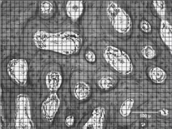
captured with a digital camera (CCD IRIS/RGB, Sony Corp., Tokyo, Japan) coupled with the micro-scope and connected to a computer and were ana-lyzed using Image J 1.34s image-analysis software (National Institute of Health, Bethesda, Mary-land, USA). A 1,200-point grid with a total area of 694 x 520 µm was positioned onto each digitized image. The number of intersection points of the hor-izontal and vertical lines of the grid on the image of the bone tissue (not in the medullar spaces) was counted. The ratio between the number of points in the bone tissue and the total number of points of the grid (1,200) allowed for the determination of the bone density of the analyzed tissue (Figure 2).
The bone density data from the control and alen-dronate-treated groups were compared and statisti-cally analyzed by the Student’s t-test at 5% signii-cance level using SAS software (v.3.43; SAS Institute Inc., Cary, North Carolina, USA).
Results
The mean bone density values for each ani-mal and for each group are represented in Table 1. There was no statistically signiicant difference (p = 0.3754) in the alveolar bone density between the control and alendronate-treated animals (Table 1 and Graph 1). The groups were homogeneous with an F = 0.89 (homogeneity is considered if F > 0.05).
Figure 2 - 1,200-point grid positioned onto each digitized image of a histological section. The number of intersection points of the horizontal and vertical lines of the grid in the bone tissue (not in the medullar spaces) was counted.
Discussion
The effects of the bisphosphonates occur at both tissue and cellular levels.4 At a tissue level, these compounds promote a decrease of bone turnover,4 as demonstrated by biochemical markers,16,17,18 show-ing a reduction in the extension of the bone resorp-tion areas and the depth of the resorbed areas. At a cellular level, bisphosphonates act inhibiting the activity and recruitment of osteoclasts to the bone surface, inducing the apoptosis of these cells and al-tering the mineral exchange.4 The bisphosphonates incorporated into the bone are released in How-ship’s lacunae and internalized in the osteoclasts by endocytosis, affecting these cells directly. After this phenomenon, the osteoclasts lose their rufled bor-der and their cytoskeleton is progressively disrupt-ed, inactivating the resorptive activity of these cells and inducing apoptosis(Figure 1).19 This process is due to the inhibition of the enzyme farnesyl diphos-phate synthase,20,21 especially by the administration
of nitrogen-containing bisphosphonates, such as alendronate. This enzyme is necessary for the pre-nylation of small GTPases,22 which are responsible for the signaling proteins that control morphology, cytoskeleton arrangement, rufled borders, vesicle transportation and apoptosis of osteoclasts.23
It is known that the systemic effects of bisphos-phonates are not immediate4 and that, in spite of their recognized inhibitory effect of calciication in
vitro, a prolonged administration of these drugs is
necessary to produce an effective inhibition of bone resorption.
Bisphosphonates are incorporated into miner-alized structures. After bone uptake, the bisphos-phonates are liberated again only when the bone in which they are deposited is resorbed. Under patho-logical conditions, alveolar bone loss is related to alterations in the normal balance between bone for-mation and resorption. Therapeutic agents that are capable of avoiding bone resorption and accelera-tion of bone deposiaccelera-tion may act in preventing bone loss in patients with conditions such as periodontal disease.
The effects of bisphosphonates in controlling alveolar bone loss in advanced periodontal disease and their action in the inlammatory processes asso-ciated with periodontal breakdown have been inves-tigated in rat experimental models.24 The effects of bisphosphonates in controlling alveolar bone loss in advanced periodontal disease and their action in the inlammatory processes associated with periodontal breakdown have also been examined with the induc-tion of periodontal disease by Porphyromonas
gingi-valis25 in monkeys26 and dogs.27 The results of these
studies conirm that bisphosphonates were more effective preventing alveolar bone loss in animals with experimentally induced periodontal disease. When large bone tissue destruction was present, the groups treated with bisphosphonates exhibited less bone loss and a greater amount of remaining alveo-Table 1 - Mean bone density values (bone areas/non-bone areas ratio) in each animal and each group.
Group 1 2 3 4 5 6 7 8 9 Total
Control 92.22 81.43 85.31 72.21 77.10 76.86 83.56 59.49 83.28 79.05 ± 9.31
Alendronate 73.70 61.87 79.96 84.67 82.08 61.94 79.30 64.80 86.24 74.95 ± 9.75
B
o
ne
dens
ity
100%
90% ± 9.31
80%
70%
60%
50%
40%
30%
20%
10%
0%
79.05 74.95
Control Alendronate
Experimental groups ± 9.75
lar bone. However, the microscopic results showed no differences in the mandibles of the animals with little or no periodontal disease compared to the healthy control animals. In the same way, a previous study evaluating the post-extraction bone repair in rat teeth showed that the administration of dichlo-romethylene bisphosphonate (clodronate) inhibited the occurrence of rapid alveolar bone loss after ex-traction. This effect was observed exclusively in the surgical wounds and no differences were found be-tween control and experimental animals in the in-tact bone areas.28
In this way, studies investigating the effects of bisphosphonates on inlammatory process and al-veolar repair seem to demonstrate that these drugs do not microscopically alter the bone structure in healthy areas, corroborating the results of the pres-ent study in which no difference was found in the bone density of control and experimental animals (Table 1 and Graph 1). Bisphosphonates bind pref-erentially to bones that have high turnover rates, and their distribution in the bone tissue is not ho-mogeneous.4,5 Therefore, it may be inferred that the incorporation of bisphosphonates to maxillary bones with no pathological processes would provide a longer period of time for a structural organiza-tion, without compromising or altering their archi-tecture.
The analysis by light microscopy did not show any effects of the alendronate on the composition of the mineralized bone matrix in either the control or experimental groups. Morphologically, the char-acteristics of the bone tissue were similar in both groups with respect to the distribution and number of reversion lines, proportion of medullar spaces, bone trabeculae and cortical bone. Similar distribu-tion and number of osteoclasts and bone remodeling units in the analyzed bone structures was also ob-served in both groups (Figure 3).
The therapeutic effects of bisphosphonates do not depend on the inhibition of normal bone re-modeling, but is associated, rather, with pathologi-cal bone resorption or with regions with high bone turnover.29 Therefore, based on the indings of the present study, no complications in dental treatments, such as orthodontic movement, dental extractions or implant placement, are expected because these procedures are usually performed in healthy bone tissue.16
In the present study, there was no difference in the bone density between the animals that received alendronate and the non-treated control animals. It was not possible to determine, using optical micros-copy, any effect of the drug in the composition of the mineralized bone matrix in either of the groups. Morphologically, the alveolar bone in the animals
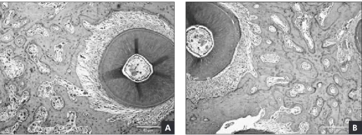
Figure 3 - Alveolar bone in the alendronate-treated (A) and control (B) group. The bone tissue characteristics were similar in both groups with respect to the distribution and number of reversion lines, and proportion of medullar spaces, bone trabeculae and cortical bone. The distribution and number of osteoclasts and bone remodeling units in the analyzed bone structures was
also similar in both groups (Hematoxylin and eosin - HE; ×40 magnification).
treated with alendronate had the same proportions of medullar spaces and bone trabeculae as those of the control animals.
Conclusion
No statistically signiicant difference in alveo-lar bone density between the animals that received bisphosphonate (alendronate) and control animals was found.
Histomorphometric analysis using an optical mi-croscope was unable to determine any effect of so-dium alendronate in the composition of mineralized bone matrix between the control and experimental groups. Morphologically, the bone in the experi-mental and control groups showed comparatively
the same proportion between medullar spaces and bone tissue.
Acknowledgements
This study was supported by a grant-in-aid from the São Paulo State Research Foundation (FAPESP) and a research postgraduate scholarship granted by the Brazilian Government Research Funding Agen-cy CAPES. The authors are indebted to the Depart-ment of Physiology of Ribeirão Preto Dental School, University of São Paulo, Brazil, and to the histotech-nician MS. Fátima Aparecida Silveira from the Ser-vice Pathology of Bauru Dental School, University of São Paulo, Brazil.
References
1. Fleisch H, Russell RG, Straumann F. Effect of pyrophosphate on hydroxyapatite and its implication in calcium homeostasis. Nature. 1966 Nov 26; 212(5065):901-3.
2. Bisaz S, Russell RG, Fleisch H. Isolation of inorganic pyro-phosphate from bovine and human teeth. Arch Oral Biol. 1968 Jun;13(6):683-96.
3. Fleisch H, Russell RG, Francis MD. Diphosphonates inhibit hydroxyapatite dissolution in vitro and bone resorption in tis-sue culture and in vivo. Science. 1969 Sep 19;165(899):1262-4.
4. Fleisch H. Bisphosphonates: mechanisms of action. Endocr Rev. 1998 Feb;19(1):80-100.
5. Rodan GA. Mechanisms of action of bisphosphonates. Annu Rev Pharmacol Toxicol. 1998;38:375-88.
6. Lipton A, Theriault RL, Hortobagyi GN, Simeone J, Knight RD, Mellars K, et al. Pamidronate prevents skeletal compli-cations and is effective palliative treatment in women with breast carcinoma and osteolytic bone metastases: long term follow-up of two randomized, placebo-controlled trials. Can-cer. 2000 Mar 1;88(5):1082-90.
7. Berenson JR, Rosen LS, Howell A, Porter L, Coleman RE, Morley W, et al. Zoledronic acid reduces skeletal-related events in patients with osteolytic metastases. Cancer. 2001 Apr 1;91(7):1191-200.
8. Woo SB, Hellstein JW, Kalmar JR. Narrative [corrected] re-view: bisphosphonates and osteonecrosis of the jaws. Ann Intern Med. 2006 May 16;144(10):753-61.
9. Roelofs AJ, Thompson K, Gordon S, Rogers MJ. Molecular mechanisms of action of bisphosphonates: current status. Clin Cancer Res. 2006 Oct 15;12(20 Pt 2):6222s-30s.
10. Hughes DE, Macdonald BR, Russell RG, Gowen M. Inhibi-tion of Osteoclasts-like cell formaInhibi-tion by bisphosphonates
in long-term cultures of human bone marrow. J Clin Invest. 1989 Jun;83(6):1930-5.
11. Vitté C, Fleisch H, Guenther HL. Bisphosphonates induce osteoblasts to secrete an inhibitor of osteoclast-mediated re-sorption. Endocrinology. 1996 Jun;137(6):2324-33. 12. Anbinder AL, Prado FA, Prado MA, Balducci I, Rocha RF.
The influence of ovariectomy, simvastatin and sodium alen-dronate on alveolar bone in rats. Braz Oral Res. 2007 Jul-Sep;21(3):247-52.
13. Chaiamnuay S, Saag KG. Postmenopausal osteoporosis. What have we learned since introduction of bisphosphonates? Rev Endocr Metab Disord. 2006 Jun;7(1-2):101-12.
14. Farris EJ, Griffith JQ. The rat in laboratory investigation. 2nd ed. Philadelphia: Lippincott Company; 1963. Chapter 1,
Breeding of the rat;p. 1-17.
15. Keidel WD. Physiology. 2nd ed. Barcelona: Salvat; 1971.
16. Ott SM. Clinical effects of bisphosphonates in involutional osteoporosis. J Bone Miner Res. 1993 Dec;8 Suppl 2:S597-606.
17. Storm T, Steiniche T, Thamsborg G, Melsen F. Changes in bone histomorphometry after long-term treatment with inter-mittent, cyclic etidronate for postmenopausal osteoporosis. J Bone Miner Res. 1993 Feb; 8(2):199-208.
18. Boyce RW, Paddock CL, Gleason JR, Sletsema WK, Eriksen EF. The effects of risedronate on canine cancellous bone re-modeling: three-dimensional kinetic reconstruction of the remodeling site. J Bone Minerv Res. 1995 Feb;10(2):211-21. 19. Russell RG, Rogers MJ. Bisphosphonates: from the laboratry
to the clinic and back again. Bone. 1999 Jun; 25(1):97-106. 20. Van Beek E, Pieterman E, Cohen L, Löwik C, Papapoulos S.
nitrogen-containing bisphosphonates. Biochem Biophys Res Commun. 1999 Oct 14; 264(1):108-11.
21. Russell RG. Bisphosphonates: mode of action and pharmacol-ogy. Pediatrics. 2007 Mar;119 Suppl 2:S150-62.
22. Zhang FL, Casey PJ. Protein prenylation: molecular mech-anisms and functional consequences. Annu Rev Biochem. 1996;65:241-69.
23. Zhang D, Udagawa N, Nakamura I, Murakami H, Saito S, Yamasaki K, et al. The small GTP-biding protein, Rho p21,
is involved in bone resorption by regulating cytoskeletal or-ganization in osteoclasts. J Cell Sci. 1995 Jun;108(Pt6):2285-92.
24. Fernandes MI, Gaio EJ, Oppermann RV, Rados PV, Rosing CK. Comparison of histometric and morphometric analyses of bone height in ligature-induced periodontitis in rats. Braz Oral Res. 2007 Jul-Sep;21(3):216-21.
25. Tani-Ishii N, Minamida G, Saitoh D, Chieda K, Omuro H, Sugaya A, et al. Inhibitory effects of incadronate on the
pro-gression of rat experimental periodontitis by Porphyromonas
gingivalis infection. J Periodontol. 2003 May; 74(5):603-9.
26. Weinreb M, Quartuccio H, Seedor JG, Aufdemorte TB, Brunsvold M, Chaves E, et al. Histomorphometrical analysis of the effects of the bisphosphonate alendronate on bone loss caused by experimental periodontitis in monkey. J Periodont Res. 1994 Jan;29(1):35-40.
27. Reddy MS, Weatherford TW 3rd, Smith CA, West BD, Jef-fcoat MK, Jacks TM. Alendronate treatment of naturally-occurring periodontitis in beagle dogs.J Periodontol. 1995 Mar;66(3):211-7.
28. Olson HM, Hagen A. Inhibition of post-extraction alveolar ridge resorption in rats by dichloromethane diphosphonate. J Periodontal Res. 1982 Nov;17(6):669-74.