Oral Pathology
Joelma Sousa Lima(a) Décio dos Santos Pinto Jr(a) Suzana Orsini Machado de Sousa(a)
Luciana Corrêa(b)
(a)Oral Pathology Department, School of Dentistry, Univ of São Paulo, São Paulo, SP, Brazil.
(b)General Pathology Department, School of Dentistry, Univ of São Paulo, São Paulo, SP, Brazil.
Corresponding Author: Joelma Sousa Lima E-mail:[email protected]
Oral leukoplakia manifests differently
in smokers and non-smokers
Abstract: Oral leukoplakias (OL) are potentially malignant lesions that are typically white in color. Smoking is considered a risk factor for de-veloping OL, and dysplastic lesions are more prone to malignant trans-formation. The aim of this study was to describe the clinical features observed in dysplastic and dysplastic OL in both smokers and non-smokers. A total of 315 cases of OL were retrieved and separated into either dysplastic or non-dysplastic lesions, and these cases were further categorized as originating in either smokers or non-smokers. Frequencies of the type of OL lesion, with respect to whether the patients smoked, were established. The results demonstrated that 131 cases of OL were dysplastic (74 smokers and 57 non-smokers), and 184 were non-dysplas-tic (96 smokers and 88 non-smokers). For OL cases in smokers for which information about alcohol consumption was also available (84 cases), the results revealed no signiicant difference in the amount of dysplas-tic and non-dysplasdysplas-tic lesions. Dysplasdysplas-tic lesions were more frequent in male smokers and in non-smoking females. The median age of smokers with cases of OL was signiicantly lower than in non-smokers; the low-est median ages were observed for female smokers with dysplastic OL. The most frequent anatomical sites of dysplastic lesions were the loor of the mouth in smokers and the tongue in non-smokers. Dysplastic le-sions in smokers were signiicantly smaller than non-dysplastic lele-sions in non-smokers. Being a male smoker, being female, being younger, and having smaller lesions were associated with dysplastic features in OL. These clinical data may be important for predicting OL malignant trans-formation.
Descriptors: Leukoplakia, Oral; Risk Factors; Tobacco.
Introduction
Oral leukoplakia (OL) is a clinical entity deined by the Word Health Organization (WHO) as “a white plaque of questionable risk having ex-cluded (other) known diseases or disorders that carry no increased risk for cancer.”1 This type of lesion affects 0.2% to 4.9% of the population.2 The etiology of OL is variable, and tobacco has been considered a risk factor in a large number of cases, while alcohol, as an independent risk factor, is still questionable.2-7 The concept of malignant transformation of OL is quite controversial. Studies have indicated that there is a 36% chance of malignant transformation if the lesion exhibits dysplastic fea-tures.2,7 However, when lesions with dysplastic and non-dysplastic
fea-Declaration of Interests: The authors certify that they have no commercial or associative interest that represents a conflict of interest in connection with the manuscript.
Submitted: May 17, 2012
nant transformation decreases to 0.13% to 17.5%.5,8 Recent studies have demonstrated that the inclusion of benign alveolar ridge keratosis as a type of OL causes a deviation in the reported percentage of ma-lignant transformation. However, when benign alve-olar ridge keratosis is excluded from the OL group, the frequency of dysplasia transforming into squa-mous cell carcinoma increases from 18% to 25%.9 Differences in the rates of malignant transformation are also attributed to variations in the criteria used to deine OL, such as differences in ethnic and en-vironmental factors.9,10 Risk factors for malignant transformation of OL include being female, having a long duration of OL, having a lesion size greater than 200 mm2, being a non-smoker, having a lesion located on the tongue or the loor of the mouth, hav-ing a lesion of a non-homogeneous type, presenthav-ing with dysplasia and developing DNA aneuploidy.8,11
The histopathological pattern of OL is variable, ranging from atrophy of the epithelium to hyperpla-sia, either with or without dysplasia. Epithelial dys-plasia, if present, exhibits different degrees of sever-ity.1,3,8,11
It has been shown that there are differences re-garding clinical data when comparing dysplastic and non-dysplastic lesions, mainly in relation to gen-der, age, anatomical site, alcohol consumption and smoking habits.11-13 Therefore, the aim of this retro-spective study was to describe the clinical features observed in dysplastic and non-dysplastic OL lesions in both smokers and non-smokers. The purpose of this study was to detect clinical variables observed in OL that could be associated with smoking habits and/or that might be predictors of dysplastic lesions.
Methodology
All OL cases diagnosed between 2005 and 2009 were retrieved from the iles of the Oral Pathology Service at the University of São Paulo. Research was conducted in accordance with protocol 184/2010, approved by the Ethics Committee at the University of São Paulo. Clinical criteria for OL diagnosis were the same as those adopted by the WHO.1 Lesions for which clinical history and anatomical sites were indicative of benign alveolar ridge keratosis were
excluded. Patients included in the study as smokers were those who had been smoking for at least one year and were presently still smoking. Non-smok-ers were patients who reported that they had never smoked.4 Patients were considered “alcohol consum-ers” if they presented this chronic habit by the time of the biopsy, and “non-alcohol consumers” if they reported that they never had an alcohol habit. Ad-ditionally, based on the histopathological features of the lesions, cases were divided into “dysplastic” (without distinction as to the severity of each dys-plasia) and “non-dysplastic” (Figures 1 and 2).6 This method of subdivision was adopted because the risk of malignant transformation is higher for dysplastic lesions8,11 and because there can be differences in the clinical data when comparing dysplastic and non-dysplastic lesions.12 Any OL that was histologically diagnosed as a speciic disease was not considered in this study.
The data obtained in this study included gender, age, and ethnicity of the patients. For each lesion, the diameter (mm), the duration (months), and the anatomical site were also included. The designation “White” refers to patients of Caucasian origin and their descendants, mainly Europeans; “Black” signi-ies African people and their descendants; and “Yel-low” includes Japanese, Korean, and Chinese people and their descendants. “Other ethnicity” refers to patients presenting a mixture of races. Lesions that had been present for 30 days or less were grouped as having been present for “one month.” With regard
to anatomical site, the term “tongue” was adopted for both the lateral border and the other tongue re-gions. The term “palate” includes both the soft and hard palates.
Statistical analysis
The frequencies of each variable were evalu-ated with descriptive statistics. The association between the variables in each group was analyzed using the Chi-square test. For numerical data, the Mann-Whitney U-test was applied. Estimation of the smokers-versus-nonsmokers odds ratio was performed for males and for the most frequent ana-tomical site. The calculations were performed using BioEstat 3.0 (developed by Manuel Ayres, Belém, Brazil) and Microsoft Excel (Microsoft Corpora-tion, Santa Rosa, USA). Data were considered sig-niicant when p < 0.05.
Results
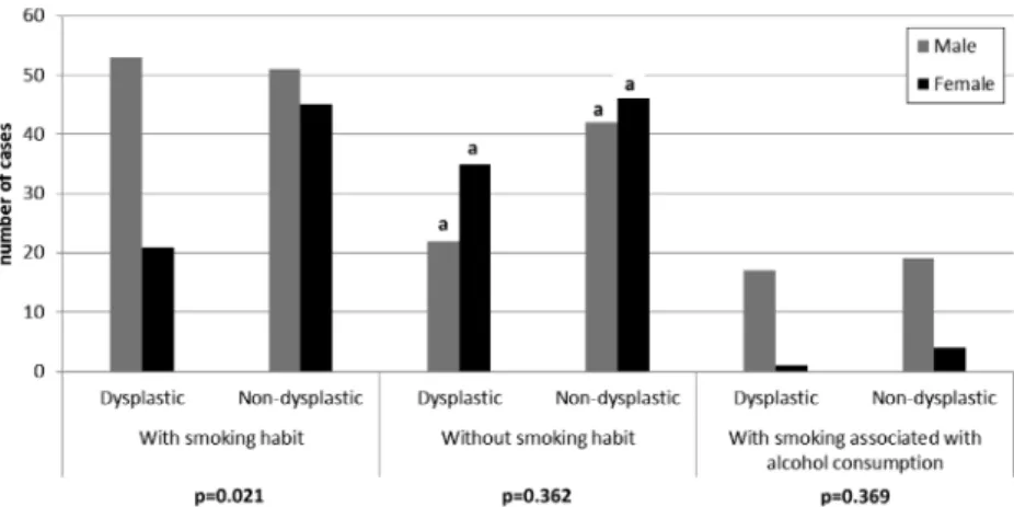
A total of 315 cases of OL were retrieved from the Oral Pathology Service at the University of São Paulo. A total of 131 cases involved dysplastic le-sions (74 smokers and 57 non-smokers), and 184 cases described non-dysplastic lesions (96 smokers and 88 non-smokers). The frequencies of lesions, according to the grade of dysplasia, were similar in both groups. The Chi-square test indicated that there was no association between the frequency of dysplastic/non-dysplastic lesions and smoking habits (p = 0.520) or alcohol consumption (p = 0.501) (Fig-ure 1). However, for some variables, such as gender, age, anatomical site of the lesion, and lesion size,
some signiicant associations were found between the smoking habits of the patient and the presence or absence of dysplasia (Figure 2).
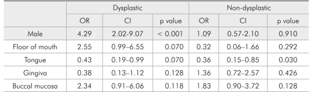
Smokers presented a signiicantly higher fre-quency of dysplastic lesions in males than in females (p = 0.021). Among non-smokers, females showed a signiicantly higher frequency of dysplastic lesions than men (p < 0.01). Considering only non-dysplas-tic lesions, there was no signiicant association be-tween a smoking habit and gender (p = 0.559). When alcohol consumption was considered, there were also no statistically signiicant differences between dysplastic and non-dysplastic lesions in women and men (Figure 2). The odds ratio for gender and smok-ing indicates that male smokers have a four-times greater chance of developing dysplastic lesions than do male non-smokers (Table 1).
The median age in the group of smokers was 55 years for non-dysplastic lesions and 56.5 years for dysplastic lesions. In the group of non-smokers, the median age was 63 years for both dysplastic and non-dysplastic lesions. The median age in smokers was signiicantly lower than in non-smokers (dysplastic lesions, p = 0.025; non-dysplastic lesions, p = 0.004). Considering the group of smokers with dysplastic le-sions, women showed a signiicantly lower median age in comparison with men (p = 0.032). In contrast, women in the group of non-smokers presented a sig-niicantly higher median age in comparison with men for both dysplastic (p = 0.001) and non-dysplastic le-sions (p = 0.010) (Table 2).
Table 3 shows the frequencies of dysplastic and non-dysplastic lesions along with the smoking
hab-Figure 2 - Frequency of dysplastic and non-dysplastic lesions according to smoking habit, alcohol consumption and gender. (Chi-Square test;
it of the patient and the anatomical site of the le-sion. Among smokers, the most frequent anatomical sites were the loor of the mouth and the gingiva for dysplastic and non-dysplastic lesions, respectively. Among non-smokers, the most frequent sites were the tongue for dysplastic lesions and the gingiva for non-dysplastic lesions. For smokers, a signiicant as-sociation between anatomical site and dysplastic/ non-dysplastic lesions was observed (p < 0.001). For non-smokers, there was no signiicant associa-tion between lesion frequency and anatomical site (p = 0.104). However, the non-smoking patients had a greater chance to develop OL on the tongue (odds
ratio = 0.364 and p = 0.03) (Table 1).
Having a smoking habit correlated with lesions on the loor of the mouth and the buccal mucosa in the cases of dysplasia, but these associations were not statistically signiicant. Non-dysplastic lesions in smokers were more frequent in the gingiva and buccal mucosa than at other sites, but this was also without signiicance (Table 3). The frequencies of dysplastic and non-dysplastic lesions with respect to smoking and ethnicity are shown in Figure 3. Whites were most affected in all groups. Due to the low frequency of other races in this study, there was no signiicant association between smoking habits,
Table 2 - Median age (minimum and maximum values) of smoking and non-smoking patients with dysplastic and non-dysplastic lesions in general, and in male and female patients.
Smoker Non-smoker
p value Smoker p value Non-smoker p value
General General Male Female Male Female
Dysplastic 56.5 (28–84) 63 (40–83) 0.025 58 (41–84) 53.5 (28–83) 0.032 50.5 (40–83) 67 (42–82) 0.001 Non-dysplastic 55 (28–87) 63 (32–89) 0.004 54 (33–87) 56 (28–79) 0.877 57 (32–86) 64 (34–89) 0.010
Mann-Whitney U-test. Statistically significant when p < 0.05.
Dysplastic Non-dysplastic
OR CI p value OR CI p value
Male 4.29 2.02-9.07 < 0.001 1.09 0.57-2.10 0.910 Floor of mouth 2.55 0.99–6.55 0.070 0.32 0.06–1.66 0.292 Tongue 0.43 0.19–0.99 0.070 0.36 0.15–0.85 0.030 Gingiva 0.38 0.13–1.12 0.128 1.36 0.72–2.57 0.426 Buccal mucosa 2.34 0.91–6.06 0.118 1.83 0.90–3.72 0.128
OR – odds ratio. CI – confidence interval. Statistically significant when p < 0.05.
smoker odds ratio for males and the most frequent anatomical sites for dysplastic and
non-dysplastic lesions.
Smoker Non-smoker
Dysplastic
(%) Non-dysplastic (%) p value Dysplastic (%) Non-dysplastic (%) p value Floor of mouth 18 (24.3) 2 (2.1)
< 0.001
8 (14.0) 6 (6.8)
0.104 Tongue 14 (18.9) 9 (9.4) 22 (38.6) 21 (23.9)
Gingiva 6 (8.1) 38 (39.6) 12 (21.1) 33 (37.5) Buccal mucosa 17 (23.0) 27 (28.1) 8 (14.0) 18 (20.5) Palate 15 (20.3) 12 (12.5) 2 (3.5) 1 (1.1) Multiple sites 4 (5.4) 3 (3.1) 3 (5.3) 4 (4.5) WI 0 (0.0) 5 (5.2) 2 (3.5) 5 (5.7) Total 74 (100.0) 96 (100.0) 57 (100.0) 88 (100.0)
WI – without information. Chi-square test. Statistically significant when p < 0.05.
dysplastic/non-dysplastic lesions, and ethnicity (Fig-ure 3).
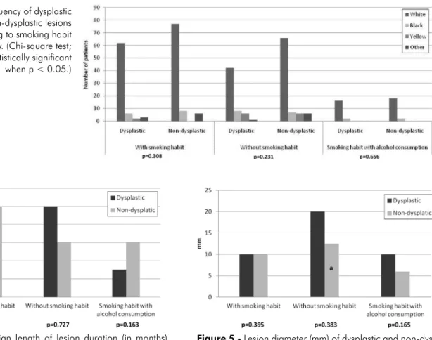
The median duration of lesions in smokers was 5 months for dysplastic lesions and 10 months for non-dysplastic lesions (Figure 4). In non-smokers, the median duration was 10 months for dysplastic lesions and 6 months for non-dysplastic lesions. The duration of the lesion had no signiicant association with smoking habits or histological type (Figure 4), most likely due to the great amount of variability in these data.
The median size of the lesions from patients in this study is shown in Figure 5. For the smoker group, the median diameter of lesions was 10 mm for both dysplastic and non-dysplastic lesions. In the non-smoker group, the median diameters were 20 mm for dysplastic and 12.5 mm for non-dys-plastic lesions. There was no signiicant difference between dysplastic and non-dysplastic lesion size in either smokers or non-smokers (Figure 5).
Statisti-cal signiicance was observed only when comparing dysplastic lesions in smokers with non-dysplastic le-sions in non-smokers; this indicated that the lele-sions in non-smokers, when non-dysplastic, were signii-cantly larger than the dysplastic lesions in smokers (p = 0.006).
Discussion
This study focused on clinical data concerning the occurrence of dysplastic and non-dysplastic OL in smokers and non-smokers. Smoking has been cor-related with a high frequency of OL in the oral cavi-ty,3,4,7 and clinical information about patient gender, the anatomical site of the lesion, the duration of the lesion, and the size of the lesion can be indicative of the potential a lesion has for undergoing malig-nant transformation.8 The most important indings of this study were the associations between
dysplas-Figure 3 - Frequency of dysplastic and non-dysplastic lesions according to smoking habit and ethnicity. (Chi-square test; statistically significant when p < 0.05.)
Figure 5 - Lesion diameter (mm) of dysplastic and non-dys-plastic lesions according to smoking habit. (Mann-Whitney U-test; statistically significant when p < 0.05; a = statis-tically significant when compared with dysplastic lesion in smokers.)
lesions and non-smoking women. Additionally, the high frequency of OL on the loor of the mouth, in the gingiva, and in the buccal mucosa of smokers was statistically signiicant. These results conirmed reports described in the literature indicating that OL in smokers is associated with male gender and with the loor of the mouth and the buccal mucosa, espe-cially when there is also a habit of alcohol consump-tion.7 However, in the present study, there was no correlation observed between alcohol consumption and dysplasia. Additionally, in non-smokers, OL without dysplasia was likely to develop in women on the tongue. These associations may be important for predicting the risk of dysplastic OL to undergo malignant transformation based only on clinical cri-teria. This method of risk assessment will prompt pathologists to conduct a careful histopathological diagnosis.
Although the main risk factors for OL are be-ing male and smokbe-ing, in this study, we noted a moderate frequency of female non-smokers with dysplastic and non-dysplastic lesions. An increas-ing frequency of OL has been described in women who do not smoke or consume alcohol,14 suggesting the existence of other risk factors for premalignant lesions that show a high potential for malignancy. In the present study, we found no signiicant asso-ciation between women, smoking, and dysplastic/ non-dysplastic lesions, except when this informa-tion was crossed with the age of the patient. In this case, smoking women with dysplastic lesions had a median age lower than that of smoking men, but non-smoking women had a higher median age than non-smoking men. These indings may indicate that, although not high, the percentage of cases in smok-ing women could predict the malignant potential of lesions. Therefore, being younger, being female, smoking, and having dysplastic lesions are consid-ered risk factors for malignant transformation of OL lesions.8,11
Regarding the patient age of lesion incidence, independent of patient gender, the median age in smokers was lower than in non-smokers for both dysplastic and non-dysplastic lesions. The median ages were between 51 and 60 years and between
spectively. This distribution of ages was similar to that described in other reports.11,14,15 However, the Asian population presents a different age distribu-tion for OL. In a Chinese survey that did not record the tobacco habits of patients, OL was considered a disease of the elderly, being present mostly in 60- to 69-year-old patients.10 It is dificult to establish an at-risk age for OL development, mainly due to the multiple independent factors that are associated with age and OL, such as chewing betel quid10,15 and whether the patient has type 2 diabetes.16,17 In our sample, patients 61 years old and above were mainly non-smoking women. As stated above, factors other than smoking may be associated with OL in this group of patients with more advanced age.
lesion size and tobacco use in oral epithelial dyspla-sia is not substantially supported in the literature. Some studies have not established an association be-tween lesion size and clinical aspects and the risk of malignant transformation.7,18
The results of this study showed that gender, age, lesion size, and anatomical site correlate with a smoking habit and dysplastic lesions. Men aged between 51 and 60 years of age and lesions on the loor of the mouth and in the buccal mucosa were the most frequent predictors of dysplasia in smok-ers. In non-smokers, dysplastic lesions were more frequent in women. Although the present study
showed that the localization of lesions on the tongue of smokers may indicate the presence of non-dysplastic lesions, it is known that white lesions lo-cated on the tongue and the loor of mouth are more likely than lesions in the buccal mucosa to undergo malignant transformation.
Conclusion
The study shows that the anatomical site of OL as well as gender and the presence of epithelial dys-plasia should be considered when treating a lesion due to the possibility of a malignant transformation of OL.
References
1. Warnakulasuriya S, Johnson NW, van der Waal I. Nomencla-ture and classification of potentially malignant disorders of the oral mucosa. J Oral Pathol Med. 2007 Nov;36(10):575-80. 2. Silverman S, Jr., Gorsky M, Lozada F. Oral leukoplakia and
malignant transformation. A follow-up study of 257 patients. Cancer. 1984 Feb 1;53(3):563-8.
3. Chiu CT, Li CF, Li JR, Wang J, Chuang CY, Huang SC, et al. Candida invasion and influences in smoking patients with multiple oral leucoplakias-a retrospective study. Mycoses. 2011 Sep;54(5):e377-83.
4. Lee JJ, Hung HC, Cheng SJ, Chen YJ, Chiang CP, Liu BY, et al. Carcinoma and dysplasia in oral leukoplakias in Taiwan: prevalence and risk factors. Oral Surg Oral Med Oral Pathol Oral Radiol Endod. 2006 Apr;101(4):472-80.
5. Cerero-Lapiedra R, Balade-Martinez D, Moreno-Lopez LA, Esparza-Gómez G, Bagán JV. Proliferative verrucous leuko-plakia: a proposal for diagnostic criteria. Med Oral Patol Oral Cir Bucal. 2010 Nov 1;15(6):839-45.
6. van der Waal I, Schepman KP, van der Meij EH, Smeele LE. Oral leukoplakia: a clinicopathological review. Oral Oncol. 1997 Sep;33(5):291-301.
7. Schepman KP, van der Meij EH, Smeele LE, van der Waal I. Malignant transformation of oral leukoplakia: a follow-up study of a hospital-based population of 166 patients with oral leukoplakia from The Netherlands. Oral Oncol. 1998 Jul;34(4):270-5.
8. van der Waal I. Potentially malignant disorders of the oral and oropharyngeal mucosa; present concepts of management. Oral Oncol. 2010 Jun;46(6):423-5.
9. Woo SB, Lin D. Morsicatio mucosae oris--a chronic oral fric-tional keratosis, not a leukoplakia. J Oral Maxillofac Surg. 2009 Jan;67(1):140-6.
10. Zhang X, Li C, Song Y, Reichart PA. Oral leukoplakia in China: a review. Oral Maxillofac Surg. 2010 Dec;14(4):195-202.
11. Jaber MA. Oral epithelial dysplasia in non-users of tobacco and alcohol: an analysis of clinicopathologic characteristics and treatment outcome. J Oral Sci. 2010 Mar;52(1):13-21. 12. Chi AC, Lambert PR 3rd, Pan Y, Li R, Vo DT, Edwards E,
et al. Is alveolar ridge keratosis a true leukoplakia? A clini-copathologic comparison of 2,153 lesions. J Am Dent Assoc. 2007 May;138(5):641-51.
13. Hamadah O, Goodson ML, Thomson PJ. Clinicopathologi-cal behaviour of multiple oral dysplastic lesions compared with that of single lesions. Br J Oral Maxillofac Surg. 2010 Oct;48(7):503-6.
14. Schepman KP, Bezemer PD, van der Meij EH, Smeele LE, van der Waal I. Tobacco usage in relation to the anatomical site of oral leukoplakia. Oral Dis. 2001 Jan;7(1):25-27.
15. Freitas MD, Blanco-Carrion A, Gandara-Vila P, Antúnez-López J, García-García A, Gándara Rey JM. Clinicopatho-logic aspects of oral leukoplakia in smokers and nonsmokers. Oral Surg Oral Med Oral Pathol Oral Radiol Endod. 2006 Aug;102(2):199-203.
16. Lee CH, Ko YC, Huang HL, Chao YY, Tsai CC, Shieh TY, et al. The precancer risk of betel quid chewing, tobacco use and alcohol consumption in oral leukoplakia and oral sub-mucous fibrosis in southern Taiwan. Br J Cancer. 2003 Fev 10;88(3):366-72.
17. Dikshit RP, Ramadas K, Hashibe M, Thomas G, Somanathan T, Sankaranarayanan R. Association between diabetes mel-litus and pre-malignant oral diseases: a cross sectional study in Kerala, India. Int J Cancer. 2006 Jan;118(7):453-7. 18. Meisel P, Dau M, Sumnig W, Holtfreter B, Houshmand M,