Introduction
The analysis of COP and joint position sense
in university soccer players with and without ankle instability
CDD. 20.ed. 796.023 796.024 796.33
http://dx.doi.org/10.1590/1807-55092016000300601
Antônio Francisco de ALMEIDA NETO* Alex CASTRO* Luciano Fernandes CROZARA* Márcio Fagundes GOETHEL* Pedro Vieira Sarmet MOREIRA* Mauro GONÇALVES* Adalgiso Coscrato CARDOZO*
*Instituto de Biociên-cias, Universidade Estadual Paulista, Rio Claro, SP, Brasil.
The soccer is one of the most popular sport modality around the world, with millions of practitioners of different levels, and thus, the incidence of injuries are expressive1-3. Among these injuries, the ankle sprain is the one that stands out2, 4-5.
Th e ankle sprains are very common in soccer due to the demand of changes in direction performed with high velocities, after jumps and during the sprints itself2, 4, 6. Th ese rapid changes in direction or landing on irregular surfaces produces a large supination torque, causing the ankle to perform an excessive movement, overloading the joint and its
structures, mainly the anterior talo-fi bular and the calcaneo-fi bular ligaments7.
Repeated episodes of ankle sprains may negatively aff ect the proprioception, which correspond to the perception of position and movement (i.e., synesthesia) of the body and its segments8-9. Th ese information are provided by mechanoreceptors in the muscles, tendons, skin, joint capsules and ligaments10-12. Th ese mechanoreceptors are sensitized by mechanics energy imposed to the joint and it is transmitted to the central nervous system by aff erents impulses13. As a consequence to sprains, the aff erent fi bers of the
Abstract
The aim of the study was to compare the behavior of COP and passive ankle position sense in subjects with and without functional ankle instability. Took part in this study 20 subjects, divided into two groups: stable group (SG) and unstable group (UG). The COP evaluation was made with the single-leg balance test, with eyes opened and closed, on a force plate. The passive ankle position sense test was performed with subjects blindfolded. The ankle was positioned in a target angle (10° and 20°) and the dynamometer moved passively the ankle, then the subjects were instructed to push the stop button when they feel that the ankle was on the target angle, obtaining the absolute angular error (AAE). The following variables were obtained: total displacement (TD); antero-posterior (SDap) and medio-lateral standard deviation (SDml); total mean velocity (TMV); antero-posterior (MVap) and medio-lateral mean velocity (MVml). The comparison between the data with normal distribution was made with the Student’s t test, while to the TD and SDml was used the Mann-Whitney test. The correlations were performed with the Pearson and Spearman tests. We adopted α < 0.05. We observed difference between AAE-10º (p < 0.05). Strong
correlations were found between: AAE-10° and TMV (p < 0.01 r = -0.867); AAE-10° and MVap (p < 0.01 r = -0.854); AAE-10° and MVml (p < 0.01 r = -0.771), with eyes opened, and AAE-10° and TD (p < 0.05 r = -0.666); AAE-10º and SDap (p < 0.05 r = -0,685) and AAE-10° and MVml (p < 0.05 r= -0.766) with eyes closed. Ankle sprains harm the joint position sense without affecting the balance.
mechanoreceptors joints become damaged, impairing particularly the joint position sense, resulting in a situation known as the functional ankle instability (FAI)14-16, which is defi ned by Freeman17 as a complain of “false subjective perception”. Hertel18 attributed its causes to defi cits in the joint position sense, reduced muscle strength, delay in fi bular muscles activation, equilibrium defi cits, alterations in the activity of the fi bular nervous and decrease in the dorsifl exion range of the movement, and its residuals symptoms may remain for long periods19.
Due to its proximity with the base of support, the ankle is essential to the balance maintenance, and the proprioceptive defi cit evoked by the FAI tends to
Method
Sample
worst the postural stability control, as a consequence to the larger displacement of the center of pressure (COP) and also resulting in a longer time to recovery the stability8, 20-23. Garn and Newton24 observed that individuals with FAI present losses in the joint position sense, which could be one of the reasons for the lower performance in the COP stabilization.
Th e aims of the present study were to compare the behavior of the COP displacement and the passive ankle position sense between practitioners of fi eld- and indoor-soccer with and without FAI, as well as to verify the relationship/correlation between the passive ankle position sense and the displacement of the COP variables.
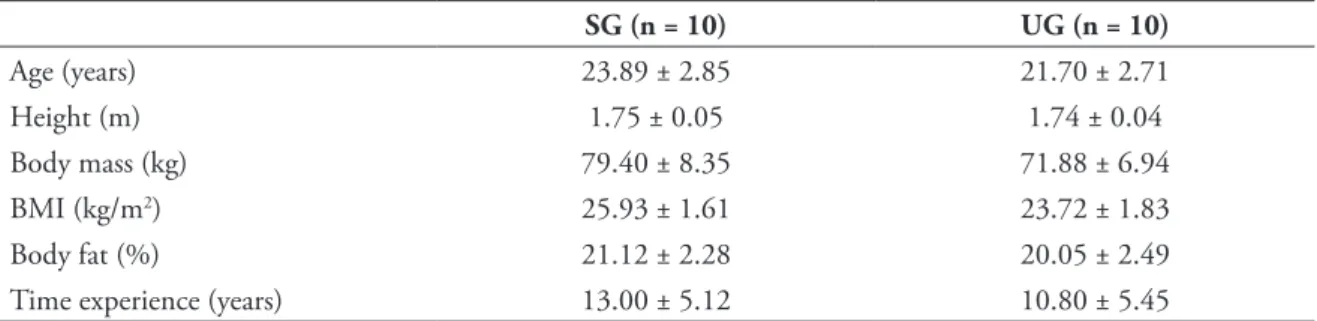
TABLE 1 - Groups’ characteristics.
SG: stable group; UG: unstable group; BMI: body mass index.
Twenty male individuals that take part in fi eld- and indoor-soccer at the university level, with a minimum of three years of experience, were allocated in one of two groups: without functional ankle instability (stable group - SG) or with functional ankle instability (unstable group - UG). For the SG the ankle were classifi ed as dominant (D) and non-dominant (ND); for the UG the ankle were classifi ed as stable (E) or instable (I), despite dominance. For between groups
comparison purpose the dominant ankle of the SG were paired with the unstable ankle of the UG, given that previous studies did not demonstrate signifi cant diff erence on COP behavior between the dominant and non-dominant lower limbs of healthy individuals25-26. Both groups had a training frequency of three times a week, and participated in three championship during the whole year. Th e individuals’ characteristics are presented in TABLE 1. Th e perceptual of body fat were measured with the skinfolds method27 and the body density was calculated accordingly28.
SG (n = 10) UG (n = 10)
Age (years) 23.89 ± 2.85 21.70 ± 2.71
Height (m) 1.75 ± 0.05 1.74 ± 0.04
Body mass (kg) 79.40 ± 8.35 71.88 ± 6.94
BMI (kg/m2) 25.93 ± 1.61 23.72 ± 1.83
Body fat (%) 21.12 ± 2.28 20.05 ± 2.49
Time experience (years) 13.00 ± 5.12 10.80 ± 5.45
The groups were divided according to the Cumberland Ankle Instability Tool (CAIT) score, proposed by Hiller et al.29, which was adapted to the Brazilian population by Noronha et al.30. Th e questionnaire is composed by nine multiple-choice
24 was adopted as a cut point, whereas individuals with values lower or equal to 24 were classifi ed as unstable. Th e scores for both groups are presented in TABLE 2.
Initially, the anamnesis, ankle sprain historic, physical characteristics and anthropometric data were collected. Additionally, it was performed the TABLE 2 - Cumberland Ankle Instability tool (CAIT)
score for both groups.
SG: stable group; UG: unstable group; D/U: dominant ankle/unstable, ND: non-dominant ankle/stable.
Groups D/U ND/S
SG (n = 10) 27.50 ± 1.84 27.10 ± 1.60
UG (n = 10) 20.30 ± 4.03 26.00 ± 2.87
Evaluations
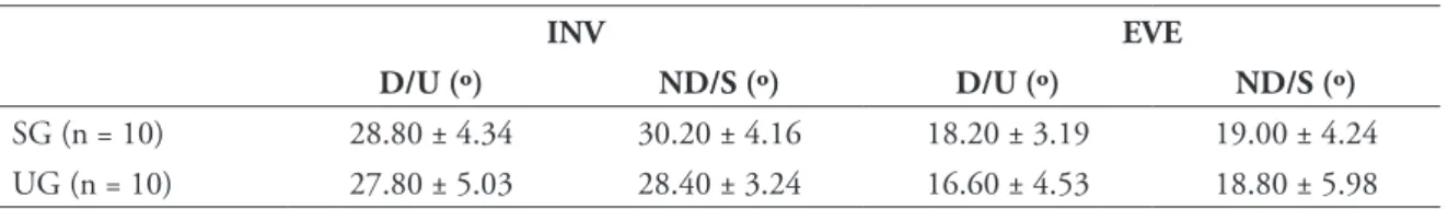
TABLE 3 - Goniometry (degrees) of inversion and eversion for both groups.
SG: stable group; UG: unstable group; INV: inversion; EVE: eversion; D/U: dominant/unstable ankle;
ND/S: non-dominant/ stable ankle.
INV EVE
D/U (º) ND/S (º) D/U (º) ND/S (º)
SG (n = 10) 28.80 ± 4.34 30.20 ± 4.16 18.20 ± 3.19 19.00 ± 4.24
UG (n = 10) 27.80 ± 5.03 28.40 ± 3.24 16.60 ± 4.53 18.80 ± 5.98
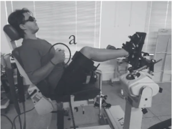
After anamnesis, the subject was familiarized with the single-leg balance test. Th e subject was positioned in the center of the force platform (OR6-6; AMTI®), with an acquisition frequency of 2000 Hz, and it was instructed to hold on the single-leg position for 20 seconds. Data were collected throughout the ForceNet (AMTI®) software. Th e lower limb that maintained contact with the force platform was held with a small knee fl exion and a neutral position for the ankle, whereas the lower limb suspended hold on with the hips and knee fl exed (FIGURE 1). Th e single-leg balance test was performed with the eyes open and with eyes closed (blindfolded), with both the lower limbs. During the test with the eyes open a circular target were positioned in front of the subjects34-35. Th ree attempts was performed for each condition with a 20 seconds rest interval between them. If the subject performed any kind of jump, or touched the fl oor/platform with the suspended limb the test was repeated36-38. Th ree attempts were allowed in order to familiarize the subjects to the balance test protocol.
FIGURE 1 - Equilibrium test with single-leg support.
After that, the passive ankle position sense test was performed. The test was performed in a dynamometer isokinetic Byodex System 4 Pro (Biodex®), with a sample frequency of 100 Hz. Th e data were collected using the Biodex Advantage dominance test for lower limbs, as well as the test of the ankle inversion and eversion goniometry. Th en, subjects performed a single-leg support test on the force platform and the passive ankle-repositioning test in the isokinetic dynamometer.
Th e dominance tests were composed of kicking a ball in a target of one meter of width positioned at 10 meters away; climb a step with 20 cm of height; and recovery the balance after a hard push applied in the middle point between the shoulder blades in an anterior-posterior way, causing the subject to give a step forward to maintain balance.
FIGURE 2 - Passive reposition joint test. a: device to stop the
movement of the dyna-mometer.
Data processing
Results
Statistical analysis
Data were analyzed with the SPSS Statistic 18.0 (SPSS®) software. Firstly, all data were tested for normality, after that, the statistical test was used accordingly.
All data considered normal according to the Shapiro-Wilk test were analyzed with the Student t test. Only the variables TD and SDml did not met the criteria for normality, thus, they were analyzed with the Mann-Whitney test. Similarly, the relationship between variables for the normal data was performed with Pearson’ correlation; for the non-normal data the Spearman’s correlation was used. Th e signifi cant level for all variables was set as α < 0.05.
For the analysis of the single-leg balance test, the signal of the force platform was processed with the 4º order Butterworth low-pass with a cut-off fi lter of 95 Hz, defi ned by the residual analysis43. Th e
following variables related to the displacement of the center of pressure were observed:
- Total displacement (TD): sum of the root mean square of the squares of the displacement in the anterior-posterior displacement and medium-lateral during the 20 seconds of test;
- Standard deviation anterior-posterior (SDap): standard deviation of the mean of the displacement in the anterior-posterior direction during the 20 seconds of test;
- Standard deviation medium-lateral (SDml): standard deviation of the mean of the displacement in the medium-lateral during the 20 seconds of test;
- Total mean velocity (TMV): mean of the velocity of the displacement in the anterior-posterior and medium-lateral direction during the 20 seconds of test;
- Anterior-posterior mean velocity (MVap): mean of the velocity of the displacement in the anterior-posterior direction during the 20 seconds of test;
- Medium-lateral mean velocity (MVml): mean of the velocity of the displacement in the medium-lateral direction during the 20 seconds of test;
Th e data regarding the passive joint reposition were obtained with the Biodex Advantage (Biodex®) software. Th e AAE values were acquired for the angle of 10º (AAE-10º) and 20º (AAE-20º). software (Biodex®). Firstly, the researcher positioned
the ankle of the subject passively, starting from a neutral position, with angular velocity of 1º/s, with 10º of inversion and held for 10 seconds. After that, the dynamometer was adjusted to perform the movement with an angular velocity of 1º/s. Th en, the subject was instructed to reposition the segment at the same angle in which it was held for 10 seconds, stopping the dynamometer manually by pressing the stop button. Th e same procedure was repeated for the 20º of inversion position39-42. Th e diff erence
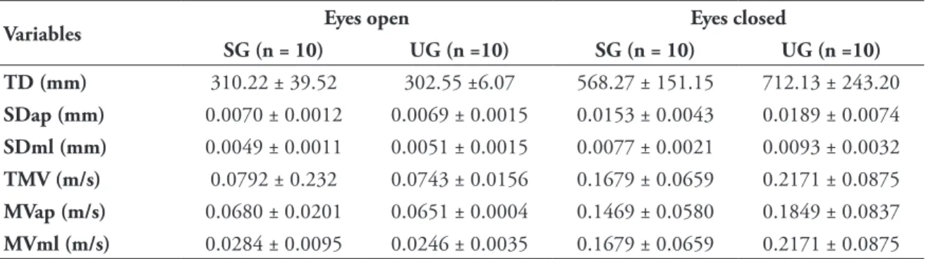
TABLE 4 - Variables of the center of pressure during the single-leg balance test with the eyes open and eyes closed.
SG: stable group; UG: unstable group; TD: total displacement; SDap: anterior-posterior standard deviation; SDml: medium-lateral standard deviation; TMV: mean total ve-locity;
MVap: anterior-posterior mean velocity; MVml: medium-lateral mean velocity.
Th e present study compared the displacement of the center of pressure and the joint position sense of university students that practiced soccer with and without FAI.
Th e variables related to COP did not present signifi cant diff erences between the SG and UG in the present study. Ross et al.37 observed that individuals with FAI exhibited higher values for TD, SDml, MVap and MVml. However, these authors demonstrated that the most sensible variables to TABLE 5 depicted the absolute angular errors at angles 10º (AAE-10º) and 20º (AAE-20º) during the passive joint reposition test, performed by the SG and UG, in which it was observed statistical diff erences between the AAE-10º in the dominant/unstable ankle.
Th e results regarding the correlations between AAE and COP variables are depicted in TABLE 6. It were observed strong correlations only in the UG between: 10º and TMV (p = 0.001 and r = -0.867); AAE-10º and MVap (p = 0.002 and r= -0.854); AAE-AAE-10º and MVml (p = 0.009 and r = -0.771), in the eyes open condition, and AAE-10º and TD (p = 0.036
Variables Eyes open Eyes closed
SG (n = 10) UG (n =10) SG (n = 10) UG (n =10) TD (mm) 310.22 ± 39.52 302.55 ±6.07 568.27 ± 151.15 712.13 ± 243.20
SDap (mm) 0.0070 ± 0.0012 0.0069 ± 0.0015 0.0153 ± 0.0043 0.0189 ± 0.0074
SDml (mm) 0.0049 ± 0.0011 0.0051 ± 0.0015 0.0077 ± 0.0021 0.0093 ± 0.0032
TMV (m/s) 0.0792 ± 0.232 0.0743 ± 0.0156 0.1679 ± 0.0659 0.2171 ± 0.0875
MVap (m/s) 0.0680 ± 0.0201 0.0651 ± 0.0004 0.1469 ± 0.0580 0.1849 ± 0.0837
MVml (m/s) 0.0284 ± 0.0095 0.0246 ± 0.0035 0.1679 ± 0.0659 0.2171 ± 0.0875
TABLE 6
TABLE 5
-Correlation coeffi cient between the absolute angular error and the center of pressure variables. Between-groups comparison for absolute angular error (AAE) in the passive joint reposition test.
AAE: absolute angular error;
EO: eyes open; EC: eyes closed; SG: stable group; UG: unstable group; TD: total displacement; SDap: anterior-posterior standard deviation; SDml: medium-lateral standard deviation; TMV: total mean ve-locity;
MVap: anterior-posterior mean velocity; MVml: medium-lateral mean velocity. SG: stable group, UG: unstable group. * Signifi cant different to SG (p<0.05.
Angle SG (n = 10) UG (n = 10)
AAE-10º (º) 1.29 ± 0.90 2.82 ± 1.70* AAE-20º (º) 1.89 ± 1.33 1.73 ± 1.17
AAE x
10° 20°
EO EC EO EC
SG r UG r SG r UG r SG r UG r SG r UG r
TD 0.700 -0.595 0.632 -0.321 0.872 0.590 0.036* -0.666 0.680 0.149 0.235 -0.413 0.656 0.162 0.463 -0.263
SDap 0.099 -0.551 0.248 -0.517 0.996 -0.002 0.029* -0.685 0.234 0.414 0.258 -0.395 0.947 -0.024 0.447 -0.272
SDml 0.583 -0.198 0.126 -0.201 0.875 0.057 0.103 -0.546 0.290 -0.372 0.427 -0.284 0.639 -0.17 0.574 -0.203
TMV 0.362 -0.362 0.001** -0.867 0.995 0.002 0.082 -0.575 0.137 0.137 0.949 0.023 0.598 0.19 0.403 -0.298
MVap 0.399 -0.301 0.002** -0.854 0.963 0.017 0.179 -0.462 0.160 0.160 0.990 -0.004 0.672 0.153 0.743 -0.119
MVml 0.32 -0.351 0.009** -0.771 0.825 -0.081 0.01* -0.776 0.135 0.135 0.618 0.180 0.385 0.309 0.587 -0.196
Discussion
diff erences between these individuals. In the present study, we believed that the lack of diff erence between groups might be related to the characteristic of the single-leg test used. Ankle sprains are associated to fast movements, such as jumps, sprints and changes in direction2, 4-6, 8; however, the measures used herein, stabilization time and COP reposition in the support, are more accurate to diff erentiate between individuals with and without stability35, 37.
Additionally, another factor that may have infl uenced our results is the period of the sprain event35, 37. Holme et al.44, McKeon and Hertel45
and Hertel et al. 46 claimed that the negative eff ects
associated with this type of injury on the postural control may be more apparent in acute phases. Holme et al.44, showed that the postural control
returned to normal condition after four months of the injury incident (i.e., ankle sprain), even though no treatment has been carried out. However, a 12 months follow-up demonstrated that only 7% of the athletes that performed the rehabilitation presented another sprain, while 29% that did not perform any treatment exhibited another injury incident. McKeon and Hertel45 also observed
a lower incidence risk for injury after six weeks
of rehabilitation treatment. Hertel et al. 46
demonstrated that two weeks after the ankle sprain incident there is an acute degradation in the postural control, which returned to normal condition after four weeks of rehabilitation. Although, the results of these studies are not able to explain the FAI occurrence in chronic cases, as for example, in the Konradsen et al.19 study, whereas the residual
symptoms remained for as long as seven year after the injury incident.
In order to diagnose the instability47 and to prevent
ankle sprains, evaluate the position sense and the joint movement, both passively or actively, seems to be crucial48-49. Despite of diff erences between studies’
protocols, our results demonstrated a greater error in the joint reposition sense in individuals with ankle instability. In the present study, these errors were more apparent in the joint reposition sense test performed passively at 10º (AAE-10º) between the SG and UG, with no diff erence at 20º.
Th e association of the AAE at 10º and 20º with the COP variables showed strong correlations for the UG between AAE-10º and TD, AAE-10º and SDap, AAE-10º and MVml with the eyes closed. Th e injury caused in the mechanoreceptors impairs the responses and the mechanisms of adaptations related to perturbation in the postural balance and, as expected, these results indicates that the proprioceptive defi cit are related to the control of the posture stability50-51.
In spite of the reduced sample size, the results from the present study demonstrated important indicatives that the FAI impairs the joint position sense in the initial inversion angles. Th e greater stratifi cation of the angles as well as the balance tests with similar movements to that performed by soccer practitioners may contribute to better understand how the FAI increases the susceptibility to new sprains and the prevalence of residuals symptoms.
Th e ankle sprain, even if it is old, may infl uence the position joint sense of soccer player’s university students with and without ankle instability, even without impairment in balance. These results point out for a greater attention to training and rehabilitation of this joint and the continuity of a measure with the aim to prevent a new sprain.
Resumo
Análise do COP e sentido de posição em jogadores universitários de futebol com e sem instabilidade de tornozelo
References
1. Russel M, Benton D, Kingsley M. Reliability and construct validity of soccer skills tests that measure passing, shooting and dribbling. J Sport Sci. 2010;28:1399-408.
2. Rein S, Fabian T, Weindel S, Schneiders W, Zwipp H. Th e infl uence of playing level on functional ankle instability in soccer players. Arch Orthop Trauma Surg. 2011;131:1043-52.
3. Siegle M, Lames M. Game interruptions in elite soccer. J Sports Sci. 2012;30:619-24.
4. Le Gall F, Carling C, Reilly T. Injuries in young elite female soccer players: an 8-season prospective study. Am J Sports Med. 2008;36:276-84.
5. Oztekin HH, Boya H, Ozcan O, Zeren B, Pinar P. Foot and ankle injuries and time lost from play in professional soccer players. Foot (Edinb). 2009;19:22-8.
6. Ekstrand J, Tropp H. Th e incidence of ankle sprains in soccer. Foot Ankle. 1990;11:41-4.
7. Wright IC, Neptune RR, Van Den Bogert AJ, Nigg BM. Th e infl uence of foot positioning on ankle sprains. J Biomech. 2000;33:513-19.
8. Lee AJY, Lin WH. Twelve-week biomechanical ankle platform system training on postural stability and ankle proprio-ception in subjects with unilateral functional ankle instability. Clin Biomech. 2008;23:1065-72.
9. Mohammadi F. Comparison of 3 preventive methods to reduce the recurrence of ankle inversions sprains in male soccer players. Am J Sports Med. 2007;35:922-26.
10. McCloskey DI. Kinesthetic sensibility. Physiol Rev. 1978;58:763-820.
11. Newton RA. Joint receptor contributions to refl exive and kinesthetic responses. Phys Th er. 1982;62:22-9.
12. Proske U, Wise AK, Gregory JE. Th e role of muscle receptors in detection of movements. Prog Neurobiol. 2000;60:85-96. 13. Ribeiro F, Oliveira J. Efeito da fadiga muscular local na propriocepção do joelho. Fisioter Mov. 2008;21:71-83. 14. Freeman MAR, Dean MRE, Hanham IWF. Th e etiology and prevention of functional instability of the foot. J Bone
Joint Surg Br. 1965;47:678-85.
15. Willems T, Witvrouw E, Verstuyft J, Vaes P, De Clercq D. Proprioception and muscle strength in subjects with a history of ankle sprains and chronic instability. J Athl Train. 2002;37:487-93.
16. Noronha M, França LC, Haupenthal A, Nunes GS. Intrinsic predictive factors for ankle sprain in active university students: a prospective study. Scand J Med Sci Sports. 2013;23:541-7.
17. Freeman MAR. Instability of the foot after injuries to the lateral ligament of the ankle. J Bone Joint Surg Br. 1965;47:669-77.
18. Hertel J. Functional instability following lateral ankle sprain. Sports Med. 2000;29:361-71.
19. Konradsen L, Bech L, Ehrenbjerg M, Nickelsen T. Seven years follow-up after ankle inversion trauma. Scand J Med Sci Sports. 2002;12:129-35.
20. Hertel J. Functional anatomy, pathomechanics, and pathophysiology of lateral ankle instability. J Athl Train. 2002;37:364-75.
21. Blackburn T, Guskiewicz KM, Petschauer MA, Pretince WE. Balance and joint stability: the relative contributions of proprioceptive and muscular strength. J Sport Rehabil. 2000;9:315-28.
22. Ross SE, Guskiewicz KM, Yu B. Single-leg jump-landing stabilization times in subjects with functionally unstable ankles. J Athl Train. 2005;40:298-304.
23. Brown CN, Bowser B, Orellana A. Dynamic postural stability in females with chronic ankle instability. Med Sci Sports Exerc. 2010;42:2258-63.
que apresentaram distribuição normal foi feita com o teste t de Student, enquanto que para DT e DPml foi utilizado o teste de Mann-Whitney. Da mesma forma, foram usados os testes de Pearson e Spearman para correlacionar as variáveis. Foi adotado α < 0,05. Houve diferença entre EAA-10º (p < 0,05). Foram
encontradas fortes correlações entre: EAA-10° e VMT (p < 0,01 r = -0,867); EAA-10° e VMap (p < 0,01 r = -0,854); EAA-10° e VMml (p < 0,01 r = -0,771), na condição olhos abertos, e EAA-10° e DT (p < 0,05 r = -0,666); EAA-10º e DPap (p < 0,05 r = -0,685) e EAA-10° e VMml (p < 0,05 r = -0,766) na condição olhos fechados. Entorses de tornozelo prejudicam o sentido de posição, sem afetar o equilíbrio.
24. Garn SN, Newton RA. Kinesthetic awareness in subjects with multiple ankle sprains. Phys Th er. 1988;68:1667-71. 25. Hrysomallis C, McLaughlin P, Goodman C. Relationship between static and dynamic balance tests among elite
Aus-tralian footballers. J Sci Med Sport. 2006;9:288-91.
26. Teixeira LA, Oliveira DL, Romano RG, Correa SC. Leg preference and interlateral asymmetry of balance instability in soccer players. Res Q Exerc Sport. 2011;82:21-7.
27. Jackson AS, Pollock ML. Generalized equations for predicting body density of men. Br J Nutr. 1978;40:497-504. 28. Lohman TG. Skinfolds and body density and their relation to body fatness: a review. Hum Biol. 1981;53:181-225. 29. Hiller CE, Refshauge KM, Bundy AC, Herbert RD, Kilbreath SL. Th e Cumberland Ankle Instability Tool: a report
of validity and reliability testing. Arch Phys Med Rehabil. 2006;78:1235-41.
30. Noronha M, Refshauge KM, Kilbreath SL, Figueiredo VG. Cross-cultural adaptation of the Brazilian-Portuguese version of the Cumberland Ankle Instability Tool (CAIT). Disabil Rehabil. 2008;30:1959-65.
31. Hiller CE, Refshauge KM, Herbert RD, Kilbreath SL. Balance and recovery from a perturbation are impaired in people with functional ankle instability. Clin J Sport Med. 2007;17:269-75.
32. Sawikins K, Refshauge K, Kilbreath S, Raymond J. Th e placebo eff ect of ankle taping in ankle instability. Med Sci Sports Exerc. 2007;39:781-7.
33. Hoff man M, Schrader J, Applegate T, Koceja D. Unilateral postural control of the functionally dominant and non-dominant extremities of healthy subjects. J Athl Train. 1998;33:319-22.
34. Trojian TH, Mckeag DB. Single leg balance test to identify risk of ankle sprains. Br J Sports Med. 2006;40:610-3. 35. Hertel J, Olmsted-Kramer LC. Defi cits in time-to-boundary measures of postural control with chronic ankle instability.
Gait Posture. 2007;25:33-9.
36. Ross SE, Guskiewicz KM, Gross MT, Yu B. Assessment tools for identifying functional limitations associated with functional ankle instability. J Athl Train. 2008;43:44-50.
37. Ross SE, Guskiewicz KM, Gross MT, Yu B. Balance measures for discriminating between functionally unstable and stable ankles. Med Sci Sports Exerc. 2009;41:399-407.
38. Hamlyn C, Docherty CL, Klossner J. Orthotic intervention and postural stability in participants with functional ankle instability after an accommodation period. J Athl Train. 2012;47:130-5.
39. Sahin N, Baskent A, Cakmak A, Salli A, Ugurlu H, Berker E. Evaluation of knee proprioception and eff ects of proprioception exercise in patients with benign joint hypermobility syndrome. Rheumatol Int. 2008;28:995-1000. 40. Sekir U, Yildiz Y, Hazneci B, Ors F, Saka T, Aydin T. Reliability of a functional test battery evaluating
functionali-ty, proprioception, and strength in recreational athletes with functional ankle instability. Eur J Phys Rehabil Med. 2008;44:407-15.
41. Goble DN, Coxon JP, Wenderoth N, Van Impe A, Swinnen SP. Proprioceptive sensibility in the elderly: degeneration, functional consequences and plastic-adaptive processes. Neurosci Biobehav Rev. 2009;33:271-8.
42. Ju YY, Wang CW, Cheng HYK. Eff ects of active fatiguing movement versus passive repetitive movement on knee proprioception. Clin Biomech (Bristol, Avon). 2010;25:708-12.
43. Winter AD. Biomechanics and motor control of human movement. 2nd ed. Waterloo: Wiley-Interscience; 1990. 44. Holme E, Magnusson SP, Becher K, Bieler T, Aagaard P, Kjaer M. Th e eff ects of supervised rehabilitation on strength,
postural sway, position sense and re-injury risk after acute ankle ligament sprain. Scand J Med Sci Sports. 1999;9:104-9. 45. McKeon PO, Hertel J. Systematic review of postural control and lateral ankle instability, part I: can defi cits be detected
with instrumented testing? J Athl Train. 2008;43:293-304.
46. Hertel J, Buckley WE, Denegar CR. Serial testing of postural control after acute lateral ankle sprain. J Athl Train. 2001;36:363-8.
47. Hartsell HD. Th e Eff ect of external bracing on joint position sense awareness for the chronically unstable ankle. J Sport Rehabil. 2000;9:279-89.
48. Bernier JN, Perrin DH. Eff ect of coordination training on proprioception of the functionally instable ankle. J Orthop Sports Phys Th er. 1998;27:264-75.
ADDRESS Antônio Francisco de Almeida Neto Laboratório de Biomecânica Departamento de Educação Física Instituto de Biociências Universidade Estadual Paulista Av. 24 A, 1515 13506-900 - Rio Claro - SP - BRASIL e-mail: [email protected]