From the Department of Internal Medicine and the Pathology Post-Graduation Course, Triângulo Mineiro Medical School, Minas Gerais.
CASE REPORT
WIDESPREAD HEMATOGENOUS METASTASES AND
TROUSSEAU’S SYNDROME IN GASTRIC
ADENOCARCINOMA
Vitorino Modesto dos Santos, Denise Bertulucci Rocha Rodrigues, Eumênia Costa da Cunha Castro, João Carlos Saldanha, Sheila Soares, Vicente de Paula Antunes Teixeira and Marlene Antônia dos Reis
RHCFAP/3042
SANTOS VM dos et al. - Widespread hematogenous metastases and Trousseau’s syndrome in gastric adenocarcinoma. Rev. Hosp.
Clín. Fac. Med S. Paulo 56(3):91-96, 2001.
A case of widespread hematogenous metastases and Trousseau’s syndrome is reported in a 40 year-old white housewife with gastric cancer, presenting subdural hematoma, ecchymoses, epistaxis, stomach and uterine bleeding. After undergoing hematoma drainage, she was unsuccessfully treated with platelets, red blood cells, plasma cryoprecipitate transfusions, and antibiotics. Necropsy disclosed gastric ring-signet adenocarcinoma invading the serous layer, with massive disseminated intravascular coagulation and systemic neoplastic embolism. Multiple old and recent hyaline (rich in fibrin and platelets) microthrombi, and tumor emboli were observed in the bone marrow, meninges, liver, lungs, kidneys, lymph nodes, adrenals, thyroid, heart, pancreas, and ovaries (Krukenberg tumor).
DESCRIPTORS: Trousseau’s syndrome. Disseminated intravascular coagulation. Subdural hematoma. Gastric cancer.
Krukenberg tumor.
Haemostatic abnormalities, mani-fested as hemorrhage or thrombosis, often occur in patients with advanced cancer of the stomach, pancreas, liver, ovary, breast, lung, and prostate. Clini-cal features of disseminated intravascu-lar coagulation (DIC) may be acute or chronic, usually following venous thromboembolism1-4.
The diagnosis of gastric cancer of-ten occurs first, but there are an in-creasing number of associations with DIC and microangiopathic hemolytic anemia (MHA), a condition that im-plies the worst prognosis1–11. Although bone metastases are uncommon in gas-tric cancer, this solid tumor has been the cause of DIC following diffuse bone marrow metastases1–3,6,8–12. Extra
gastric signs and symptoms may pre-cede the diagnosis of gastric cancer, including acanthosis nigricans, neuromyopathy, central nervous sys-tem disturbances, and Trousseau’s syn-drome. In 1865, the French physician Armand Trousseau (1801-1867) de-scribed migratory venous thromboses occurring in the course of his own pan-creatic cancer.
The term Krukenberg tumor (Friedrich Krukenberg, German pa-thologist) refers to bilateral metastases of mucin producing gastrointestinal
cancer (stomach, biliary tree, pancreas) into the ovaries.
DIC is often observed in advanced cancer patients due to acute or insidi-ous development of disseminated fibrin microthrombi, and may simulate pri-mary hematological disease2,4,6,13. Oc-casionally, acute DIC may be one of the earliest manifestations of gastric cancer2, and the course of the disease is rapidly fatal in patients not receiv-ing chemotherapy2,3.
CASE REPORT
A 40-year-old white housewife came to the hospital on May 10, 1999 complaining of a continuous 3-week headache, which had become more se-vere in the last 48 hours. She had been using meloxicam, without any im-provement. She also presented dizzi-ness, vomiting, ecchymosis, epistaxis, melena, metrorrhagia, mental confu-sion, and lethargy. Physical examina-tion showed a bad general status, pale mucosa, and no peripheral edema. There was anisocoria (right > left) and lack of photo motor reflex and nuchal stiffness. Anthropometric data revealed body mass index 31.6 kg/m2. Body temperature was 36.5 oC. The heart was normal, without murmurs, with 78 bpm, and blood pressure was 140/70 mmHg. The lungs, liver, and lymph nodes were normal on initial examina-tion.
The patient´s blood group was B, Rh-positive. The erythrocyte count was 3.56 106/mm3, with polychromasia and anisocytosis, erythroblasts 6%, hemo-globin 10 g/dl, hematocrit 32%, MCV 89.8fL, leukocyte count 10,000/mm3 (neutrophils: bands 11%, segmented 65%, eosinophils 2%, lymphocytes 13%, monocytes 9%), platelets 71,000/ mm3, glucose 110 mg/dL, urea 42 mg/ dL, creatinine 0.8 mg/dL, sodium 138 mEq/L, potassium 3.8 mEq/L, chloride 113 mEq/L, calcium 9 mg/dL, phos-phorous 3.7 mg/dL, magnesium 2.1 mg/dL, total bilirubin 2.3 mg/dL, indi-rect bilirubin 1.2 mg/dL, AST 42 U/L, ALT 50 U/L, gGT 161 U/L, alkaline phosphatase 347 U/L, fibrinogen 1.15 g/L, and prothrombin activity 36%. Antinuclear antibody and LE cells tests and rheumatoid factors were negative, and C-reactive protein was positive. The bone marrow aspirate showed in-tense erythroid hyperplasia, dyserythropoiesis, several mitosis, cy-toplasmic bridges, megaloblasts and micro erythroblasts, relative myeloid
hypoplasia and cells in all phases of maturation, moderate megakaryocytic hyperplasia, and normal histiocytic and lymphoplasmocytic elements. A com-puted tomography of the brain revealed a chronic subdural hematoma in the right parietal region, and surgical drainage was performed. In spite of treatment with plasma cryoprecipitate, platelets, and red blood cell transfu-sions, plus ceftriaxone and gentamicin, the clinical status worsened. There was fever (37.8 oC), more intense jaundice (bilirubins: direct 3.25 and indirect 1.53 mg/dL), and higher liver enzymes (AST 144, ALT 51 and gGT 218 U/L). On the fourth day after admission, af-ter severe epistaxis and hematemesis, she died due to irreversible circulatory shock.
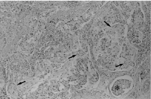
Pathologic study. An ulcerated
ad-enocarcinoma (diameter 3 cm) infiltrat-ing the serosa layer, with cells show-ing intense atypia and several mitoses, and some containing mucicarmine-positive material (Fig. 1), were de-tected in pyloric antrum. There was widespread embolism in vessels from the stomach wall and esophagus sub-mucosa, in addition to submucosa and serosa vessels from the small intestine.
Hemorrhage through multiple puncti-form lesions in the gastric and intesti-nal mucosa and diffuse hypotrophic gastritis were also noted.
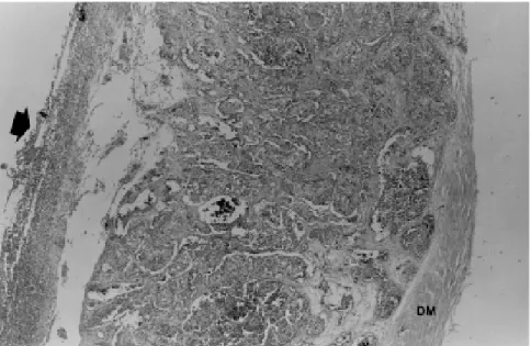
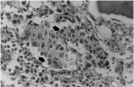
The dura mater examination showed signs of drained subdural hematoma, hemosiderin impregnation, metastatic emboli, and hemorrhage (Figs. 2A and 2B). The encephalon weighed 1,300 g, showing recent cortical hemorrhage in the right parietal lobe and diffuse edema plus hyperemia. In bone marrow, there was neoplastic infiltration (Fig. 3) and areas of fibrosis. The heart weighed 310 g (0.37% of body weight) with neoplas-tic emboli in the myocardium and mul-tiple petechial hemorrhages in epicar-dium. Marantic endocarditis was not found. The lungs weighed 500 g (right) and 530 g (left), with multiple fibrin emboli and small metastatic focuses around the vessel walls. There was evi-dence of some hyaline thrombi canali-zation and several hemorrhagic foci, with edema and parenchyma collapse, in addition to subpleural petechia and bilateral bloody pleural effusion. Tra-chea and bronchi were full of bloody material. The thyroid showed multiple metastases. The liver weighed 3,670 g, with several whitish nodes, massive
DISCUSSION
This 40-year-old female with mu-cin-producing gastric adenocarcinoma (signet-ring cells) presented DIC, pete-chiae, ecchymoses, epistaxis, stomach and uterine bleeding, bloody pleural effusion, anisocoria, and subdural he-matoma, in addition to hemorrhagic foci and neoplastic embolism, mainly in the subarachnoidal vessels.
In necropsies of cancer patients, the frequency of thromboses increases with age, especially in cases of tumors producing thromboplastic substances, such as mucin (esophagus, stomach, pancreas, and colon)2,4,5,14. However, similar to this case, DIC and MHA in younger patients and precocious bone marrow metastases have been reported in gastric cancer2,3,6,8–11,15.
Neoplastic cells may generate thromboembolic phenomena through direct lesion of the endothelium, acti-vation of the coagulation cascade, pro-duction of procoagulant substances, or reduction in the synthesis or activity of anticoagulant factors2,5,8. The tumor-derived procoagulant factor that con-verts factor X to Xa, favoring throm-bin formation, fibrinogen consumption, and fibrin microthrombi production, has been reported to respond well to chemotherapy with 5-fluorouracil8,11. In this patient, the subdural hematoma could have had its origin in venous thrombi of the dense external layer of the dura mater, following the rupture of capillaries in the internal areolar layer, and the hemorrhage may have been fa-vored by coagulation disturbances due to DIC4,6,9–11,15.
In 4,906 consecutive necropsies it was found only 88 cases (1.8%) of DIC; forty were patients with cancer (82.5% had metastases in 2 or more sites), and 11 were cases of gastric can-cer (27.5%)4. In 109 patients there was a clinical suspicion of DIC, but the di-agnosis was confirmed in 25 cases (23.0%); while among the remaining Figure 2A - Subdural hemorrhage (arrow), in addition to neoplastic embolus (*) and dura mater
(DM) infiltration (hematoxylin-eosin X 50).
Figure 2B - Detail of neoplastic emboli (*) into dura mater (DM) vessels (hematoxylin-eosin X 200).
boli in portal vein branches (Fig. 4), hi-lar lymph node metastases, discrete foci of macro vesicular steatosis, diffuse congestion, and jaundice. Microscopic metastases and cytosteatonecrosis were observed in the pancreas. The kidneys weighed 180 g each, with bilateral neo-plastic emboli, fibrin thrombi in small vessels and glomerular capillaries, re-duction of cortical thickness, and med-ullar congestion. In the retroperitoneum,
4,797 patients without clinical suspi-cion of DIC, in 63 cases (1.3%) microthrombi were present in 3 or more sites. Fibrin microthrombi were more frequently found in the kidney, followed by the lungs, spleen, adrenals, heart, brain, and liver4.
Either massive or disseminated microthrombi may cause infarctions in vital organs, as well as respiratory, re-nal, hepatic, and/or heart failure7,13 pre-disposing to irreversible circulatory shock14.
Histopathologic criteria to confirm the diagnosis of DIC consist of dem-onstration of fibrin microthrombi in ar-terioles, capillaries, and venules, in at least 3 different organs4.
In the present case, skin manifesta-tions suggesting migratory or recurrent thrombophlebitis were absent; never-theless, the low platelet count and pro-longed prothrombin time, in addition to the unequivocal demonstration of disseminated fibrin microthrombi in more than 3 different organs, allowed us to establish the diagnosis of DIC4,13. As occurred in this case, the asso-ciation of disseminated cancer with multiple venous and arterial thombo-embolic phenomena, including DIC, has been considered as a variant of the classic Trousseau’s syndrome5.
Another concern could be the nor-mal plasma fibrinogen; however, in some patients with DIC, the fibrinogen levels have been described to lower more slowly, in part due to higher he-patic production under influence of se-vere stress13.
The patient did not undergo chemo-therapy; in spite of treatment with platelets, red blood cells, and plasma cryoprecipitate transfusions, she pre-sented signs of respiratory, renal, and hepatic failure and circulatory shock, and she died in 4 days. In this case, the nonsteroidal anti-inflammatory drug (meloxican) may have contributed to the severity of bleeding associated with multiple punctiform lesions observed Figure 3 - Neoplastic infiltrate (arrows) in bone marrow (hematoxylin-eosin X 500).
Figure 4 - Neoplastic embolism (arrow) in portal vein branches (hematoxylin-eosin X 250).
In adults, the sudden appearance of DIC in the absence of infection or bone marrow primary disease should stimu-late a strong suspicion of gastric
can-cer2,4, and if diagnosis is confirmed, an initial schedule of non-myelosuppressive chemotherapy with 5-fluorouracil must be started as soon as possible. By increasing the survival
time of patients with gastric cancer and DIC, chemotherapy with 5-fluorouracil allows further employment of comple-mentary antineoplastic therapies11.
RESUMO RHCFAP/3042
SANTOS VM dos e col. - Metástases hematogênicas disseminadas e síndrome de Trousseau em adeno-carcinoma gástrico. Rev. Hosp.
Clín. Fac. Med. S. Paulo
56(3):91-96, 2000.
Relata-se caso de metástases hema-togênicas disseminadas e síndrome de Trousseau em mulher branca de 40 anos, portadora de câncer gástrico, apresentando hematoma subdural,
equimoses, epistaxes, hemorragia gás-trica e metrorragia. Inicialmente sub-metida à drenagem do hematoma, foi tratada sem sucesso com transfusões de plaquetas, hemácias e plasma criopre-cipitado, além de antibióticos. O estu-do de necropsia revelou adenocarci-noma gástrico com células em anel de sinete invadindo a serosa, com maciça coagulação intravascular disseminada e embolismo neoplásico sistêmico. Microtrombos hialinos (constituídos de
fibrina e plaquetas) antigos e recentes, além de êmbolos tumorais, foram ob-servados na medula óssea, meninges, fígado, pulmões, rins, linfonodos, adrenais, tireóide, coração, pâncreas e ovários (tumor de Krukenberg).
DESCRITORES: Síndrome de
Trousseau. Coagulação intravascular disseminada. Hematoma subdural. Câncer gástrico. Tumor de Krukenberg.
REFERENCES
1. NIEL F, GAUSSEM P, ANDREUX MH et al. - Cancer généralisé chez une femme de 82 ans: envahissement médullaire et coagulation intravasculaire disséminée. Ann Biol Clin 1998; 56: 580-3. 2. PASQUINI E, GIANNI L, AITINI E et al. - Acute disseminated
intravascular coagulation syndrome in cancer patients. Oncology 1995; 52: 505-8.
3. SUSANO R, CAMINAL L, FERRO J et al. - Anemia hemolítica microangiopática asociada a neoplasias: análisis de cinco casos y revisión de la literatura. Rev Clin Esp 1994; 194: 603-6. 4. TANAKA K & IMAMURA T - Incidence and clinicopathological
significance of DIC in autopsy cases. In: ABE T & YAMANAKA M - Disseminated Intravascular Coagulation. Bibliotheca Haematologica Series. Bern, Hässig A, 1981. p. 79-83.
5. CAFAGNA D & PONTE E - L’embolia polmonare da causa paraneoplastica. Minerva Med 1997; 88:523-30.
6. COLOMBO E, GIORGI S, SONZINI E et al. - Disseminated intravascular coagulation and bone marrow metastases as presenting manifestations of gastric carcinoma. Haematologica 1985;70: 187. 7. ISHIKAWA M, SUSUKI S, AKUTU Y et al. - An autopsy case of AIDS with hemophilia A who died of DIC and gastrointestinal bleeding associated with gastric carcinoma (signet ring cell carcinoma). Jpn J Clin Hematol 1994; 35: 886-91.
9. MORIMATSU M, SHIROUZU K, IRIE K et al. - Gross and microscopic characteristics of stomach cancer with microangiopathic hemolytic anemia and/or disseminated intravascular coagulopathy. Acta Pathol Jpn 1985; 35: 809-22. 10. TAKEUCHI R, KUTO M, KATAYAMA N et al. - A case of
carcinomatosis associated with microangiopathic hemolytic anemia and disseminated intravascular coagulation occurring after 12 years of operation for gastric cancer. Jpn J Clin Hematol 1983; 24: 1423-9.
11. YEH K-H & CHENG A-L - Gastric cancer associated with acute disseminated intravascular coagulation: successful initial treatment with weekly 24-hour infusion of high-dose 5-fluorouracil and leucovorin. Br J Hematol 1998; 100: 769-72.
12. NODA N, SANO T, SHIRAO K et al. - A case of bone marrow recurrence from gastric carcinoma after a nine-year disease-free interval. Jpn J Clin Oncol 1996; 26: 472-5.
13. HARDAWAY RM & WILLIAMS CH- Disseminated intravascular coagulation: an update. Compr Ther 1996; 22: 737-43. 14. BRESCIANI C, BORGES PCM, GAMARODRIGUES JJ et al.
-Fatal pulmonary thromboembolism in gastrectomy intraoperative procedures by gastric adenocarcinoma: Case report. Rev Hosp Clín Fac Med S Paulo 1999; 54: 115-20.
15. FURUI T, ICHIHARA K, IKEDA A et al. - Subdural hematoma associated with disseminated intravascular coagulation in patients with advanced cancer. J Neurosurg 1983; 58: 398-401.