%!
From the Pediatric Surgery Division andLaboratory (LIM-30), Hospital das Clínicas, Faculty of Medicine, University of São Paulo.
HEART HYPOPLASIA IN AN ANIMAL MODEL OF
CONGENITAL DIAPHRAGMATIC HERNIA
Uenis Tannuri
RHCFAP/3057
TANNURI U - Heart hypoplasia in an animal model of congenital diaphragmatic hernia. Rev. Hosp. Clín. Fac. Med. S. Paulo 56(6):173-178, 2001.
Purpose: In previous papers, we described a new experimental model of congenital diaphragmatic hernia in rabbits, and we also reported noninvasive therapeutic strategies for prevention of the functional and structural immaturity of the lungs associated with this defect. In addition to lung hypoplasia, pulmonary hypertension, biochemical, and structural immaturity of the lungs, the hemodynamics of infants and animals with congenital diaphragmatic hernia are markedly altered. Hence, cardiac hypoplasia has been implicated as a possible cause of death in patients with congenital diaphragmatic hernia, and it is hypothesized to be a probable consequence of fetal mediastinal compression by the herniated viscera. Cardiac hypoplasia has also been reported in lamb and rat models of congenital diaphragmatic hernia. The purpose of the present experiment was to verify the occurrence of heart hypoplasia in our new model of surgically produced congenital diaphragmatic hernia in fetal rabbits.
Methods: Twelve pregnant New Zealand rabbits underwent surgery on gestational day 24 or 25 (normal full gestational time
- 31 to 32 days) to create left-sided diaphragmatic hernias in 1 or 2 fetuses per each doe. On gestational day 30, all does again underwent surgery, and the delivered fetuses were weighed and divided into 2 groups: control (non-surgically treated fetuses) (n = 12) and congenital diaphragmatic hernia (n = 9). The hearts were collected, weighed, and submitted for histologic and histomorphometric studies.
Results: During necropsy, it was noted that in all congenital diaphragmatic hernia fetuses, the left lobe of the liver herniated
throughout the surgically created defect and occupied the left side of the thorax, with the deviation of the heart to the right side, compressing the left lung; consequently, this lung was smaller than the right one. The body weights of the animals were not altered by congenital diaphragmatic hernia, but heart weights were decreased in comparison to control fetuses. The histomorphometric analysis demonstrated that congenital diaphragmatic hernia promoted a significant decrease in the ventricular wall thickness and an increase in the interventricular septum thickness.
Conclusion: Heart hypoplasia occurs in a rabbit experimental model of congenital diaphragmatic hernia. This model may be utilized for investigations in therapeutic strategies that aim towards the prevention or the treatment of heart hypoplasia caused by congenital diaphragmatic hernia.
DESCRIPTORS: Congenital diaphragmatic hernia. Heart. Hypoplasia. Fetus.
In previous papers, we have de-scribed a new experimental model of congenital diaphragmatic hernia (CDH) in rabbits, and we also have re-ported on studies of noninvasive thera-peutic strategies for prevention of func-tional and structural immaturity of lungs associated with this defect. These strategies consist of prenatal intra-am-niotic administration of porcine
sur-factant or dexamethasone and prenatal tracheal ligation (TL), an invasive method that effectively treats or pre-vents pulmonary hypoplasia of CDH1,2.
Based on functional, morphological,
histomorphometric and biochemical studies, we proved that in this model, CDH is associated with hypoplastic lungs3, and the preventive effects of
in-tra-amniotic administration of drugs on lung maturation were comparable to the changes induced by TL4-6.
%"
the hemodynamics of infants and ani-mals with CDH are markedly altered7,8.
Cardiac insufficiency has also been ob-served in CDH patients, and it has been suggested that this failure and systemic circulatory problems may be of more importance than pulmonary hyperten-sion9. Siebert et al. quantitatively
docu-mented cardiac hypoplasia in 8 infants who had died of the complications of left CDH10. Consequently, cardiac
hy-poplasia has been implicated as a pos-sible cause of death in patients with CDH, and it is hypothesized to be a probable consequence of fetal medi-astinal compression by the herniated viscera11. Cardiac hypoplasia has also
been reported in the primary animal models of CDH, i.e. lambs12 and rats13.
The purpose of the present experiment is to verify the occurrence of heart hy-poplasia in a new model of surgically produced CDH in fetal rabbits.
MATERIAL AND METHODS
Experimental Design
Twelve pregnant New Zealand rab-bits underwent surgery on gestational day 24 or 25 (normal full gestational time - 31 to 32 days) to create left-sided diaphragmatic hernias in 12 fe-tuses as previously described3-5.
Anesthesia was initiated with intramus-cular ketamine (45 mg/kg of body weight) and maintained with 0.5% so-dium thiobarbiturate intravenously. Briefly, using sterile technique, a ma-ternal laparotomy was performed, and the gravid bicornuate uterus was deliv-ered out of the abdomen. There were usually 6 to 9 fetuses in the uterus of pregnant rabbits. The most distal fetus of each horn was chosen for surgical treatment, and only 1 or 2 fetuses per each doe underwent surgery. During surgical manipulations, the proximal portion of the horn undergoing surgery and the other horn were maintained
in-side the maternal abdomen. The surgi-cal field was continuously moistened with warmed sterile saline solution to prevent fetal hypothermia.
The head and left foreleg of the fe-tus were palpated, and using microsur-gical instruments, a 1 cm transversal hysterotomy was performed at the re-gion overlaying the palpated foreleg. This incision was made as far as pos-sible from the mesometrial border of the uterus. The subjacent chorion and amnion were opened with scissors, and the amniotic fluid was aspirated from the uterus. Only the left foreleg was then exteriorized through the hyster-otomy to permit the exposure of the corresponding chest wall (Fig. 1). A low left lateral thoracotomy was per-formed, and the diaphragm was par-tially excised by microsurgical scissors. The chest incision was closed in two layers of simple interrupted monofila-ment 6-0 nylon sutures.
As the hysterotomy was closed with monofilament nylon sutures, warmed sterile saline solution was in-fused into the amniotic cavity until uterine repletion to reconstitute the amniotic fluid. Finally, the maternal laparotomy was closed. After the
sur-gical manipulations, each doe received 400 mg of cephalothin intravenously and Depo-Provera (5 mg intramuscularly) (NV – Upjohn, Bel-gium). On gestational day 30, all does were again placed under general anesthesia and the 9 surviving surgi-cally treated fetuses were delivered by cesarean section and sacrificed, body weights were recorded. All the un-treated fetuses were also delivered, and 12 of them formed the control group. Through a medial longitudinal inci-sion, the lungs and hearts of all fetuses were collected.
Histomorphometric studies
The hearts were separated from the great vessels, weighed on a precision balance, and fixed in 10% formalin. Finally, each heart was transversely sectioned in an equatorial plane be-tween the apex and the aortic root, and stained with hematoxylin and eosin. Ventricular wall and interventricular septum thickness were measured by using a Nikon microscope equipped with a 5X magnification objective and a 20X magnification eyepiece that con-tained a test scale of 1 mm. The
%#
thologist, who was masked regardinganimal groups, performed at least 20 measurements per each heart.
Statistical Analysis
Results were reported as mean ± standard deviation (sd) or mean ± stand-ard error of the mean (sem) , and they were compared using analysis of variance. The level of significance was P£ .05.
RESULTS
During necropsy, it was noted that in all CDH fetuses, the left lobe of the liver herniated through the surgically created defect and largely occupied the left side of the thorax, with a deviation of the heart to the right side (Fig. 2) and compression over the left lung; consequently, the left lung was smaller than the right one (Fig. 3).
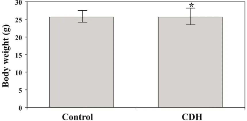
The body weights of the animals were not altered by CDH (P > .05) (Fig. 4). However, CDH promoted a significant decrease in heart weights in comparison to control fetuses (P < .05) (Fig. 5).
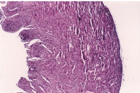
The myocardial structure appeared histologically normal on microscopic examination in control and CDH groups (Figs. 6 and 7), and the histomorphometric studies demon-strated that CDH promoted a signifi-cant decrease in the ventricular wall thickness and an increase in the inter-ventricular septum thickness (Tables 1 and 2).
DISCUSSION
Despite all refinements of the cur-rent management, survival rates of pa-tients with CDH remains frustratingly low because of pulmonary hypoplasia, biochemical immaturity of lungs, and arterial thickening and constriction with persistence of the fetal circulation
Figure 2 - Macroscopic appearance of the fetal lungs and heart. Note that the left lobe of the liver is herniated throughout the surgically created defect indicated by the microscissors, compresses the left lung, and the heart is deviated to the right side (bar corresponds to 1 cm).
Figure 3 - Macroscopic appearance of both lungs and trachea. Note that the left lung is smaller than the right one, due to the compression promoted by the herniated left lobe of the liver (bar corresponds to 1 cm).
Figure 5 - Heart weights of the fetuses (mean ± standard error of the mean). Note that CDH promoted a significant decrease of heart weights (*P < .05).
%$
pattern6. In addition, associated
anoma-lies—primarily cardiac anomalies— account for many of the deaths. The role of hypoplastic heart in the poor prognosis of patients with CDH has been recognized; therefore, hypoplas-tic heart has been used as an ultrasonographic predictor of poor prognosis in prenatally diagnosed fe-tuses14-16.
The primary focus of the present study was to demonstrate whether the herniated viscera to the thorax pro-duced compression over the develop-ing heart and consequently promoted cardiac hypoplasia. First, since the body weights of the animals were not altered by CDH, we concluded that the decrease in the heart weights promoted by CDH was a clear evidence of heart hypoplasia. Second, the histomor-phometric parameters also demon-strated that CDH promoted cardiac hy-poplasia. We suggest that the compres-sion of the herniated viscera in CDH and the resulting bilateral pulmonary hypoplasia, as we demonstrated previ-ously in this model, are responsible for the decreased pulmonary blood flow and decreased right ventricular wall thickness. As a result, the decreased pulmonary venous return to the left side of the heart is also responsible for the left ventricular hypoplasia and de-creased wall thickness. However we could not explain why CDH promoted an increase in the interventricular septum thickness.
There is some evidence that the compressive mechanism of the herni-ated viscera to the lungs and heart is the primary reason for the underdevel-opment and hypoplasia of both organs, mainly in surgically produced CDH. However, in the nitrofen model of CDH, it was shown that heart hypopla-sia was present also in the fetuses with-out hernia13. So we can hypothesize
that in that model, other factors are im-plicated in the hypoplasia other than the anatomic changes or the hypopla-Figure 7 - Microscopic field of ventricular wall from a heart of CDH fetus. Note that the wall is
thinner in comparison to a control fetus, showed in Fig. 6 (Hematoxylin and eosin, original magnification X100).
Table 1 - Ventricular wall thickness (mm).
GROUP CONTROL CDH
Ventricular wall (mean ± sd) (240 measurements) (400 measurements) 1.043 ± 0.349 0.888 ± 0.321* (*P < .05)
Table 2 - Interventricular septum thickness (mm).
GROUP CONTROL CDH
(120 measurements) (200 measurements) Septum (mean ± sd) 1.054 ± 0.262 1.247 ± 0.409* (*P < .05)
%%
sia of the lungs and mechanicalcom-pression. On the other hand, in the nitrofen model of CDH, the heart-re-lated indices utilized to evaluate left ventricular hypoplasia are not signifi-cantly altered, although a global heart hypoplasia is observed17.
The reduction in the heart size cor-responds to hypoplasia rather than to atrophy, because wet-dry weight and DNA-to-protein ratios were similar in CDH and control groups, according to studies in the lamb model of CDH18.
Furthermore, it was shown that prena-tal dexamethasone rescues in part heart hypoplasia in fetal rats with CDH19,
and intrauterine correction of surgically created CDH late in gestation reversed
cardiac hypoplasia in the of fetal lamb model12,20. However, tracheal ligation in
this animal model could not reverse the left ventricular hypoplasia, and this was interpreted as probably caused by the ongoing heart compression pro-moted by the expanding lung in the tra-cheal ligated fetus, as we also showed in this rabbit model of CDH4,5.
We believe that human CDH and other digestive organ atresias are more complex malformations than the surgi-cally produced CDH, although animal models accurately mimic hypoplasia of both lungs and heart.
In summary, the current study is the first report concerning the deleterious effects of the herniated viscera on the
developing heart in a rabbit model of CDH. Certainly this model may be uti-lized for future investigations concern-ing the utilization in the human species of strategies that aim for the prevention or the treatment of heart hypoplasia caused by CDH.
ACKNOWLEDGMENTS
The authors are very grateful to Mrs. Maria Cecilia Mendonça Coelho, Dulcineia Aparecida da Silva, Neide Aparecida da Silva Rosendo dos Santos and to the medical student Rocio M. Gimenez Segovia for their excellent technical assistance.
RESUMO RHCFAP/3057
TANNURI U – Hipoplasia cardíaca em modelo animal de hérnia diafragmática congênita. Rev. Hosp. Clín. Fac. Med. S. Paulo 56(6):173-178, 2001.
Objetivo: Em trabalhos anteriores investigamos um novo modelo experi-mental de hérnia diafragmática congê-nita em coelhos e estudamos também métodos terapêuticos não invasivos para prevenir a imaturidade estrutural e funcional dos pulmões decorrente deste defeito. Além da hipoplasia pul-monar, hipertensão pulpul-monar, imaturi-dade bioquímica e estrutural dos pul-mões, ocorrem alterações hemodi-nâmicas significativas em crianças com hérnia diafragmática congênita. Desta forma, hipoplasia cardíaca tem sido implicada como provável causa de óbi-to em crianças com hérnia diafrag-mática congênita, e interpretada prova-velmente como conseqüência da com-pressão exercida pelas vísceras herniadas durante o desenvolvimento do feto. Este fenômeno tem sido
rela-tado também em modelos experimen-tais de hérnia diafragmática congênita em fetos de ovelhas e ratos. O objeti-vo da presente experiência é o de ve-rificar a ocorrência de hipoplasia car-díaca em nosso novo modelo de hér-nia diafragmática congênita produzida com cirurgia em fetos de coelho.
Métodos: Doze coelhas prenhes foram operadas no 24o ou 25o dia de
gestação (duração total da gestação – 31 a 32 dias), com o objetivo de pro-duzir hérnia diafragmática esquerda em um ou dois fetos em cada mãe. No 30o
dia as coelhas foram novamente ope-radas para retirada dos fetos, que foram pesados e divididos em dois grupos: controle – fetos não operados (n=12) e grupo com hérnia diafragmática (n=9). Os corações foram retirados, pesados e submetidos a estudos histo-lógicos e histomorfométricos.
Resultados: Durante a necropsia verificou-se que em todos os fetos com hérnia diafragmática o lobo esquerdo do fígado sofreu herniação através do defeito produzido cirurgicamente e
ocupou o lado esquerdo do tórax com desvio do coração para a direita, com-pressão do pulmão esquerdo e em con-seqüência, este pulmão encontrava-se menor do que o direito. O peso total dos animais não sofreu alteração em decorrência da hérnia diafragmática, mas os pesos dos corações estavam di-minuídos em comparação aos dos ani-mais do grupo controle. Os estudos histomorfométricos demonstraram que a hérnia diafragmática provocou signi-ficativa redução na espessura da pare-de dos ventrículos e aumento da espes-sura do septo interventricular.
Conclusão: Hipoplasia cardíaca ocorre em modelo de hérnia diafrag-mática congênita. Este modelo pode ser utilizado em investigações sobre métodos terapêuticos que tenham por objetivo prevenção ou tratamento da hipoplasia cardíaca decorrente da hér-nia diafragmática congênita.
%&
REFERENCES
1. DIFIORE JW, FAUZA DO, SLAVIN R et al. - Experimental fetal tracheal ligation reverses the structural and physiological effects of pulmonary hypoplasia in congenital diaphragmatic hernia. J Pediatr Surg 1994;29:248-257.
2. HEDRICK MH, ESTES JM, SULLIVAN KM et al. - Plug the lung until it grows (PLUG): A new method to treat congenital diaphragmatic hernia in utero. J Pediatr Surg 1994;29:612-617. 3. FAUZA DO, TANNURI U, AYOUB AAR et al. - Surgically produced
congenital diaphragmatic hernia in fetal rabbits. J Pediatr Surg 1994;29:882-886.
4. TANNURI U, MAKSOUD-FILHO JG, SANTOSMM et al. - The effects of prenatal intraamniotic surfactant or dexamethasone administration on lung development are comparable to changes induced by tracheal ligation in an animal model of congenital diaphragmatic hernia. J Pediatr Surg 1998;33:1198-1205. 5. TANNURI U, RODRIGUES CJ, MAKSOUD-FILHO JG et al. - The
effects of prenatal intraamniotic surfactant or dexamethasone administration on lung development are comparable to changes induced by tracheal ligation in an animal model of congenital diaphragmatic hernia: Studies of lung glycogen content, elastic fiber density and collagen content. J Pediatr Surg 1998;33 :1776-1783.
6. RODRIGUES CJ, TANNURI U, TANNURI ACA et al. - Prenatal tracheal ligation or intraamniotic administration of surfactant or dexamethasone prevent the structural changes in the pulmonary arteries of surgically created diaphragmatic hernia in rabbits. Rev Hosp Clín Fac Med S Paulo 2002; 57(in press).
7. DIBBINS AW & WIENER ES - Mortality from neonatal diaphragmatic hernia. J Pediatr Surg 1974,9:653-662.
8. KENT GM, OLLEY PM, CREIGTHON RE et al. - Hemodynamic and pulmonary changes following creation of a diaphragmatic hernia in fetal lungs. Surgery 1972;72:427-433.
9. HALLER A – Apud DIBBINS AW & WIENER ES7 .
10. SIEBERT JR, HAAS JE & BECKWITH JB - Left ventricular hypoplasia in congenital diaphragmatic hernia. J Pediatr Surg 1984;19:567-571.
11. SCHWARTZ SM, VERMILLION RP & HIRSCHL RB - Evaluation of left ventricular mass in children with left congenital diaphragmatic hernia. J Pediatr Surg 1994;125:447-451. 12. KARAMANOUKIAN HL, O’TOOLE SJ, ROSSMAN JR et al. - Fetal
surgical interventions and the development of the heart in congenital diaphragmatic hernia. J Surg Res 1996;65:5-8.
13. MIGLIAZZA L, XIA H, ALVAREZ JI et al. - Heart hypoplasia in experimental congenital hernia. J Pediatr Surg 1999;34:706-711. 14. KARAMANOUKIAN HL, O’TOOLE SJ, ROSSMAN JR et al. - Can cardiac weight predict lung weight in patients with congenital diaphragmatic? J Pediatr Surg 1996;31:823-825.
15. BAUMGART S, PAUL JJ, HUHTA JC et al. - Cardiac malposition , redistribution of fetal cardiac output and left heart hypoplasia reduce survival of neonates with congenital diaphragmatic hernia requiring extracorporeal membrane oxygenation. J Pediatr 1998;133 :57-62.
16. KAVVADIA V, GREENOUGH A, LAUBSCHER B et al. -Perioperative assessment of respiratory compliance and lung volume in infants with congenital diaphragmatic hernia: Prediction of outcome. J Pediatr Surg 1997;32:1665-1669.
17. CORREIAPINTO J, BAPTISTA MJ, ESTEVÃOCOSTA J et al. -Heart-related indices in experimental congenital diaphragmatic hernia. J Pediatr Surg 2000;38:1449-1552.
18. KARAMANOUKIAN HL, GLICK PL, WILCOXDT et al. -Pathophysiology of congenital diaphragmatic hernia. 11 Anatomic and biochemical characterization of the heart in the fetal lamb CDH model. J Pediatr Surg 1995;30:925-929.
19. MIGLIAZZA L, XIA HM, ARNAIZ A et al. - Prenatal dexamethasone rescues heart hypoplasia in fetal rats with congenital diaphragmatic hernia. J Pediatr Surg 2000;35:1757-1761.
20. ROSSMAN AS, HOLM BA, AZIZKHAN RG et al. - Tracheal ligation does not reverse the selective left ventricular hypoplasia in fetal lamb model of CDH. Pediatr Res 1995;37:63A.