0103 - 5053 $6.00+0.00
A
r
ti
c
le
*e-mails: [email protected]
Mass Spectrometry of 1,2,5-Oxadiazole N-Oxide Derivatives. Use of Deuterated Analogues in
Fragmentation Pattern Studies
Hugo Cerecettoa, Mercedes González*,a, Gustavo Seoanea, Carola Stankoa, Oscar E. Pirob
and Eduardo E. Castellanoc
a
Departamento de Química Orgánica, Facultad de Química – Facultad de Ciencias, Universidad de la República. CC 1157, Gral. Flores 2124, 11800 Montevideo, Uruguay
b
Departamento de Física, Facultad de Ciencias Exactas, Universidad Nacional de La Plata, CC 67, 1900 La Plata, Argentina
c
Instituto de Física de São Carlos, Universidade de São Paulo, CP 369, 13560 São Carlos - SP, Brazil
Reportamos neste trabalho o estudo sistemático de fragmentação dos derivados de N-óxidos de
1,2,5-oxadiazóis por espectroscopia de massa, usando análogos marcados com deutério para identificar algumas fragmentações críticas. Foi confirmada a perda neutra de CH2O a partir do N2-óxido de
3-hidroximetil-4-fenil-1,2,5-oxidiazol, usando o análogo mono-deuterado. A perda de OH, a partir do oxigênio do N-óxido, por um rearranjo β-H e δ-H, foi claramente verificada a partir do N2-óxido de
3-(4-metilpiperazina-1-metil)-4-fenil-1,2,5-oxidiazol, usando-se o analogo tetra-deuterado adequado. O isômero N-óxido e análogos desoxigenados foram também usados para confirmar a participação
do fragmento óxido no processo de defragmentação.
This paper reported on the study of fragmentation pattern in mass spectrometry of 1,2,5-oxadiazole
N-oxide derivatives involving deuterium-labeled analogues to identify some critical fragmentations. A neutral CH2O loss from 3-hydroxymethyl-N2-oxide-4-phenyl-1,2,5-oxadiazole was confirmed
with the corresponding mono-deuterated analogue. An OH loss, involving the oxygen of N-oxide,
via β-H and δ-H rearrangement, was clearly revealed from 3-(4-methylpiperazine-1-ylmethyl)-N2
-oxide-4-phenyl-1,2,5-oxadiazoleusing the adequate tetra-deuterated analogue. N-oxide isomer and
deoxygenated analogues were also used to confirm the participation of the oxide moiety in the fragmentation process.
Keywords: 1,2,5-oxadiazole N-oxide, D-labeled furoxan, mass fragmentation, EI/MS
Introduction
As part of an ongoing research program on the chemistry and biological characterization of N-oxide containing molecules, a number of 1,2,5-oxadiazole N-oxide, benzo[1,2-c]1,2,5-oxadiazole N-oxide, quinoxaline N,N’ -dioxide, and 1,2,4-triazine N-oxide derivatives were synthesized and evaluated against different biological targets.1 In the course of our synthetic chemical approach we developed 1,2,5-oxadiazole N-oxide derivatives as potential herbicides,2 bioreductive compounds,3-6 and anti-trypanosomal drugs,7,8 using previously described synthetic methods.9 During the structural elucidation of
233 Mass Spectrometry of 1,2,5-Oxadiazole N-Oxide Derivatives
Vol. 15, No. 2, 2004
In this context, the initial aim of the present study was to characterize the fragmentation pattern in mass spectrometry of the 1,2,5-oxadiazole N-oxide derivatives as an alternative structural determination technique. On the other hand, this work describes the elucidation of the fragmentation patterns of these compounds through the use of deuterated derivatives,11 N-oxide isomer and deoxygenated (without N-oxide) analogues.12
Results and Discussion
The spectra for selected derivatives 1-13 (Figure 1) revealed different fragmentation patterns, resulting from different side chains of the 1,2,5-oxadiazole heterocycle at C-3. The most relevant mass spectral data for derivatives 1-13 are presented in Table 1 and Figure 2 rationalizes important fragmentation pathways.
The molecular ion was detected in all cases. However, the abundances were very low for derivatives 4-6, which contain a residue proven to undergo retro-Diels-Alder-fragmentation (morpholine, thiomorpholine, and methylpiperazine).13 The same was observed for derivative 12, that bears an aliphatic side chain on the semicarbazone moiety. The [M-16u]+• ion, corresponding to an oxygen loss, was observed in all cases as a relatively small peak. However, the peak corresponding to m/z [M-17u]+, became increasingly important for derivatives 4-7 and 11 as result of an OH• loss. This kind of fragmentation has been previously described for similar moieties, e.g., a NO2 group losses OH• in o-nitrotoluenes and o-nitroanilines,13 or for other adequately substituted N-oxide heterocycles.14,15 In these compounds, two different rearrangement processes could explain this radical loss, β-H and/or δ-H rearrangement (Figure 3). The high abundance of this fragment ion for
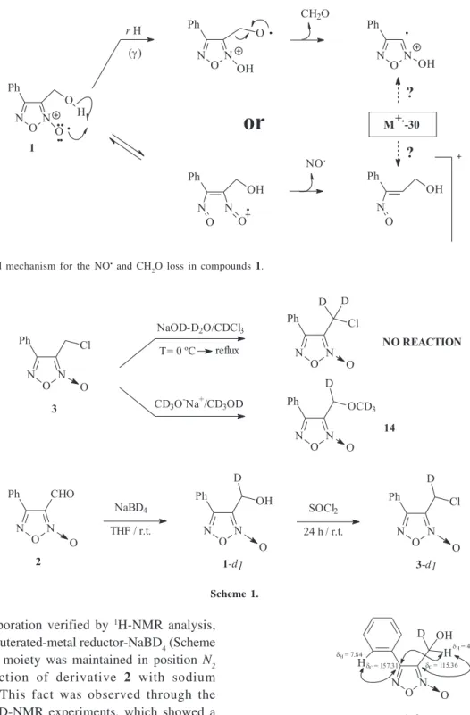
derivatives 4-7 (non-aromatic cyclic amine derivatives) compared to fragment ion abundance’s for derivatives 8-10 (phenylamino derivatives) made us to think that a δ-H rearrangement was the main process in this kind of structures. With a conventional EI/MS equipment it is not possible to study this fact and others, e.g., to determine whether the [M-30u]+ fragment ion in derivative 1 corresponds to a NO loss (as for the other derivatives) or a CH2O loss from the hydroxymethyl substituent in 3-position of the 1,2,5-oxadiazole heterocycle is due to an initial γ-H transference, which produces the stable neutral-product formaldehyde (Figure 4).
In order to explain these critical fragmentations in mass spectrometry, using a EI/MS equipment, we centered our
Table 1. Abundance of the most characteristic fragment ions in the corresponding 1,2,5-oxadiazole N-oxide derivatives EI mass spec-trum
Compound Abundance (%) a,b
No M+• M+•-16 M+•-17 M+•-30 M+•-60 M+•-61
1 15.7 0.5 0.2 20.7 58.0 100.0
2 27.6 0.5 0.2 25.5 62.5 100.0
3 12.0 0.3 0.3 10.6 35.3 1.7
4 0.8 0.9 4.1 0.2 0.3 0.7
5 0.8 1.9 11.8 1.3 0.2 0.3
6 0.3 8.5 50.4 0.2 1.0 0.8
7 10.0 7.1 33.8 0.6 0.2 0.3
8 10.6 1.1 0.1 - c 1.1 2.6
9 12.1 2.1 - - 1.1 2.5
1 0 21.2 3.9 1.7 - 1.6 1.6
1 1 10.6 3.5 11.3 57.6 55.5 6.8
1 2 0.2 0.2 0.8 31.8 7.3 0.3
1 3 2.3 - - 7.5 13.3
-a Analytical conditions for EI/MS: direct injection, ion source
tem-perature 250 ºC, energy 70 eV. b The results are the averages for
three independent experiments. c The “-” denotes that the fragment
ion was not observed.
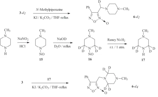
efforts on the synthesis of deuterium analogues of some selected furoxan derivatives.16,17 Initially, we tried to prepare di-deuterium analogue, at the benzylic position, of derivative 6. The synthesis of the di-deuterium chloride 3 was attempted by trying to exchange the “acidic” benzylic-protons using a biphasic system NaOD-D2O/ CDCl3 at different temperatures (room temperature to reflux for 24 h) (as shown in Scheme 1). Unfortunately, 1 H-NMR monitoring of the reaction mixture showed no significant
exchange under these conditions. When CD3O-Na+ / CD3OD was used, exchange took place, but the ether 14 was obtained as the result of the substitution by the powerful nucleophile deuterated methoxide (Scheme 1). These synthetic problems led us to undertake the preparation of the mono-deuterium analogue of derivative 6, at the benzylic position, with 3-d1 as starting material (Scheme 1). Using the aldehyde 2, the mono-deuterium alcohol 1-d1 was produced with more than 95% of
Figure 2. Most characteristic fragment ions in the 1,2,5-oxadiazole N-oxide derivatives EI mass spectrum.
Figure 3. Postulated mechanism for the OH• loss in compounds 4, 5, 6 and 7 (Note: we are gratefully thank to one of the referee for suggesting
235 Mass Spectrometry of 1,2,5-Oxadiazole N-Oxide Derivatives
Vol. 15, No. 2, 2004
deuterium incorporation verified by 1H-NMR analysis, employing the deuterated-metal reductor-NaBD4 (Scheme 1). The N-oxide moiety was maintained in position N2 during the reaction of derivative 2 with sodium borodeuteride. This fact was observed through the corresponding 2D-NMR experiments, which showed a quaternary carbon (HMQC experiment) at 115.36 ppm correlating with a proton at 4.73 ppm (HMBC experiment) and another quaternary carbon (HMQC experiment) at 157.31 ppm correlating with protons at 4.73 ppm and 7.84 ppm (HMBC experiment). These HETCOR experiments allowed us to assign unambiguously the heterocyclic carbon atoms (dC-3 = 115.36 ppm, dC-4= 157.31 ppm) (Figure 5). The product 1-d
1was then transformed in a good yield
into the corresponding chloride 3-d1using thionyl chloride (Scheme 1).
Finally, the amine derivative 6-d1 was obtained by the reaction between chloride 3-d1 and N-methylpiperazine (Scheme 2). The molecular structure of derivative 6-d1 has been determined by X-ray diffraction methods (Figure 6).18,19 In addition, we prepared 6-d
4, the tetra-deuterium
analogue in the 2,6-position of piperazine ring, through reaction between the chloride 3 and the heterocyclic amine Figure 4. Postulated mechanism for the NO• and CH
2O loss in compounds 1.
Figure 5. HMBC correlation for derivative 1-d1.
17 (Scheme 2). This amine was deuterated via the nitrosamine 15, prepared following the Ravindran et al. procedure,20 which was converted into the tetra-deuterium analogue 16 following the Keefer-Fodor methodology.21 To transform nitrosamine 16 into amine 17 we tried a procedure by Kano et al. (reduction with NaBH4:TiCl4 (2:1) in diglyme) with bad results.22 The reduction process did not occur and probably, a complex between the methylpiperazine nitrosamine and TiCl4 was obtained. The use of H2 in Raney-Nickel at room temperature and atmospheric pressure led to compound 17 in an adequate yield.23 The deuterium incorporation in compound 16 was more than 95% (by 1H-NMR analysis).
To know how the N-oxide group position affect on the furoxans’ mass spectrometry behavior, we prepared the N -oxide-positional isomer 3-i. This compound was obtained
via the alcohol 1-i, which was prepared following the Gasco et al. methodology (Scheme 3).10 To study the fragmentation patron of the molecule “N-oxide-free”, we prepared the deoxygenated analogues 1-deoxy and 7-deoxy using Zn in NH4Cl solution as the reduction reagent (Scheme 3).6,24 These products were clearly confirmed through HETCOR experiments (HMQC and HMBC).
Mass spectrometry was carried out on all the analogues developed. The most characteristic peaks in the mass spectrum (EI/MS) for derivatives 1-d1, 1-deoxy, 3-d1,3-i, 6-d
1, 6-d4 and 7-deoxy, together with those for parent
compounds 1, 3, 6 and 7, are presented in Table 2. The results clearly indicate that the fragmentation process of compound 1-d1 does not occur exclusively through a NO• loss, the [M-31u]+• ion (not present in the parent compound 1) probably arise from a CDHO loss. On the other hand, comparing the relative abundance of the M+• and [M-30u]+• ions in compounds 1-deoxyand 7-deoxy, 9.9% and 1.6% respectively, we could notice that the [M-30u]+• ion was more abundant in the first one, this fact is in accordance with the neutral CH2O loss fragmentation process in derivative 1.
The mass spectrum of positional isomer 3-i did not show the [M-17u]+•and [M-31u]+• ions, this fact could be indicative that the parent compound 3 losses OH. and HNO as a β-H participation. Deuterium labeling of compound 6 indicated that the [M-17u]+• ion of this derivative was the result of β-H and δ-H rearrangements (see Table 2). While derivative 6 showed an [M-17u]+• ion abundance of 50.4%, derivatives mono- and tetra-deuterated (6-d1 and 6-d4), that could present the β-H and the δ-H rearrangement phenomena, showed the abundances of the corresponding Scheme 2.
Figure 6. Molecular plot of derivative 6-d1. The ORTEP drawing of
237 Mass Spectrometry of 1,2,5-Oxadiazole N-Oxide Derivatives
Vol. 15, No. 2, 2004
ion ([M-18u]+•) near to 20% (23.0 and 17.0%, respectively). Unfortunately, the mono-deuterium analogue, 6-d1, did not allow us to conclude about the kind of the initial H-rearrangement, because the benzylic position contain one hydrogen and one deuterium. On the other hand, the tetra-deuterium analogue, 6-d4, permitted us to confirm both initial rearrangement pathways, yielding two possible stable radical cations. The high abundance of the [M-18u]+• ion in derivative 6-d4, and the very low abundance in the parent compound, indicated the participation of the deuterium atom in a fragmentation process.
Further the deoxygenated derivatives 1-deoxy and
7-deoxy allowed us to establish that in the parent compounds the loss of oxygen, as expected, principally occurs from the N-oxide moiety. The deoxy-derivatives showed the [M-16u]+• ion in a very low abundance, i.e. abundance ([M-16u]+•) / abundance (M+•) = 0.002 for 1-deoxy and 0.031 for 7-deoxy (compare with the corres-ponding abundance ([M-16u] +•) / abundance (M+•) values for 1 and 7, 0.032 and 0.71 respectively), probably due to an oxygen loss from the heterocycle system 1,2,5-oxadiazole. In the deoxy-analogues the [M-17u]+• ion could be the result of a further H• loss.
Scheme 3.
Table 2. Abundance of the critical fragment ions in the corresponding 1,2,5-oxadiazole N-oxide analogues EI mass spectrum
Abundance (%) a,b
Compound No M+• M+• M+• - 16 M+• - 17 M+• - 18 M+• - 30 M+•- 31 M+• - 61 M+• - 62
1 192 15.7 0.5 0.2 0.6 20.7 - c 100.0 0.8
1-d1 193 25.6 1.7 0.6 1.1 27.8 3.0 100.0 20.4
1-deoxy 176 100.0 0.2 1.2 0.3 9.9 3.5 8.2 6.0
3 210 12.0 0.3 0.3 - 10.6 0.1 1.7 0.1
3-d1 211 12.2 0.2 0.3 0.1 11.4 1.2 4.7 0.3
3-i 210 8.6 0.1 - - 6.9 - 1.6
-6 274 0.3 8.5 50.4 0.4 0.2 0.2 0.8 0.3
6-d1 275 1.2 7.7 45.6 23.0 0.3 - 0.7 0.5
6-d4 278 0.3 8.7 46.2 17.0 - - 0.4 0.1
6-d4
d 278 4.6 17.1 100.0 35.5 0.3 - 2.6 0.9
7 338 10.0 7.1 33.8 0.2 0.6 0.2 0.3
-7-deoxy 322 12.7 0.4 1.7 0.1 0.2 0.1 0.5 1.1
a MS experiments were performed using a Shimadzu MS QP 1100 EX equipment, with EI at 70 eV, with direct insertion probe, the ion source
temperature 250 ºC and the mass range was 40-500 amu. b The results are the averages for three independent experiments. c The “-” denotes that
the fragment ion was not observed. d At 20 eV, ion source temperature 150 ºC. These conditions were used in order to increase the abundance
Experimental
All starting materials were commercially available research-grade chemicals and used without further purification. All solvents were dried and distilled prior to use. All the reactions were carried out in a nitrogen atmosphere. The typical work-up included washing with brine and drying the organic layer with sodium sulphate. Compounds 1-3, 1-i, 1-deoxy, 7, 7-deoxy, 15 and 16 were prepared as previously described.4,6,9,20,21 Elemental analyses were obtained from vacuum-dried samples (over phosphorous pentoxide, 24 h at room temperature) and performed on a Fisons EA 1108 CHNS-O analyzer, and were within ± 0.4% of theoretical values. Infrared spectra were recorded on a Perkin Elmer 1310 apparatus, using potassium bromide tablets; the frequencies are expressed in cm-1. 1H-NMR spectra and HETCOR experiments were recorded on a Bruker DPX-400 (at 400 MHz and 100 MHz) instrument, with tetramethylsilane as the internal reference; the chemical shifts are reported in ppm. MS experiments were performed using the Shimadzu MS QP 1100 EX equipment, with EI at 20 or 70 eV, with direct insertion probe, the ion source was set a 150 ºC or 250 ºC and the mass range was 40-500 amu.
3-(1-Deuterio-1-hydroxymethyl)-N
2
-oxide-4-phenyl-1,2,5-oxadiazole (1-d1). A solution of 2 (1.0 g, 5.3 mmol) in THF (5 mL) was stirred at 0 °C. Sodium borodeuteride (220 mg, 5.3 mmol) was then added, and the resulting solution was stirred for 3 h at room temperature. The solvent was removed in vacuo and the residue was dissolved in EtOAc. After the work-up process the residue was purified by column chromatography (SiO2, petroleum ether:EtOAc (10 to 30%)), to yield 600 mg (59%), mp 65.0-67.0 °C; IR
νmax/cm-1: OH 3400, 1560, 1410, 1050, 770, 680 cm-1; 1H NMR (deuteriochloroform): δ 2.30 (bs, 1H, OH), 4.73 (s, 1H, -CDHOH), 7.57 (m, 3H, phenyl protons), 7.84 (m, 2H, phenyl protons); 13C NMR (HMQC and HMBC experiments) (deuteriochloroform): δ 53.40 (-CDH), 115.36 (-C=N+-O-), 126.55 (-C-phenyl), 128.18 (-C-phenyl), 129.80 (-C-phenyl), 131.76 (-C-phenyl), 157.31 (-C=N). Anal. Calc for C9H7DN2O3: C, 55.96; H, 3.63; N, 14.51%. Found: C, 56.00; H, 3.92; N, 14.35%.
3-(1-Chloro-1-deuteriomethyl)-N
2
-oxide-4-phenyl-1,2,5-oxadiazole (3-d1). A mixture of 1-d1 (300 mg, 1.6 mmol) and SOCl2 (0.19 mL) was stirred at room temperature for 24 h. The reaction mixture was treated with ice, sodium bicarbonate saturated solution (until basic pH), and extracted three times with EtOAc (20 mL). Then of the work-up process the residue was purified by chroma-tography (Al2O3, petroleum ether:EtOAc (0 to 5%)) to yield 300 mg (89%), colorless oil; IR νmax/cm-1: 1601, 1462,
1435, 772, 696 cm-1; 1H NMR (deuteriochloroform): δ 4.57 (s, 1H, -CDHCl), 7.56 (m, 3H, phenyl protons), 7.80 (m, 2H, phenyl protons); 13C NMR (HMQC and HMBC experiments) (deuteriochloroform): δ 32.94 (-CDH), 113.31 (-C=N+-O-), 126.20 (-C-phenyl), 127.84 (-C-phenyl), 129.81 (-C-phenyl), 131.51 (-C-phenyl), 156.40 (-C=N). Anal. Calc for C9H6ClDN2O2: C, 51.06; H, 2.84; N, 13.24%. Found: C, 50.69; H, 2.99; N, 13.00%.
4-(Chloromethyl)-N
2-oxide-3-phenyl-1,2,5-oxadiazole
(3-i). A mixture of 1-i (150 mg, 0.8 mmol) and SOCl2 (0.1 mL) was stirred at room temperature for 24 h. The reaction mixture was treated with ice, sodium bicarbonate saturated solution (until basic pH), and extracted three times with EtOAc (20 mL). Then of the work-up process the residue was purified by chromatography (Al2O3, petroleum ether:EtOAc (0 to 5%)) to yield 110 mg (65%), colorless oil%; IR νmax/cm-1: 1595, 1450, 1415, 770, 690 cm-1; 1H NMR (deuteriochloroform): δ 4.72 (s, 2H, -CH2Cl), 7.58 (m, 3H, phenyl protons), 7.83 (m, 2H, phenyl protons). Anal. Calc for C9H7ClN2O2: C, 51.31; H, 3.33; N, 13.30%. Found: C, 51.07; H, 3.05; N, 12.92%.
3-[1-Deuterio-1-(4-methylpiperazine-1-yl)methyl]-N2 -oxide-4-phenyl-1,2,5-oxadiazole (6-d
1). A mixture of 3-d1
(150 mg, 0.7 mmol), N-methylpiperazine (65 mg, 0.7 mmol), K2CO3 (100 mg, 0.7 mmol), KI (10 mg, 0.07 mmol) and THF as solvent was heated at reflux until absence of chloride (Al2O3, 20% EtOAc in petroleum ether). The solvent was removed in vacuum and the residue was purified by column chromatography (Al2O3, CH2Cl2) to yield 100 mg (51%), mp 97.5-99.5 °C; IR νmax/cm-1: 2926, 2797, 1595, 1574, 1456, 768, 700 cm-1; 1H NMR (deuteriochloroform): δ 2.29 (s, 3H, CH3-N), 2.45 (m, 4H, -CH2N), 2.58 (m, 4H, -CH2N), 3.54 (s, 1H, -CDHN), 7.52 (m, 3H, phenyl protons), 7.92 (m, 2H, phenyl protons); 13C NMR (HMQC and HMBC experiments)
(deuteriochloroform): δ 46.27 (CH3-N), 50.04 (-CDH), 53.10 (-CH2-N), 55.23 (-CH2-N), 113.04 (-C=N+-O-), 127.35 (-C-phenyl), 128.52 (-C-phenyl), 129.47 (-C-phenyl), 131.45 (-C-phenyl), 158.00 (-C=N). Anal. Calc for C14H17DN4O2: C, 61.09; H, 6.18; N, 20.36%. Found: C, 60.96; H, 6.30; N, 20.22%.
239 Mass Spectrometry of 1,2,5-Oxadiazole N-Oxide Derivatives
Vol. 15, No. 2, 2004
the residue was the product 17 (60 mg, 40%), which was used in the next reaction without further purification. 1H NMR (deuteriochloroform): δ 2.00 (s, 1H, NH), 2.35 (s,
3H, CH3-N), 2.36 (m, 4H, -CH2N).
3-[(4-Methyl-2,2,6,6-tetradeuteriopiperazine-1-yl)methyl]-N2-oxide-4-phenyl-1,2,5-oxadiazole (6-d4). A mixture of 3 (121 mg, 0.57 mmol), 17 (60 mg, 0.57 mmol), K2CO3 (80 mg, 0.57 mmol), KI (8 mg, 0.06 mmol) and THF as solvent was heated at reflux until absence of chloride (Al2O3, 20% EtOAc in petroleum ether). The solvent was removed in vacuum and the residue was purified by column chromatography (Al2O3, CH2Cl2) to yield 40 mg (25%), colorless oil; IR νmax/cm-1: 2917, 2849, 1599, 1575, 1456, 767, 699cm-1; 1H NMR (acetone-d
6): δ 2.30 (s, 3H, CH3-N), 2.51 (m, 4H, -CH2N), 3.63 (s, 2H, Ar-CH2N), 7.59 (m, 3H, phenyl protons), 8.02 (m, 2H, phenyl protons); 13C NMR (HMQC and HMBC experiments) (acetone-d
6): δ 45.90
(CH3-N), 50.00 (Ar-CH2-N), 51.80 (-CD2N), 54.00 (-CH2-N), 113.50 (-C=N+-O-), 126.00 (-C-phenyl), 128.50 (-C-phenyl), 129.50 (-C-phenyl), 131.50 (-C-phenyl), 157.50 (-C=N). Anal.Calc for C14H14D4N4O2: C, 60.43; H, 5.04; N, 20.14%. Found: C, 60.08; H, 5.44; N, 20.02%.
Crystallography
Suitable needles shaped single crystals of 6-d1 were obtained by slow evaporation from AcOEt. Derivative 6-d
1
crystallizes in the monoclinic P21/c space group with a = 10.906(3), b = 14.581(3), c = 10.100(2) Å, β = 114.83(2)° and Z=4. The structure were solved from 1455 reflections with I>2s(I) and refined to agreement R1-factors of 0.044. Most H-atoms were detected in a difference Fourier map. However, they were positioned stereochemically and refined with the riding model. The program used to solve and refine the structure was SHELXS.25 The program used to generate the ORTEP graphics was ORTEP-II.19
Conclusions
In summary, by using labeling experiments and adequate analogues, we were able to interpret EI/MS data to elucidate the fragmentation patterns in mass spectrometry of 1,2,5-oxadiazole N-oxide derivatives.
Electronic Supplementary Information
Listings of interatomic bond distances, selected angles, atomic anisotropic displacement parameters, hydrogen atoms positions and isotropic displacement parameters for derivative 6-d1 X-ray studies. Available at http:// jbcs.sbq.org.br as a PDF file.
Acknowledgement
We thank the financial support from CONICYT-Uruguay (Grants 347/95 and 1019/95), Comisión Honoraria de Lucha contra el Cáncer (Uruguay), CONICET-Argentina and by FAPESP-Brazil. Part of the X-ray diffraction experiments were carried out at the National Diffraction Laboratory (LANADI), La Plata, Argentina. We also thank Dr. Ana Denicola and Dr. Alvaro Diaz for critical reading of the manuscript.
References
1. Cerecetto, H.; González, M.; Onetto, S.; Risso, M.; Saenz, P.; Seoane, G.; Bruno, A.M.; Alarcon, J.; Olea-Azar, C.; López de Ceráin, A.; Ezpeleta, O.; Monge, A.; Med. Chem. Res. 2001,
10, 328.
2. Cerecetto, H.; Dias, E.; Di Maio, R.; González, M.; Pacce, S.; Saenz, P.; Seoane, G.; Suescun, L.; Mombrú, A.; Fernández, G.; Lema, M.; Villalba, J.; J. Agric. Food Chem. 2000, 48, 2995.
3. Monge, A.; López de Ceráin, A.; Ezpeleta, O.; Cerecetto, H.; Dias, E.; Di Maio, R.; González, M.; Onetto, S.; Risso, M.; Seoane, G.; Zinola F.; Olea-Azar, C.; Pharmazie 1998, 53, 698. 4. Monge, A.; López de Ceráin, A.; Ezpeleta, O.; Cerecetto, H.; Dias, E.; Di Maio, R.; González, M.; Onetto, S.; Seoane, G.; Suescun, L.; Mariezcurrena, R.; Pharmazie 1998, 53, 758.
5. Cerecetto, H.; González, M.; Risso, M.; Seoane, G.; López de Ceráin, A.; Ezpeleta, O.; Monge, A.; Suescun, L.; Mombrú, A.; Bruno, A.M.; Arch. Pharm. Pharm. Med. Chem. 2000,
333, 387.
6. Boiani, M.; Cerecetto, H.; González, M.; Risso, M.; Olea-Azar, C.; Piro, O.E.; Catellano, E.E.; López de Ceráin, A.; Ezpeleta, O.; Monge-Vega, A.; Eur. J. Med. Chem. 2001, 36, 771.
7. Cerecetto, H.; Di Maio, R.; González, M.; Risso, M.; Saenz, P.; Seoane, G.; Denicola, A.; Peluffo, G.; Quijano, C.; Olea-Azar, C.; J. Med. Chem. 1999, 42, 1941.
8. Aguirre, G.; Cerecetto, H.; Di Maio, R.; González, M.; Porcal, W.; Seoane, G.; Ortega, M.A.; Aldana, I.; Monge, A.; Denicola, A.; Arch. Pharm. Pharm. Med. Chem. 2002, 335, 15.
9. Fruttero, R.; Ferrarotti, B.; Serafino, A.; Di Stilo, A.; Gasco, A.;
J. Heterocyclic Chem. 1989, 26, 1345; Gasco, A.M.; Fruttero, R.; Sorba, G.; Gasco, A.; Liebigs Ann. Chem. 1991, 1211;
Gasco, A.; Mortarini, V.; Ruà, G.; Nano, G.M.; Menziani, E.;
J. Heterocyclic Chem. 1972, 9, 577.
10. Hesse, M.; Meier, H.; Zeeh, B.; Spectroscopic Methods in Organic Chemistry; Thieme: New York, 1997.
11. Suwinski, J.; Svierczek, K.; J. Labelled Compd. Radiopharm.
2002,45, 795.
13. McLafferty, F.D.; Turecek, F.; Interpretation of Mass Spectra,
University Science Books: California, 1993.
14. Paudler, W.W.; Chen, T-K.; J. Org. Chem. 1971, 36, 787.
15. Sasaki, T.; Minamoto, K.; Nishikawa, M.; Shima, T.; Tetrahe-dron 1969, 25, 1021.
16. Hagen, D.F.; Haddad, L.C.; Marhevka, J.S.; Spectrochimica Acta 1987, 42, 253.
17. Mikaya, A.I.; Trusova, E.A.; Zaikin, V.G.; Volinsky, N.P.; Karaulova, E.N.; Galpern, G.D.; Org. Mass Spectrom. 1984,
19, 428.
18. As expected, the 1,2,5-oxadiazole heterocycle is planar to within experimental accuracy, with the oxygen atom of
N-oxide on the ring plane. N-O bond distances in the
hetero-cycle clearly reflect the asymmetry in the bonding structure imposed by the N-oxide moiety. In fact, the single N1-O2
bond is appreciably longer (1.434(3) Å) than the other, par-tially double, N2-O2 bond (1.371(3) Å). The phenyl ring subtends a dihedral angle of 39.20(9)° with the heterocycle.
19. Johnson, C.K.; ORTEP-II. A Fortran Thermal-Ellipsoid Plot Program. Report ORNL-5138, Oak Ridge National Labora-tory: Tennessee, USA, 1976.
20. Ravindran, T.; Jeyaraman, R.; Murray, R.W.; Singh, M.; J. Org. Chem. 1991, 56, 4833.
21. Keefer, L.K.; Fodor, C.H.; J. Am. Chem. Soc. 1970, 92, 5747. 22. Kano, S.; Tamaka, Y.; Sugino, E.; Hibino, S.; Synthesis 1980,
741.
23. Enders, D.; Pieter, R.; Renger, B.; Seebach, D.; Org. Syn.
1978, 58, 113.
24. Aoyagi, Y.; Abe, T.; Ohta, A.; Synthesis 1997, 8, 891.
25. Sheldrick, G.M.; SHELXS-97. Program for Crystal Structure Resolution, University of Göttingen: Göttingen, Germany,
1997.
Received: January 30, 2003
Published on the web: February 27, 2004