0103 - 5053 $6.00+0.00
A
r
ti
c
le
*e-mail: [email protected], [email protected]
Complexes of Thallium(I) and Cadmium(II) with Dipeptides of
L-phenylalanylglycine and Glycyl-L-phenylalanine
Sasan Sharifi,a Davood Nori-shargha,b and Azar Bahadory*,a
a
Chemistry Department, Islamic Azad University, Arak Branch, Arak, Iran
b
Chemistry Department, Islamic Azad University, Science and Research Branch, Tehran, Hesarak, Iran
As constantes de estabilidade dos complexos de íons tálio(I) e cádmio(II) com dipeptídeos de glicil-L-fenilalanina e L-fenilalanilglicina foram determinadas em solução aquosa a 25 °C e meio iônico 0,1 mol dm-3 usando uma combinação de técnicas potenciométricas e
espectrofotométricas. Perclorato de sódio foi usado para manter a força iônica. A composição dos complexos formados foi determinada e observou-se que tálio(I) e cádmio(II) formam duas espécies mononucleares 1:1 com os ligantes, do tipo [Tl(HL)]+, TlL, [Cd(HL)]2+ e [CdL]+ no
intervalo do pH de estudo (1,5-10,5), onde L representa um ligante completamente dissociado. Os logaritmos das contantes de estabilidade cumulativas, βxyz, dos complexos, [(íon metálico)x(H+)
y(ligante)z], são log β111 e log β101: 12,15 e 3,39 (para Tl+ com L-fenilalanilglicina),
11,36 e 2,13 (para Tl+ com glicil-L-fenilalanina), 12,06 e 2,82 (para Cd2+ com L-fenilalanilglicina)
e 10,70 e 1,70 (para Cd2+ com glicil-L-fenilalanina), respectivamente.
The stability constants of the complexes of thallium(I) and cadmium(II) ions with dipeptides of glycyl-L-phenylalanine and L-phenylalanylglycine were determined in aqueous solution at 25 °C and 0.1 mol dm-3 ionic medium using a combination of potentiometric and
spectrophotometric techniques. Sodium perchlorate was used to maintain the ionic strength. The composition of the formed complexes was determined and it was shown that thallium(I) and cadmium(II) forms two mononuclear 1:1 species with the ligands, of the type [Tl(HL)]+,
TlL, [Cd(HL)]2+ and [CdL]+ in the pH range of study (1.5-10.5), where L represents a fully
dissociated ligand. The logarithms of the cumulative stability constants,βxyz, of the complexes, [(Metal ion)x(H+)
y(ligand)z], are log β111 and log β101: 12.15, 3.39 (for Tl+ with
L-phenylalanylglycine), 11.36, 2.13 (for Tl+ with glycyl-L-phenylalanine), 12.06, 2.82 (for Cd2+
with L-phenylalanylglycine), 10.70 and 1.70 (for Cd2+ with glycyl-L-phenylalanine), respectively.
Keywords: L-phenylalanylglycine, glycyl-L-phenylalanine, cadmium(II) complexes, thallium(I) complexes
Introduction
The recent increased use of peptides in biomedical therapy is a result of their large range of activity and specificity, usually with low toxicity and rapid meta-bolization. For the near future, an increasing activity in design and synthesis of new peptide-based drugs is expected, as a result of combined advances in proteomic research and biotechnology. Thus, separation, analysis of peptides and peptide hormones and determination of stability constants of metal complexes with peptides has become increasingly important for an ever-widening range of research disciplines.1
On the other hand, research results have clearly demonstrated that certain transition metal ions play a basic role in directing a number of biochemical processes.2
Many biological processes involve hydrolysis of proteins and peptides. Metal ions can promote the hydrolysis of peptides and related compounds in both homogeneous and heterogeneous systems. Hydrolysis of proteins and peptides has been studied more from a biochemical than from chemical point of view, and since certain proteolytic enzymes require metal ions for activity, hydrolysis reactions and complexes of peptides with metal ions have attracted the attention of chemists.3
contaminated soils and wastes.4 Also, thallium has been
recognized as a toxic element for many years. It produces a variety of adverse effects in human being. This element acts on the central nervous system and induces inflammatory response. However, the metabolic mechanism and fate of thallium toxicity is still not well understood. Since thallium(I) shows marked similarities to that of potassium cation, its interaction with nucleotides, the monomeric units of DNA and RNA, in aqueous would be of a major biochemical interest.5
This revealed the need for comprehensive studies of metal ion-bioligand interactions, as model systems; within this, the investigation of the complex-forming properties of amino acids and peptides is of particular importance.2
The first studies were on metal complexes of polyglycines, the behaviour of the peptide group and, in particular, its acid dissociation being the most interesting problem at that stage.6
Transition metal complexes of amino acids and peptides in aqueous solution were studied by various techniques.6-20
Potentiometry and UV-Vis spectrophotometry are the most widely used methods in the investigation of metal-peptide complexes.6 Many analytical methods use molecular
absorption spectrophotometry of coordination compounds for the determination of metal ions in solution. Consequently, the spectroscopic parameter of molar absorptivity (ε) is of great importance in the practice of a chemical laboratory. Obviously, the interest is focalized on the research of coordination compounds that result in spectrophotometric measurements of great sensitivity, and therefore, high εmax
values. The spectroscopic parameters of molecular absorption
λmax (wavelength at absorption maximum) and εmax (molar absorptivity at absorption maximum) are largely used in chemistry. There are generally three scopes for which the measurement of the absorption spectrum of a substance occurs: the qualitative recognition, the structural examination (these two objectives are in reality connected) and the quantitative determination. It is important to keep in mind, particularly when the study is performed in solution, that the spectra do not only depend on structural conditions of the measurant, but also on the environmental elements (nature of the solvent, pH value of the aqueous solutions, etc.), which make the acquired spectroscopic data to assume a conditional character. Many analytical methods base themselves on the molecular absorption spectrophotometry of coordination compounds for the determination of metal ions in solution; generally, it deals with relatively fast and economic methods, suitable for routine measurements on known matrixes, for which their sensitivity is linked to the εmax of the compound that is made to form in view of the spectrophotometric measurement.21
Therefore has been decided to carry out an equilibrium study of the interaction of peptides with metal ions to determine stability of species formed. These complexes may serve to determine the interactions leading to metal promoted hydrolysis. Also, the knowledge of the distribution of species with pH is a prerequisite for future kinetic studies.3
This work deals with the study of Tl(I) and Cd(II) complexes by L-phenylalanylglycine and glycyl-L-phenylalanine, and determining their stability constants at 25 °C and constant ionic strength 0.1 mol dm-3 sodium
perchlorate. The formation constants of the formed complexes have been compared with similar systems and interpreted.
Experimental
Chemicals
Glycyl-L-phenylalanine was obtained from Merck, while the L-phenylalanylglycine was provided by Fluka (purity ≥ 99%). Both of the ligands were employed without further purification and the aqueous stock solutions of the ligands were freshly prepared daily. The NaOH solution was prepared from a titrisol solution (Merck) and its concentration was determined by several titrations with standard HCl. Perchloric acid, sodium perchlorate, cadmium(II) nitrate and thallium(I) nitrate were from Merck as analytical reagent grade materials and were used without further purification. Dilute perchloric acid solutions were standardized against standard NaOH solution. All the standard solutions were prepared using deionized and twice-distilled water with specific conductance equal to (1.8 ± 0.1)
µΩ-1 cm-1. Ionic strength was adjusted to 0.1 mol dm-3 by
addition of NaClO4.
Apparatus
An Horiba pH-meter, M-12 was used for pH measurements. The hydrogen ion concentration was measured with an Ingold UO 3234 glass electrode and Ingold UO 3236 calomel electrod. Spectrophotometric titrations were performed on a UV-Vis PerkinElmer, Lambada 25 double beam spectrophotometer from 200 to 350 nm and optical path 1.000 cm.
Measurements
calibrated for the relevant H+ concentration with solution
of 0.01 mol dm-3 perchloric acid solution containing 0.09
mol dm-3 sodium perchlorate (for adjusting the ionic
strength to 0.1 mol dm-3). For this standard solution, we
set –log[H+] = 2.0.22 Junction potential corrections have
been calculated from equation 1
– log [H+]
real = – log[H+]measured + a + b[H+]measured (1)
a and b were determined by measuring hydrogen ion concentration for two different solutions of HClO4 with sufficient NaClO4 to adjust the ionic medium.
Procedure
Volumes of 25 cm3 acidic solution of Tl+ and Cd2+ in
the concentrations range 5 × 10⁻⁶ to 5 × 10⁻⁵ mol dm-3
(respectively) were titrated with an alkali solution of each ligand containing a large excess of each ligand (the ratios of metal ion to ligand 1:100). Ionic strength was maintain at 0.1 mol dm-3, in the presence of NaClO
4. The –log[H +]
and absorbance were measured after addition of a few drops of titrant, and this procedure extended up to required –log[H+]. A purified nitrogen atmosphere was
maintained in the vessel during the titrations. In all cases, the procedure was repeated at least three times. The resulting average values and corresponding standard deviation are shown in the Tables 1 and 2.
Results and Discussion
The complex MxHyLz(nx+y-z)+ formed is characterized
by its stoichiometry (x:y:z), where M and L represent the metal ion and each ligand, respectively. To determine the stability constant of the complexation or the protonation, equation 2 is defined by βxyz22
xMn+ + yH+ + zL– M
xHyLz(nx+y-z)+ (2)
) ] [L ] [H ] ]/([M L
H
[Mx y z(nx yz) n x y z
xyz
+ + + − +
=
β (3)
The protonation constants of the ligands have been used for computation of the stability constant, βxyz, of the metal-ligand. The protonation constants of the ligands
have been extensively studied in different kinds of background electrolytes, and the results were reported in the literature. To illustrate, there are L-phenylalanylglycine and glycyl-L-phenylalanin dipeptides in solution to three species: molecular, cationic and anionic.23 For these
dipeptides, we have two stages of the equilibriums
H2L+(aq) + H2O(l) H3O+(aq) + HL(aq)
L ]+ [H
][HL] O [H K
2 3 1
a
+
= (4)
HL(aq) + H2O(l) H3O+(aq) + L
-(aq)
[HL] ] ][L O [H K
-3 2
a
+
= (5)
where Ka1 and Ka2 are stepwise dissociation constants. By used of equations 4 and 5 are obtained Henderson equations. Henderson,s equations shows that, pH is a linear
function to of log[H[HLL]] 2
+ and ] HL [
] L [ log
in acidic and basic titrations, respectively. From the intercept of these equations, the values of pKa1 and pKa2 can be obtained, respectively. In this work, the protonation constants of the ligands have been determined using potentiometric techniques and calculated using a computer program which employs a least-squares method.22
These values are listed in Table 1 together with the values reported in the literature, which are in good agreement with those reported in the literature and curves are given in Figures 1 and 2. The method of determination of the stability constant is based on the relation A = f(pH) on account of the high stability of the complexes studied. Asborbance, A, and –log[H+], were measured for a solution
containing Tl+, Cd2+ with a large excess of each ligand.
Treatments of the spectrophotometric data (each 1nm)
y = 0.6702 x + 3.7037
R2= 0.9994
3.30 3.40 3.50 3.60 3.70 3.80
-0.6 -0.5 -0.4 -0.3 -0.2 -0.1 0.0 0.1
log[HL]/[H2L
+
] pH
Figure 1. Curve pH vs. log [HL]/[H2L+] for L-phenylalanylglycine.
Table 1. Protonation constants of glycyl-L-phenylalanine and L-phenylalanylglycine at 25 °C and ionic strengths, I, 0.1 mol dm-3 (NaClO
4)
Species log β011 log β021 Experimental Conditions Ref.
obtained during the titrations as a function of the H+
concentration were conducted on the computer program. The program allows calculation of stability constant for different models of stoichiometries. The degree of refinement then guides to thechoice between models.
Considering the protonation constants of the ligands, in acidic pH the predominant species for complexation is H2L+
for glycyl-L-phenylalanine and L-phenylalanylglycine. In this case the spectrophotometric titration data were analysed by using the absorbance of Tl+ with each ligand at
wavelengths in UV range that is given by
[HL] ε ] L [H ε ] [TlHL ε ] [Tl ε
A Tl C HL 2 HL
2 +
+ +
= + + +
+ (6)
where εT1 and εC are the molar absorptivities of Tl+ and
each ligand, respectively. For the mass balance
] [TlHL ] [Tl
FTl= + + + (7)
and if α1 and α0 be the fractions of the total concentration HL in the H2L+ and HL, respectively
HL 2 1
F ] L [H α
+
= ,
HL 0
F [HL]
α = (8) , (9)
where FTl and FHL are the total concentrations of Tl and each ligand, respectively. Substituting equations 3 and 7-9 into equation 6 gives the final equation for fitting. The method of determining εTl+ was previously described25 and its values at
different wavelengths are used in this work. Using a suitable computer program22 the data were fitted to the final equation
for estimating the formation constant of equation 3. We used
the Gauss-Newton nonlinear least-squares method in computer program to refine the absorbance by minimizing the error squares sum from equation 8
2 ) (ai bi
U= − (i = 1, 2, 3,...) (10)
where ai is a quasi-experimental and bi is a calculated one. The computer program consisted of two different kind of fitting, (A) graphical, (B) numerical. The final selection of the species was based on both graphical and numerical methods, considering in addition the various statistical criteria, i.e. sums of squared residuals, differences of FTl (exp) and FHL(exp) from those of calculated one. Figure 3 is shown as an example of graphical fitting for the observed and calculated absorbances (from the computer program, by fitting method) of Tl(I)-L-phenylalanylglycine system against –log[H+].
Different models including ML, MHL and several polynuclear and protonated species were tested by the program. As expected, polynuclear complexes were systematically rejected by the computer program, as also were MH2L, MHL2, ML2 and MH2L2. Values for some species were calculated by the program, but the species were not further considered because the estimated error in their formation constants are unacceptable, and their inclusion do not improve the goodness of the fit.
The models finally chosen, formed by Tl(HL)+ and TlL,
for L-phenylalanylglycine and glycyl-L-phenylalanine, resulted in a satisfactory numerical and graphical fitting. The average values for various wavelengths calculated for the stability constants are listed in Table 2.
Figure 2. Curve pH vs. log [L-]/[HL] for L-phenylalanylglycine.
y = 0.9395 x + 7.5236
R2= 0.9998
6.70 6.90 7.10 7.30 7.50 7.70 7.90 8.10
-0.8 -0.6 -0.4 -0.2 0.0 0.2 0.4
log [L-]/[HL] p H
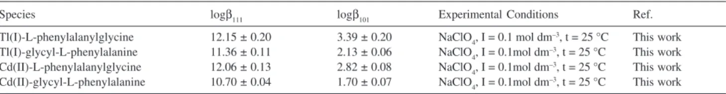
Table 2. Average values of log b111 and log b101 for different wavelengths at 25 oC and ionic strengths, I, 0.1 mol dm-3 (NaClO
4)
Species logβ111 logβ101 Experimental Conditions Ref.
Tl(I)-L-phenylalanylglycine 12.15 ± 0.20 3.39 ± 0.20 NaClO4, I = 0.1 mol dm–3, t = 25 °C This work Tl(I)-glycyl-L-phenylalanine 11.36 ± 0.11 2.13 ± 0.06 NaClO4, I = 0.1mol dm–3, t = 25 °C This work Cd(II)-L-phenylalanylglycine 12.06 ± 0.13 2.82 ± 0.08 NaClO4, I = 0.1mol dm–3, t = 25 °C This work Cd(II)-glycyl-L-phenylalanine 10.70 ± 0.04 1.70 ± 0.07 NaClO4, I = 0.1mol dm–3, t = 25 °C This work
Figure 3. A typical graphical fitting Tl(I)-L-phenylalanylglycine system
at 25 oC, 220 nm, and ionic strength 0.1 mol dm-3 (NaClO
4), (A) experi-mental absorbance, (B) calculated absorbance from the computer pro-gram by fitting method.
1.20 1.40 1.60 1.80 2.00 2.20
1.20 3.20 5.20 7.20 9.20
pH A
Benzyl-containing ligands, L-phenylalanylglycine and glycyl-L-phenylalanine, studied in this work may coordinate thallium(I) via the -COO– and -NH2 donor groups. In the case of Tl(I)-L-phenylalanylglycine system, the computer program suggests that the species MHL is possibly the only one present. This assumption was confirmed by offering some other species, as mentioned before, that all were rejected by the computer program. In Figures 4 and 5 the equilibrium distribution of various species in Tl(I)-L-phenylalanylglycine and glycyl-L-phenylalanine systems are shown as a function of -log[H+],
respectively. The calculations are based on the stability constant values given in Table 2. In the Tl(I)-L-phenylalanylglycine and glycyl-L-phenylalanine systems, Figures 4 and 5, TlL is the dominant species in the –log[H+] ≥ 10, while TlHL+, is formed in the –log[H+]
range 2.4-8.7 with a maximum near –log[H+], 5.4, always
accounts for less than 5% of the total metal ion concentration.
Considering Table 2, complexes formed by Tl(I)-L-phenylalanylglycine are much more stable than those formed by glycyl-L-phenylalanine, and its log β111 is higher than those of glycyl-L-phenylalanine by about 1.1 log units. There is a benzyl group in the ligands, the complexation of thallium(I) by the ligands indicates influence benzyl group on the donor sites -COO– and
-NH2 in L-phenylalanylglycine and glycyl-L-phenylala-nine for complex formation, i.e. -COO– in the
L-phenylalanylglycine preference of thallium(I) for the -COO– donor group in the glycyl-L-phenylalanine
because benzyl group decreases Lewis basicity and nucleophilicity donor sites.
The similar results were obtained for Cd(II)-L-phenylalanylglycine and glycyl-L-phenylalanine systems, Figures 6 and 7, by potentiometric studies. Also, in this report we showed that the complexes formed by Tl(I)-L-phenylalanylglycine and glycyl-L-phenylalanine systems are much more stable than those formed by Cd(II)-L-phenylalanylglycine and glycyl-L-phenylalanine, and this could be due to thallium(I) have much more standard reduction potential and effective nuclear charge than cadmium(II), so we should expect a higher stability constant value for complex formation with thallium(I).
In this work, the study of thallium(I) and cadmium(II) with dipeptides of L-phenylalanylglycine and glycyl-L-phenylalanine interaction was attempted in order to better understand the influence of the presence of a benzyl group in the stability of dipeptides to bind thallium(I) and cadmium(II), also influence type of metal for complex formation.
These assumptions have been confirmed by the finding that the stability constant values of MHL and ML species formed by L-phenylalanylglycine are much higher than the corresponding values of glycyl-L-phenylalanine and the stability constant values thallium(I) complexes with
Figure 4. The equilibrium distribution of the species in
Tl(I)-L-phenylalanylglycine system as a function of –log[H+] at 25 oC and ionic strength 0.1 mol dm-3 sodium perchlorate.
0.00 0.25 0.50 0.75 1.00
TI+
TIHL+
TIL
0.00
mol
fraction
2.00 4.00 6.00 8.00 10.00 12.00
log[H+]
Figure 5. The equilibrium distribution of the species in
Tl(I)-glycyl-L-phenylalanine system as a function of –log[H+] at 25 oC and ionic strength 0.1 mol dm-3 sodium perchlorate.
mol
fraction
0.00 0.25 0.50 0.75 1.00
TI+
TIHL+
TIL
0.00 2.00 4.00 6.00 8.00 10.00 12.00
log[H+]
Figure 6. The equilibrium distribution of the species in
Cd(II)-L-phenylalanylglycine system as afunction of –log[H+] at 25 oC and ionic strength 0.1 mol dm-3 sodium perchlorate.
mol
fraction
0.00 0.25 0.50 0.75 1.00
Cd2+ CdHL
2+
CdL+
0.00 2.00 4.00 6.00 8.00 10.00 12.00
log[H+]
Figure 7. The equilibrium distribution of the species in
Cd(II)-glycyl-L-phenylalanine system as a function of –log[H+] at 25 oC and ionic strength 0.1 mol dm-3 sodium perchlorate.
0.00 0.25 0.50 0.75 1.00
0.00 2.00 4.00 6.00 8.00 10.00 12.00
log[H+]
mo
l
fr
a
cti
o
n Cd2+ CdHL
2+
both of ligands are much higher than the corresponding values for cadmium(II) complexes.
Acknowledgments
Thanks are gratefully extended to the Chemistry Department of Islamic Azad University, Arak Branch, for its invaluable help to this work.
References
1. Sanz-Nebot, V.; Benavente, F.; Toro, I.; Barbosa, J.; J. Chromatogr., A2001, 921, 69.
2. Sóvágó, I.; Petocz, G.; J. Chem. Soc. Dalton Trans. 1987, 7, 1717.
3. Bordignon Luiz, M. T.; Szpoganicz, B.; Rizzoto, M.; Martell, A. E.; Basallote, M. G.; Inorg. Chim. Acta1997, 254, 345.
4. Mavropoulos, E.; da Rocha, N. C. C.; Moreira, J. C.; Bertolino, L. C.; Rossi, A. M.; J. Braz. Chem. Soc.2005, 16, 62.
5. Gharib, F.; Monajjemi, M.; Ketabi, S.; Main Group Met. Chem.
2004,27,71.
6. Daniele, P. G.; Zerbinati, O.; Aruga, R.; Ostacoli, G.; J. Chem. Soc. Dalton Trans. 1988, 5, 1115.
7. Nair, M. S.; Santappa, M.; Natarajan, P.; J. Chem. Soc. Dalton Trans. 1980, 11, 2138.
8. Lau, S.; Sarkar, B.; J. Chem. Soc. Dalton Trans. 1981, 2, 491.
9. Farkas, E.; Sóvágó, I.; Kiss, T.; Gergely, A.; J. Chem. Soc. Dalton Trans. 1984, 4, 611.
10. Daniele, P. G.; Zerbinati, O.; Zelano, V.; Ostacoli, G.; J. Chem. Soc. Dalton Trans.1991, 10, 2711.
11. Luiz, M. T. B.; Szpoganicz, B.; Rizzoto, M.; Martell, A. E.; Basallote, M. G.; Inorg. Chim. Acta1997, 254, 345.
12. Várnagy, K.; Bóka, B.; Sóvágó, I.; Sanna, D.; Marras, P.; Micera, G.; Inorg. Chim. Acta1998, 275, 440.
13. Nold, M. J.; Cerda, B. A.; Wesdeiotis, C.; J. Am. Soc. Mass Spectrom.1999,10, 1.
14. Harrison, A. G.; Csizmadia, I. G.; Tang, T.; J. Am. Soc. Mass Spectrom.2000, 11, 427.
15. Bruni, S.; Cariati, F.; Daniele, P. G.; Prenestil, E.; Spectrochim. Acta, Part A2000, 56, 815.
16. Szabó-Plánka, T.; Nagy, N.; Rockenbauer, A.; Korecz, L.;
Polyhedron2000, 19, 2049.
17. Szabó-Plánka, T.; Árkosi, Z.; Rockenbauer, A.; Korecz, L.;
Polyhedron2001, 20, 995.
18. Argirova, M. D.; Argirova, O. K.; Spectrochim. Acta, Part A
1999, 55, 245.
19. Garib, F.; Zare, K.; Habibi, M.; Tahghvamanesh, A.; Main Group Met. Chem.2002,25, 283.
20. Garib, F.; Zare, K.; Tahghvamanesh, A.; Shamel, A.; Shafiee, G.; Main Group Met. Chem.2002, 25, 647.
21. Prenesti, E.; Daniele, P. G.; Toso, S.; Anal. Chim. Acta2002,
459, 323.
22. Monajjemi, M.; Garib, F.; Aghaei, H.; Shafiee, G.; Thghvamanesh, A.; Shamel, A.; Main Group Met. Chem.2003, 26, 39.
23. Lurie, Ju.; Handbook of Analytical Chemistry, 1st ed., Mir: Moscow, 1975.
24. Gharib, F.; Nasiri, R.; Rev. Inorg. Chem. 2005, 25, 79.
25. Gharib, F.; Zare, K.; J. Sci., Isl. Azad Univ. 1992,2, 397.
Received: December 5, 2006

![Figure 7. The equilibrium distribution of the species in Cd(II)-glycyl-L- Cd(II)-glycyl-L-phenylalanine system as a function of –log[H + ] at 25 o C and ionic strength 0.1 mol dm -3 sodium perchlorate.](https://thumb-eu.123doks.com/thumbv2/123dok_br/18991908.460943/5.892.95.435.897.1052/figure-equilibrium-distribution-species-phenylalanine-function-strength-perchlorate.webp)
