J. Braz. Chem. Soc., Vol. 18, No. 5, 924-927, 2007.
Printed in Brazil - ©2007 Sociedade Brasileira de Química 0103 - 5053 $6.00+0.00
Article
*e-mail: [email protected]
Synthesis and Characterization of ZnS Nanotubes with Crossed-channels
Yun Chen,a Qing-sheng Wu*,a and Ya-ping Dingb
a
Department of Chemistry, Tongji University, Shanghai 200092, P. R. China
b
Department of Chemistry, Shanghai University, Shanghai 200444, P. R. China
Nanotubos de ZnS em forma de X foram sintetizados usando um método hidrotérmico. Uma nova reação hidrotérmica envolvendo hidrólise e oxidação simultâneas do solvente foi desenvolvido usando zinco em pó, água e NH2CSNH2 como reagentes. As imagens de microscopia de transmissão eletrônica mostram que cada segmento dos canais tem cerca de 400 a 500 nm de comprimento. O diâmetro interno é 50 nm e o externo 70 nm e o espaço entre as camadas é cerca de 7 nm. Dados de difração de raios (XRD) mostram que o nanotubo obtido apresenta boa cristalização na fase Esfalerita cúbica. A banda em 293 nm no espectro de absorção no ultravioleta-visível encontra-se deslocado para o azul em cerca de 50 mm em relação ao ZnS
bulk. O espectro de fotoluminescência dos nanotubos de ZnS mostra que o pico de emissão
ocorre em 409 nm quando excitado a 365 nm.
ZnS nanotubes with crossed-channels have been synthesized successfully using a hydrothermal method. A novel hydrothermal reaction of simultaneous solvent-oxidation-hydrolysis has been developed using Zn powder, H2O and NH2CSNH2 as reagents. TEM images show that each branch of the channels is about 400-500 nm long. The inner diameter is 55 nm, the outer diameter is 70 nm, and the interlayer spacing is about 7 nm. XRD data show that the product is well-crystallized in the cubicSphalerite phase. The peak appears at 293 nm in the UV-Vis absorption spectrum, which is about 50 nm blue shifted from the bulk counterpart. The photoluminescence spectrum of ZnS nanotubes shows that the emission peak occurs at 409 nm when excited at 365 nm.
Keywords: ZnS, nanotubes, crossed-channels, simultaneous reaction, hydrothermal
Introduction
There have been great developments in the research of nanomaterials in the recent decades. In contrast, developments in nanotubes seem to be slower, especially in non-carbon nanotubes. The tubular form is particularly attractive because it provides access to three different contact regions: inner and outer surface and tube ends. There are many investigations on carbon nanotubes, including synthesis, physical properties, application etc.1-3 Some
non-carbon nanotubes have been prepared, such as oxides (vanadium oxide, TiO2, RuO2, Al2O3, ZrO2, MoO3, etc.),4-9
layered structures (WS2, MoS2, MoTe2, NiCl2, NbS2, TaS2, InS, Bi, Bi2O3, etc.),10-12 nitrides (GaN, B
24N24),13-14 Si, Te,
noble-metal nanotubes (Ag, Au, Pt, Pd),15-17 titanate
nanotubes18 and copolymer nanotubes,19 but reports on the
synthesis of ZnS nanotubes are scarce.20,21 Most nanotubes
that have been synthesized are open-ended linear tubes. There are many methods to prepare nanotubes, e.g.: arc discharge, plasma enhanced vaporization, chemical vapor deposition, detonation of precursors, templates, hydrothermal etc.22-24 In this work, ZnS nanotubes with
crossed-channels have been synthesized successfully in a hydrothermal system, by the simultaneous solvent-oxidation-hydrolysis reaction, reported herein for the first time. The nanotubes with crossed-channels have potential applications in making nanodevices, nanoreactors, storage devices and can be used in high efficient multiple-catalysis.
Experimental
Synthesis of ZnS nanotubes
925 Chen et al.
Vol. 18, No. 5, 2007
Company, and used without any pretreatment. Deionized water was used as solvent, oxidant, as well as hydrolysis solvent.
0.01 mol of Zn powder and 0.02 mol of thiourea (NH2CSNH2) were put into a Teflon-lined autoclave of 40 mL capacity. Then the autoclave was filled with 25 mL of deionized water. 0.05 g 8-hydroxylquinoline was
added; N2 was passed to eliminate oxygen inside the
solution. The autoclave was sealed and heated at 200 oC
for 60 h, and then it was let to cool to room temperature naturally. The product was collected by filtration, washed with deionized water and absolute ethanol. An offwhite powder was obtained and was preserved in absolute ethanol for further characterization.
Characterization
Morphology analysis of the samples was conducted with H-800 transmission electron microscopy (TEM) operated at 200 kV. The sample, dissolved in ethanol, was deposited on the copper gauze, and was analyzed after the ethanol was volatilized.
X-ray diffraction (XRD) pattern of the products was recorded on a Philips PW1710 X-ray powder diffractometer. Scans were made from 3° to 80° (2θ) at the speed of 2° min-1 (CuKα:
λ=1.5418 Å). The sample was deposited on the glass substrate.
After the ethanol was volatilized, the sample was analyzed. The optoelectronic properties were measured by ultraviolet-visible absorption spectra (UV-Vis, Agilent, 8453). The photoluminescence (PL) of the samples
prepared in ethanol (1 mmol L-1) was observed under
excitation by UV light at 365 nm with a Perkin-Elmer LS-55 fluorescence spectrophotometer.
Results and Discussion
Transmission electron microscopy (TEM) characterization
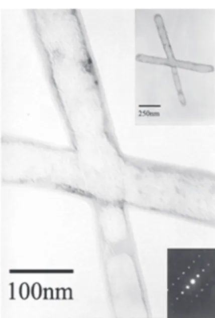
The TEM photograph of the ZnS nanotubes is shown in Figure 1. It can be seen that the nanotubes have crossed-channels. Each branch of the channels is uniform and with the length of about 400-500 nm. The inner diameter is 55 nm, the outer diameter is 70 nm, and the interlayer spacing is about 7 nm. The ends of each branch are closed and are thicker than the walls. The electron diffraction picture (inset to Figure 1) shows that the product is a single crystal with cubic phase. From the TEM figure the nanotubes have a bamboo like shape that may be explained by their formation mechanism. Under the reaction conditions described in this work, bubbles were formed first, due to the Lowest Energy Principle. Then the crystal nuclei grew along the bubbles
face. Finally these bubbles fused and the crystals assembled into nanotubes with crossed channels, induced by 8-hydroxylquinoline. So the bamboo like structure is formed by the bubbles that have not fused completely.
Though the yield was not high (about 20%), to our knowledge the nanotubes with crossed channel described herein are novel structures. Further work is on the way to optimize the experiment conditions and increase the yield.
XRD analysis
Figure 2 shows the XRD patterns of ZnS nanotubes with crossed-channels. ZnS crystalline phase appeared with 2θ = 28.8°(111), 33.3°(200), 47.7°(220), 56.5°(311),
59.3°(222), 69.6°(400), 76.9°(331), respectively, which are close to those reported in the JCPDS card (No. 5-0566). From the XRD pattern it is clear that ZnS nanotubes are well-crystallized in Sphalerite cubic phase with a lattice parameter of a=5.3926Å.
Figure 2. XRD pattern of ZnS nanotubes with crossed-channels.
926 Synthesis and Characterization of ZnS Nanotubes with Crossed-channels J. Braz. Chem. Soc.
UV-Vis and photoluminescence spectra
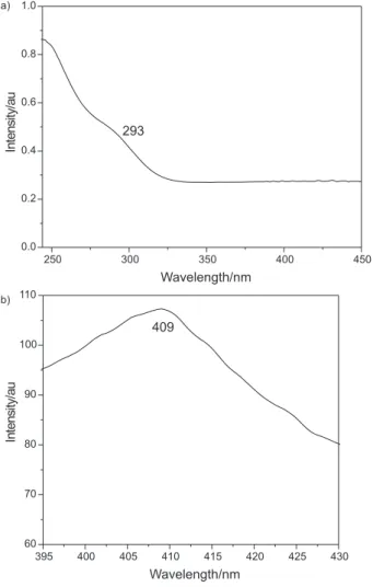
UV-Vis absorption and photoluminescence emission (PL) spectra of the ZnS nanotubes are shown in Figure 3 (a) and (b), respectively. The UV-Vis absorption spectrum (Figure 3a) exhibits an absorption peak at about 293 nm, which is blue shifted with respect to that of the bulk material (326 nm),25 due to quantum-size effects. The
PL spectrum shows a peak at (λ ≈ 409 nm) when excited
at 365 nm (Figure 3b), that corresponds to lower energy
than the ZnS band gap (λ ≈ 330 nm). This can be
attributed to the donor-acceptor (D-A) band transition.26,27
The products can be used as purple light emitting nano-devices because the emission peak is in the visible light area.
Conclusions
A new hydrothermal reaction of solvent-oxidation-hydrolysis to fabricate ZnS nanotubes was successfully
established for the first time. Nanotubes with crossed channels obtained in this work are novel structures. Although the yield is not high, about 20%, this work indicates that ZnS crossed-channeled nanotubes tend to grow under the conditions described; yields will certainly be improved by optimizing the experiment conditions. The products may have potential applications in high efficient multiple-catalysis, photochemical catalysis and multiple-medicine carriers for their novel structures. This work is a preliminary study, and further investigation is underway.
Acknowledgments
The authors are grateful to the financial support of the National Natural Science Foundation of China (Nos. 20471042, 20571051) and the Nano-Foundation of Shanghai in China (Nos. 0652nm007, 0552nm048).
References
1. Ijjima S.; Nature1991, 56, 354.
2. Che G.; Lakshmi B.B.; Fisher E.R.; Martin C.R.; Nature1998, 393, 346.
3. Valentin N. P.; Mater. Sci. Eng., R. 2004,43, 61.
4. Spahr M. E.; Bitterli P.; Nesper R.; Müller M.; Krumeich F.; Nissen, H.U.; Angew. Chem. Int. Ed.1998,37, 1263. 5. Kasuga T.; Hiramatsu M.; Hoson A.; Sekino, T.; Niihara, K.;
Adv. Mater.1999,11, 1307.
6. Min Y. S.; Bae E. J.; Jeong K. S.; Cho Y. J.; Lee J. H.; Choi W. B.; Park G. S.; Adv. Mater.2003, 15, 1019.
7. Enyashin A. N.; Ivanovskaya V. V.; Ivanovskii A. L.; Mendeleev Commun.2004, 3, 94.
8. Shin H.; Jeong D. K.; Lee J.; Sung M. M.; Kim J.; Adv. Mater.
2004, 16, 1197.
9. Pu L.; Bao X.; Zou J. P.; Feng D.; Angew. Chem.2001, 113, 1538.
10. Chen J.; Li S. L.; Gao F.; Tao Z. L.; Chem. Mater.2003, 15, 1012.
11. Yang B. J.; Li C.; Qian Y.T.; Eur. J. Inorg. Chem.2003, 3699.
12. Brorson M.; Hansen T. W.; Jacobsen C. J. H.; J. Am. Chem. Soc.2002, 124, 11582.
13. Han W. Q.; Fan S. S.; Li Q. Q.; Hu Y. D.; Science1997, 277, 1287.
14. Wu H. S.; Xu X. H.; Zhang F. Q.; Jiao H.; J. Phys. Chem. A.
2003, 107, 6609.
15. Lin H. P.; Mou C. Y.; Liu S. B.; Adv. Mater.2000, 12, 103. 16. Mo M. S.; Zeng J. H.; Liu X. M.; Yu W. C.; Zhang S. Y.; Qian Y.
T.; Adv. Mater. 2002, 14, 1658.
17. Kijima T.; Yoshimura T.; Uota M.; Ikeda T.; Fujikawa D.; Mouri S.; Uoyama S.; Angew. Chem. Int. Ed.2004, 43, 228.
Figure 3.UV-Vis (a) and photoluminescence emission (b) spectra of the ZnS nanotubes.
250 300 350 400 450
0.0 0.2 0.4 0.6 0.8 1.0 a) b) In tensi ty/ au Wavelength/nm 293
395 400 405 410 415 420 425 430
927 Chen et al.
Vol. 18, No. 5, 2007
18. Ferreira O.P.; Filho A. G. S.; Filho J. M.; Alves O. L.; J. Braz. Chem. Soc.2006, 17, 393.
19. Stewart S.; Liu G.; Angew. Chem. Int. Ed. 2000, 39, 340. 20. Zhang H.; Pan S.; Li G.; Hou J.; Zhang S.; Nanotechnology
2004, 15, 945.
21. Zhou T. Y.; Xin X. Q.; Nanotechnology2004, 15, 534. 22. Kroke E.; Schwarz M.; Buschmann V.; Miehe G.; Fuess H.;
Riedel R.; Adv. Mater.1999, 11, 158.
23. Shenton W.; Douglas T.; Young M.; Stubbs G.; Mann S.; Adv. Mater.1999, 11, 253.
24. Wang J. W.; Li Y. D.; Adv. Mater. 2003, 15, 445.
25. Yu I; Isobe T.; Senna M.; J. Phys. Chem. Solids1996, 57, 373. 26. Chestnoy N.; Harris T.D.; Hull R.; Brus L.E.; J. Phys. Chem.
1986, 90, 3393.
27. Chen Q.; Li X.; Qian Y. T.; Zhu J.; Zhang Y.; J. Mater. Sci.
1996, 68, 3582.
Received: April 18, 2006