DOI: 10.1590/0004-282X20160117
VIEW AND REVIEW
Far beyond the motor neuron: the role of glial
cells in amyotrophic lateral sclerosis
Muito além do neurônio motor: o papel das células da glia na esclerose lateral amiotrófica
Paulo Victor Sgobbi de Souza1, Wladimir Bocca Vieira de Rezende Pinto1, Flávio Moura Rezende Filho1,
Acary Souza Bulle Oliveira1
Motor neuron disease represents an important group of adult-onset progressive neurodegenerative motor
con-ditions, typiied primarily by upper and lower motor neu
-ron compromise in amyotrophic lateral sclerosis (ALS)1,2.
Amyotrophic lateral sclerosis results from progressive neu-rodegenerative processes of the central nervous system
(CNS) involving the motor cortex, brainstem motor nuclei
and anterior horn of spinal cord3. Speciic clinical, electro
-physiological and neuroimaging indings guarantee a deini
-tive diagnosis by using the revised El Escorial diagnostic
criteria for ALS1. Although alpha motor neuron and
corti-cal upper motor neuron diseases establish the motor clini -cal commitment, its pathophysiologi-cal mechanisms are
wider and involve a complex network of cell interactions,
non-neuronal cell roles and molecular mechanisms involv-ing dysfunction of glial cells. Despite great improvement in its proper clinical, neurophysiological and radiological
diagnosis, pharmacologic management is still based on
symptom exclusion therapy measures4.
Different mechanisms have been postulated as dys -functions in ALS including motor neuron oxidative stress (including endoplasmatic reticulum stress), glutamate direct excitotoxicity, dysfunction in electrolytes and vesic-ular homeostasis, disruption of axonal transport (of
pro-teins and mitochondria along microtubules), exosome and vesicular trafficking (including dysregulated endosomal trafficking), motor neuron apoptosis (including mito
-chondria-mediated mechanisms), dysfunction in the ubiq -uitin-proteasome system and in autophagy, mitochondrial
dysfunction, aberrant RNA metabolism and processing,
and glial cell pathology5,6,7,8. There is also growing evidence
that other underlying hypothetical mechanisms affecting, primarily, the glial cells should include emergent latent
virus infection and misfolded infectious protein (prion-like
1Universidade Federal de São Paulo, Departamento de Neurologia e Neurocirurgia, São Paulo SP, Brasil.
Correspondence: Wladimir Bocca Vieira de Rezende Pinto; Departamento de Neurologia e Neurocirurgia da UNIFESP; Rua Pedro de Toledo, 650; 04023-900 São Paulo SP, Brasil; E-mail: [email protected]
Conflict of interest: There is no conlict of interest to declare.
Received 28 February 2016; Received in inal form 31 May 2016; Accepted 13 June 2016. ABSTRACT
Motor neuron disease is one of the major groups of neurodegenerative diseases, mainly represented by amyotrophic lateral sclerosis. Despite wide genetic and biochemical data regarding its pathophysiological mechanisms, motor neuron disease develops under a complex network of mechanisms not restricted to the unique functions of the alpha motor neurons but which actually involve diverse functions of glial cell interaction. This review aims to expose some of the leading roles of glial cells in the physiological mechanisms of neuron-glial cell interactions and the mechanisms related to motor neuron survival linked to glial cell functions.
Keywords: motor neurons; astrocyte; microglia; amyotrophic lateral sclerosis; motor neuron disease.
RESUMO
A doença do neurônio motor constitui um dos principais grupos de doenças neurodegenerativas, representadas principalmente pela esclerose lateral amiotróica. Apesar dos amplos dados genéticos e bioquímicos em relação aos seus mecanismos isiopatológicos, a doença do neurônio motor se desenvolve sob uma complexa rede de mecanismos não restritos às funções particulares dos neurônios motores alfa, mas, na verdade, envolvendo diversas funções interativas das células da glia. Esta revisão tem como objetivo expor alguns dos principais papéis das células da glia nos mecanismos isiológicos de interações neurônio-glia e os mecanismos relacionados à sobrevivência do neurônio motor ligados a funções das células da glia.
effect)7. This review aims to summarize the most impor
-tant data established on the role of glial cell types in ALS.
PRIMARY GLIAL CELL TYPES AND FUNCTIONS RELATED TO MOTOR NEURODEGENERATION
Most information about physiopathological mecha
-nisms involved in ALS has been through post-mortem his -topathological evaluation and from mouse models related to different induced mutations, mainly the human
trans-genic mouse model with G93A mutation in the gene SOD1
(Cu-Zn superoxide dismutase-1), involved in an autosomal
inherited form of ALS, with overexpression of SOD1 pro
-tein. In motor neuron disease linked to TAR DNA-binding
protein 43 (TDP43), fused in sarcoma protein (FUS), ALS2
gene, VAPB gene, OPTN gene and C9ORF72 gene (
chromo-some 9 open reading frame 72) mechanisms7,9, few data
regarding the involvement of glial cells are available, com
-pared with cases related to SOD1 gene mutations.
here is no doubt about the importance of cellular and
non-neuronal extracellular microenvironments in the
regu-lation of mechanisms involved in neuron survival10 and in
early stage activation of astroglial and microglial cells in
ALS pathogenesis11. Changes in the adequate formation and
balance of the neuron-glia network and abnormal assembly
of its components represent one of the main mechanisms
related to diferent, slowly progressive, focal and global neu -ropsychiatric dysfunctions, such as ALS10.
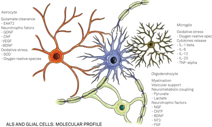
Although nearly all glial cells are involved in ALS patho-genesis, astrocytes and microglia have major roles12 (Figure 1).
Protoplasmic and ibrous astrocytes, microglial cells and oli -godendrocytes represent the major types of glial cells in the CNS and most non-neuronal pathological processes related
to neurodegeneration involve these cell types. he role of
ependymal cells and choroid plexus epithelial cells are not
yet understood, speciically in ALS pathogenesis, and have not been adequately studied10. Mechanisms involving other
glial cell types including Schwann cells, oligodendrocytes and NG2 (nerve-glia factor 2 proteglycan antibody) positive cells are also not well established12.
Neuropathological studies have shown that reactive
astrocytes, microglia activation, and macrophage and T-lymphocyte infiltration of neural tissues overcoming
classical motor neuron dysfunction in ALS3 (Figure 2).
There is a fine immune cross-talk in ALS pathogenesis in a suggestive immunopattern shown by reactive microg
-lia (with a mixture of minor expression of CX3CL1 and its
receptor and CD200 and its receptor and major expression
of IL-6, IL-12, TNF-α and reactive oxygen species);
acti-vated macrophages (with higher expression levels of IL-1β,
COX2 and glycoprotein CD68), reactive astrocytes (raised
SDF-1α); and reactive T-lymphocytes (starting with a
preponderance of regulatory T lymphocytes, and, during
Astrocyte
Gutamate clearance - EAAT2
Neurotrophic fators - GDNF
- CNF -VEGF -BDNF Oxidative stress - SOD
- Oxygen reative species
Microglia
Oxidative stress - Oxygen reative species Cytokines release - IL-1 beta - IL-6 - IL-12 - IL-23 - TNF-alpha
Oligodendrocyte
Myelination Vesicular support Neurometabolic coupling - Pyruvate
- Lactate
Neurotrophic factors - NGF
- CNTF - BDNF - NT3 - FGF
ALS AND GLIAL CELLS: MOLECULAR PROFILE
progression, with a predominance of homolog effector T lymphocytes with reduction of their neuroprotector effect with raised reactive oxygen species, inducible nitric oxide synthase and NOX2/phagocyte oxidase gp91phox)3,13.
Environmental factors and extraneuronal disturbances are
also essential in its immunopathogenesis. For example, it has
also been proven that the direct modulatory efect of vitamin D in glial cell function in patients with ALS afects diferent cell
and extracellular matrix mechanisms: cell-signaling mecha-nisms (glutamate, matrix metalloproteinases,
mitogen-acti-vated protein kinase pathways, Wnt/β-catenin signaling
path-way, prostaglandins and reactive oxygen species release, and nitric oxide synthase), major histocompatibility complex class II molecules, toll-like receptors, poly (ADP-ribose) polymerase-1, heme oxygenase-1, calcium-binding proteins, and a reduced
form of nicotinamide adenine dinucleotide phosphate14.
a1.
b.
a2.
d1.
c1.
c2.
d2.
e2.
f. e1.
ALS Healthy
Figure 2. Glial cells functioning in healthy subjects and in ALS patients. a1. Astrocyte: nourishes motor neurons through the synthesis of amino acids, neurotransmitters and neurotrophic molecules; clears extracellular glutamate via speciic transporters (EAAT2); offers antioxidant defenses. a2. Reactive astrocyte: reduced production of neurotrophic factors; ineficient glutamate clearance; induces metabolic pathways that favor oxidative stress. b. Motor neuron. c1. Microglia: expresses ligands and receptors with neuroprotective roles (CX3CL1/ CX3CL1R, CD200/CD200R); minimal production of oxygen and nitrogen reactive species. c2. Reactive microglia: increased synthesis and release of oxidant agents (H2O2, NO2) and cytokines (IL-6, IL-12, IL-23, TNF-α).
At diferent stages of evolution, there are remarkable changes in neuroinlammation patterns and cell activation in ALS. Briely, there is no doubt that the balance of astro
-cyte neuroprotective or microglia pro-inlammatory func -tions generate a progression, or decrease, in the rate of
pri-mary intrinsic motor neuron degeneration15,16. During the
late stages of ALS, there is a wide increase in cytotoxic T cell iniltration of the spinal cord leading to a pro-neurotoxic pro
-ile of cytokines and chemokines and decreases in local lev
-els of neuroprotective neurotrophic factors, such as GLT1 (SLC1A2) and GLAST (SLC1A3)16. Next, we will sumarize
the most relevant physiopathological functions described in
each glial cell type.
ASTROCYTE ROLE IN ALS PATHOGENESIS
Although classically related to neuronal regulation func-tions, astrocytes are involved in regulation of the extracel-lular microenvironment in neurotransmitters at synapses and during their developmental stages; control of signal-ing of CNS vascular development (includsignal-ing modulation of
blood-brain-barrier development); neurotrophic support and
stimulation for diverse neurons, maintenance of intercellular signaling (including modulation of excitatory synaptic trans-mission via release and propagation of glutamatergic
stim-uli); and neurometabolites and ionic regulation and homeo
-stasis (acid and luid equilibrium)3,8,13,17,18,19.
he main mechanism involving astrocytes in ALS
pathophysiology is dysfunction of glutamate transporters20
with loss of the astroglial glutamate transporter EAAT2 (by aberrant RNA splicing, exon skipping and intron reten -tion) in the motor cortex and in the anterior horn of the spinal cord, disclosing its important function in excitotoxic damage in sporadic ALS. As previously mentioned, astro-cytes participate in glutamate clearance from the synaptic
clefts providing more balanced levels of extracellular excit
-atory neurotransmission, the defective reuptake of which being the key mechanism in mouse models linked to EAAT2 transporter dysfunctions. It has also been proved in mutant
SOD1 mice that astrocytes directly regulate the expression
of glutamate receptor subunit GluR2 in AMPA receptors
of motor neurons12. Other proven mechanisms included
impaired release of multiple neurotrophic factors, includ-ing glial-derived neutrotrophic factor, ciliary neurotrophic
factor, vascular endothelial growth factor and brain-derived
neurotrophic factor12. Astrocytes with SOD1 gene
muta-tion produce reactive oxygen species and soluble mole
-cules with a selective toxicity pattern to spinal cord motor
neurons. Another proven mechanism in the SOD1 mouse
model includes activation of the pro-nerve growth factor (NGF)-p75 receptor-signaling pathway involved in direct
astrocyte toxicity to motor neurons8.
It has also been established that high levels of cyclo
-oxygenase 2 is involved in prostaglandin E2 synthesis and
hyperstimulation of NMDA glutamate receptor activation
of COX2 and subsequent production of reactive oxygen spe
-cies and prostaglandin E2, and this enhances glutamate
release from astrocytes20.
THE ROLE OF MICROGLIA IN ALS PATHOGENESIS
Microglia represent the major primary resident
phago-cytic immune cells of the CNS associated with some
astrocyte immune functions, secreting pro-inflammatory
immune response molecules including cytokines and chemokines and anti-inflammatory molecules during
resolution of neural damage, and stimulating the release
of neurotrophic growth factors. Microglia result from dif
-ferentiation of precursors of the monocyte/mesodermal
lineage of hematopoietic stem cells that normally protect
against microbial infection, abnormal aggregated protein, immunoglobulin-antigen complexes and microhemor -rhagic content3,21.
Microglial activation is a common hallmark of many
neurodegenerative diseases, including ALS, despite the fact that it results mainly from proliferation of myeloid pre-cursor cells22. Activated microglia releases proinlamma
-tory cytokines (tumor necrosis factor-α, interleukin-1β,
interleukin-12, interferon-γ), mitogenic factors (monocyte chemoattractant protein 1, macrophage colony stimulating
factor), neurotrophic factors (insulin-like growth factor-1), and anti-inlammatory cytokines (tumor growth factor-β)12.
here is a direct neuropathological correlation of microg
-lial activation with severity of upper motor neuron damage. Diminishing the toxicity of mutant SOD1 transgene within microglia of mice has been shown to signiicantly slow dis -ease progression of ALS23. It has also been shown that the
CCAAT/enhancer binding protein-β is enhanced in acti-vated microglial cells of the spinal cord of ALS mouse
mod-els with SOD1 gene mutation, promoting higher expression of nitric oxide synthase-2, cyclooxygenase-2 and upregulation
of other proinlammatory gene expression24.
Reactive oxygen species and cytokines increase motor neuron susceptibility to glutamate excitotoxicity and inhibit
expression of astrocytic glutamate transporters diminishing
glutamate uptake and perpetuating this neurotransmitter’s
neurotoxicity mechanism25.
Nuclear factor-kappa B (NF-кB) is upregulated in the
spi-nal cord of mouse models and patients with ALS, although inhibitory efects of its pathway in astrocytes did not prevent neurodegeneration or rescue motor neuron death induced by microglia. However, the modulatory efect in NF-кB pathway impairs proinlammatory activation of microglia26.
Neuropathological studies have also established the acti
of ALS evolution25. It is widely known that the peripheral
immune system represents a crucial stage in the main patho-physiological mechanism: T-lymphocytes directly cross the
bood-brain barrier, and interact with resident primary microg
-lia triggering two diferent immunophenotypes depending on
the clinical and pathological stage of the ALS: an M2 protective
anti-inlammatory proile in early processes involving regula
-tory T-cells and IGF-1, or, an M1 cytotoxic proile in late pro
-cesses induced by fractalkine (CX3CL1) and CD200 involving h1 cells and interleukin, and other substances such as IL-1β, IL-6, IL-12, IL-23, reactive oxygen species (mainly H2O2) and
TNF-α15,27. Microglia production of IL-10, induced by cytokines
from leptomeningeal cells, has also been shown.
OTHER GLIAL CELLS INVOLVED IN ALS PATHOGENESIS
NG2 cells, synantocytes or pericytes, participate in CNS immune mechanisms of defense, producing new astro -cytes, oligodendrocytes and neurons, in some situations
and speciic areas of the CNS. NG2 cells become astrocytes by proinlammatory cytokine signaling in most ALS stages12.
Oligodendrocytes also regulate local neuronal microenvi
-ronments. hey represent the main myelinating cells of the
CNS28. Oligodendrocytes are involved in mechanisms of cen
-tral myelination and provide metabolic sustenance to motor
neurons. Amyotrophic lateral sclerosis does not represent a primary oligodendrocyte disease.
he efective role of NG2+ cells in the astrogliosis
pro-cess in ALS is still uncertain. NG2+ cells produce new
oligodendrocytes, neurons and perhaps astrocytes after an injury, changing the cellular microenvironment. In ALS, cell proliferation rates are enhanced in regions of motor
neuron degeneration, but myelination and remyelination
defects are also found3. Few studies have explored the role
of myelinated oligodendrocytes in ALS pathogenesis, which inluence motor neurons through neurotrophic factors
release and regeneration after neuronal injury. It has also
been shown how oligodendrocytes and their NG2+
progeni-tors mediate neuron loss in ALS, the abnormal rate of pro -liferation in mouse models secondary to oligodendrocyte
degeneration, and to the progressive neuroinlammatory and neurodegenerative processes. he role of Schwann cells in ALS pathogenesis remains undeined12.
FINAL REMARKS
Motor neuron disease represents an important group
of neurodegenerative disorders, mainly represented by ALS.
Primary alpha motor neuron involvement and degeneration and secondary mechanisms including glial cell pathological processes represent the most important features in the patho-physiology of ALS. Astrocyte and microglial dysfunctions have
been widely demonstrated in patients and animal models of ALS. More studies are needed to establish speciic molecular features linked to glial dysfunction at the microcellular level and its extracellular involvement, to allow new pharmacologi
-cal perspectives and provide data about the natural history of the diferent genetic forms and variants of ALS.
References
1. Oliveira AS, Pereira RD. Amyotrophic lateral sclerosis (ALS): three letters that change the people’s life. For ever. Arq Neuropsiquiatr. 2009;67(3A):750-82. doi:10.1590/S0004-282X2009000400040
2. Radunović A, Mitsumoto H, Leigh PN. Clinical care of patients with amyotrophic lateral sclerosis. Lancet Neurol. 2007;6(10):913-25. doi:10.1016/S1474-4422(07)70244-2
3. Rizzo F, Riboldi G, Salani S, Nizzardo M, Simone C, Corti S et al. Cellular therapy to target neuroinlammation in amyotrophic lateral sclerosis. Cell Mol Life Sci. 2014;71(6):999-1015. doi:10.1007/s00018-013-1480-4
4. Andrews J. Amyotrophic lateral sclerosis: clinical management and research update. Curr Neurol Neurosci Rep. 2009;9(1):59-68. doi:10.1007/s11910-009-0010-0
5. Shi P, Wei Y, Zhang J, Gal J, Zhu H. Mitochondrial dysfunction is a converging point of multiple pathological pathways in amyotrophic lateral sclerosis. J Alzheimers Dis. 2010;20:S311-24. doi:10.3233/JAD-2010-100366
6. Brites D, Vaz AR. Microglia centered pathogenesis in ALS: insights in cell interconnectivity. Front Cell Neurosci. 2014;8:117. doi:10.3389/fncel.2014.00117
7. Sica RE, Nicola AF, Deniselle MC, Rodriguez G, Monachelli GM, Peralta LM et al. Sporadic amyotrophic lateral sclerosis: new hypothesis regarding its etiology and pathogenesis suggests that astrocytes might be the
primary target hosting a still unknown external agent. Arq Neuropsiquiatr. 2011;69(4):699-706. doi:10.1590/S0004-282X2011000500023
8. Ferraiuolo L, Kirby J, Grierson AJ, Sendtner M, Shaw PJ. Molecular pathways of motor neuron injury in amyotrophic lateral sclerosis. Nat Rev Neurol. 2011;7(11):616-30. doi:10.1038/nrneurol.2011.152
9. Mackenzie IR, Rademakers R, Neumann M. TDP-43 and FUS in amyotrophic lateral sclerosis and frontotemporal dementia. Lancet Neurol. 2010;9(10):995-1007. doi:10.1016/S1474-4422(10)70195-2
10. Araque A. Astrocyte-neuron signaling in the brain: implications for disease. Curr Opin Investig Drugs. 2006;7(7):619-24.
11. Neusch C, Bähr M, Schneider-Gold C. Glia cells in amyotrophic lateral sclerosis: new clues to understanding an old disease? Muscle Nerve. 2007;35(6):712-24. doi:10.1002/mus.20768
12. Lasiene J, Yamanaka K. Glial cells in amyotrophic lateral sclerosis. Neurol Res Int. 2011;2011:718987. doi:10.1155/2011/718987
13. Blackburn D, Sargsyan S, Monk PN, Shaw PJ. Astrocyte function and role in motor neuron disease: a future therapeutic target? Glia. 2009;57(12):1251-64. doi:10.1002/glia.20848
14. Long K, Nguyễn LT. Roles of vitamin D in amyotrophic lateral
sclerosis: possible genetic and cellular signaling mechanisms. Mol Brain. 2013;6(1):16. doi:10.1186/1756-6606-6-16
16. Philips T, Robberecht W. Neuroinflammation in amyotrophic lateral sclerosis: role of glial activation in motor neuron disease. Lancet Neurol. 2011;10(3):253-63. doi:10.1016/S1474-4422(11)70015-1
17. Sofroniew MV, Vinters HV. Astrocytes: biology and pathology”. Acta Neuropathol. 2010;119(1):7-35. doi:10.1007/s00401-009-0619-8
18. Boillée S, Vande Velde C, Cleveland DW. ALS: a disease of motor neurons and their nonneuronal neighbors. Neuron. 2006;52(1):39-59. doi:10.1016/j.neuron.2006.09.018
19. Maragakis NJ, Rothstein JD. Mechanisms of Disease: astrocytes in neurodegenerative disease. Nat Clin Pract Neurol. 2006;2(12):679-89. doi:10.1038/ncpneuro0355
20. Seifert G, Schilling K, Steinhäuser C. Astrocyte dysfunction in neurological disorders: a molecular perspective. Nat Rev Neurosci. 2006;7(3):194-206. doi:10.1038/nrn1870
21. Graeber MB, Streit WJ. Microglia: biology and pathology. Acta Neuropathol. 2010;119(1):89-105. doi:10.1007/s00401-009-0622-0
22. Gowing G, Philips T, Van Wijmeersch B, Audet JN, Dewil M, Van Den Bosch L et al. Ablation of proliferating microglia does not affect motor neuron degeneration in amyotrophic lateral sclerosis caused
by mutant superoxide dismutase. J Neurosci. 2008;28(41):10234-44. doi:10.1523/JNEUROSCI.3494-08.2008
23. Yamanaka K, Yamashita H. [ALS and microglia: a player for non-cell-autonomous neuron death]. Brain Nerve. 2007;59(10):1163-70. Japanese.
24. Valente T, Mancera P, Tusell JM, Serratosa J, Saura J. C/EBPβ expression in activated microglia in amyotrophic lateral sclerosis. Neurobiol Aging. 2012;33(9):2186-99. doi:10.1016/j.neurobiolaging.2011.09.019
25. Henkel JS, Beers DR, Zhao W, Appel SH. Microglia in ALS: the good, the bad, and the resting. J Neuroimmune Pharmacol. 2009;4(4):389-98. doi:10.1007/s11481-009-9171-5
26. Frakes AE, Ferraiuolo L, Haidet-Phillips AM, Schmelzer L, Braun L, Miranda CJ et al. Microglia induce motor neuron death via the classical NF-κB pathway in amyotrophic lateral sclerosis. Neuron. 2014;81(5):1009-23. doi:10.1016/j.neuron.2014.01.013
27. Zhao W, Beers DR, Appel SH. Immune-mediated mechanisms in the pathoprogression of amyotrophic lateral sclerosis. J Neuroimmune Pharmacol. 2013;8(4):888-99. doi:10.1007/s11481-013-9489-x