DOI: 10.1590/0004-282X20160122 ARTICLE
Autonomic thermoregulatory dysfunction in
neurofibromatosis type 1
Disfunção autonômica termorregulatória na neurofibromatose do tipo 1
Luciana G Madeira1, Renata LF Passos1, Juliana F de Souza2, Nilton A Rezende2, Luiz O. C. Rodrigues1,2
Neuroibromatosis type 1 (NF1) is an autosomal dom -inant disorder, caused by mutations in a single gene (OMIM #162200, neuroibromin, 17 q11.2) afecting the development-maintenance-repair of neural and cutaneous tissues. Neuroibromatosis type 1 is the most common hu -man monogenetic disease (1:3000, afecting nearly 80,000 Brazilian people) and it exhibits marked phenotype expres -sion variability and an unpredictable course1,2,3.
Recently, we described decreased muscular strength4
and lower aerobic capacity in NF1 individuals5
, and both fea -tures are related to life expectancy, and quality of life. Possible mechanisms involving NF1-reduced muscle strength and
aerobic capacity could be neurological abnormalities related to the neuroibromin deiciency, such as poorer motor coor -dination/activation, as well as lower levels of daily physical activities and motivation for exercising. Both neural disor -der and reduced aerobic capacity could adversely afect ther -moregulatory capacity, leading to decreased exercise perfor -mance in hot environments and heat intolerance with higher risk for heat-related injuries6.
Despite increases in environmental temperature and/or exercise body metabolism, human internal tem -perature must be maintained within a small physiologi -cal range through heat dissipation, to prevent tissue
1Universidade Federal de Minas Gerais, Escola de Educação Física, Fisioterapia e Terapia Ocupacional, Programa de Pós Graduação em Ciências do Esporte,
Belo Horizonte MG, Brasil;
2Universidade Federal de Minas Gerais, Hospital das Clínicas, Centro de Referência em Neuroibromatoses, Belo Horizonte MG, Brasil. Correspondence: Luiz O. C. Rodrigues; R. RL Aroeira, 40; 31710-570vBelo Horizonte MG, Brasil; E-mail: [email protected]
Support: CNPq, FAPEMIG, CAPES, UFMG
Conflict of interest: There is no conlict of interest to declare.
Received 27 April 2016; Received in inal form 08 June 2016; Accepted 20 June 2016. ABSTRACT
Objective: Neuroibromatosis type 1 (NF1) causes neural and cutaneous disorders and reduced exercise capacity. Exercise/heat exposure increasing internal temperature must be compensated by eccrine sweat function and warmed skin vasodilation. We suspected NF1 could adversely affect eccrine sweat function and/or vascular thermoregulatory responses (VTR). Methods: The eccrine sweat function and VTR of 25 NF1 volunteers (14 males, 11 females; 16–57 years old) were compared with 23 non-NF1 controls matched by sex, age, height and weight (CG). Sweating was induced by 1) pilocarpine 1% iontophoresis (PILO); and 2) by passive heating (HEAT) via the lower third of the legs being immersed in 42°C water for one hour. Previously established eccrine sweat function and VTR protocols were used. Results: The NF1 group showed: a) lower sweat rate than the CG group during PILO; b) a smaller diastolic pressure decrease; and c) higher tympanic temperatures than controls during HEAT (p < 0.05). Conclusion: Reduced sweating and vascular thermoregulatory responses suggest autonomic dysfunction in NF1 individuals.
Keywords: neuroibromatosis 1; sweating; primary dysautonomias; body temperature regulation.
RESUMO
Objetivo: Neuroibromatose do tipo 1 (NF1) causa problemas neurais e cutâneos e diminuição da capacidade física. O aumento da temperatura interna durante exercício e exposição ao calor precisa ser compensada pela função sudorípara écrina (FSE) e aquecimento cutâneo por vasodilatação (RVT). Suspeitou-se clinicamente que a NF1 poderia prejudicar a FSE e a RVT. Métodos: A FSE e RVT de 25 voluntários com NF1 (14 homens, 11 mulheres; 16–57 anos) e de 23 sem-NF1, emparelhados por sexo, idade, estatura e peso corporal, foram medidas com protocolos validados anteriormente. A sudorese foi induzida por iontoforese com pilocarpina (PILO) e aquecimento passivo por imersão das pernas em água a 42°C durante uma hora (HEAT). Resultados: O grupo NF1 apresentou menor taxa de sudorese na situação PILO, menor redução da pressão diastólica e maior temperatura timpânica na situação HEAT (p < 0.05). Conclusão: As respostas sudorípara e vascular reduzidas sugerem disfunção autonômica nas pessoas com NF1.
damage, especially in the brain7. he main human physio -logical mechanisms to dissipate metabolic heat consist of increased skin temperature, through cutaneous vasodila -tion and sweat produc-tion and vaporiza-tion, both simulta -neously triggered by increased internal and skin tempera -tures. Individual characteristics may afect the magnitude of thermoregulatory responses, including sex, aerobic ca -pacity, as well as the existence of any disease6,8,9.
As an example, the neuropathy caused by progressive central demyelization in multiple sclerosis produces system -ic failure in maintaining internal temperature during heat exposure due to decreased neural control of cardiovascular responses and impaired sweat function. As a result, most multiple sclerosis patients experience transient and tempo -rary worsening of clinical signs and neurological symptoms during exposure to higher environmental temperatures and exercise10. Additionally, exercise intolerance and sweating
dysfunction are considered common manifestations of dia -betic autonomic neuropathy, even in the early stages of this disease, once the sweating response is diminished or com -pletely suppressed, especially in lower limbs11,12.
Neuropathy in NF1 patients has been related to nerve compression and/or loss of function by the growth of neu -roibromas, or to inappropriate genetic signaling between neural cells, and it is considered to be a benign condition. Polyneuropathies and neuroibromatous neuropathy have been rarely reported in 1.3% to 6% of NF1 patients, depend -ing on the investigational methods applied13,14,15. However,
to the best of our knowledge, central and peripheral ther -moregulatory responses to heat stress have not been stud -ied in NF1. Moreover, the reduced exercise functional ca -pacity5
together with heat intolerance complaints in our clinical observations led us to hypothesize a thermoregula -tory dysfunction in NF1 patients.
herefore, the aim of this study was to compare auto -nomic thermoregulatory responses of NF1 individuals with non-NF1 controls matched by sex, age, height and weight.
METHODS
We assessed 14 males and 11 females with NF1 (NF1 group), from 16 to 57 years of age, selected among patients of the Neuroibromatosis Outpatient Reference Center. hey met at least three NF1 diagnostic criteria (National Institutes
of Health Consensus Development Conference, 1988). he con
-trol group (CG) comprised 23 non-NF1 individuals (12 males and 11 females) matched by sex, age, and anthropometric features to the NF1 group.
hese experiments were conducted between February 2013 and February 2014. he Research Ethics Committee of the Universidade Federal de Minas Gerais approved this study and the procedures were performed according to the norms of Resolution 196 of the Brazilian National Health
Council (1996) on scientiic research involving humans. All volunteers provided written informed consent prior to par -ticipating in the trials.
he sweating responses were induced twice: by local stim -uli via administration of pilocarpine 1% (PILO) using a previ -ously validated protocol9, and by central stimuli, through a passive heating protocol (HEAT). In both situations, volun -teers remained seated during all tests after anthropometric (body weight, height, and body surface area) and hydration status (urine speciic gravity) evaluations were performed. Additionally, tympanic temperature (Tty) and skin tempera -tures (Tsk) were monitored every 15 and 2 minutes, respec -tively, by speciic infrared thermometers (G-TECH® IR1DB1, Brazil and FLUKE 566, Brazil).
At first, sweat secretion was locally induced at the proximal anterior face of the right forearm by local ad -ministration of pilocarpine hydrochloride (4 mL; 1%) using iontophoresis (1.5 mA, 60 μA.cm-2, 5 min), under
neutral environmental temperature conditions (~24° C). To prevent individual variations in local skin tempera -ture, the forearm was heated for 15 minutes prior to phar -macological stimulation and its temperature was main -tained (~35° C) throughout the experiment, using radiant heat from an adjustable infrared emission lamp (Quartz®, Brazil). It has been shown previously that these proce -dures induce maximal sweating to pilocarpine, irrespec -tive of sex and aerobic capacity9.
Sweat secretion was collected for 15 minutes just after stimulation using a 16 cm2 absorbent paper (J Prolab, Brazil)
placed inside a plastic chamber to prevent sweat evaporation. Active sweat glands (ASG) were printed using the iodine im -pregnated paper technique, and quantiied (gland.cm-1) via
digital processing using a free computer program (ImageJ) as previously described15. he local sweat rate (SR) was de -termined based on diferences in weight of the absorbent pa -per before and after sweat collection (Mettler Toledo AB 204 analytical scale, USA) divided by its area and time of collec -tion. Finally, sweat gland output (SGO) was calculated by di -viding SR by ASG.
Statistical analysis
In the PILO experiments, the Student’s t-test was used to assess diferences between the groups in physical character -istics, SR, ASG, and SGO, and one-way ANOVA with repeated measures was used to evaluate diferences between groups, and over time, in Tty and Tsk. In the HEAT experiments, SR, ASG and SGO diferences were assessed using two-way ANOVA, with the groups and body areas as factors.
During the entire duration of the HEAT experiments, one-way ANOVA or two-way ANOVA with repeated mea -sures was used to compare Tty among groups and Tsk, heart rate and arterial pressure parameters between the groups and body areas, respectively. ANOVA analysis was followed by Fisher’s least-signiicant diference post-hoc test when applicable. Finally, physical characteristics, in both experi -ments, and heat storage rate in HEAT, were evaluated with the Student’s t-test. Signiicance was established at α = 0.05 and results are presented as mean ± standard deviation.
RESULTS
Physical characteristics of males and females in the NF1 and CG groups are shown in Table 1. he NF1 and CG were successfully matched and there were no diferences between them in age, height, weight or body surface area. However, both NF1 and control group men were taller, heavier and pre -sented a bigger body surface area than women, as expected.
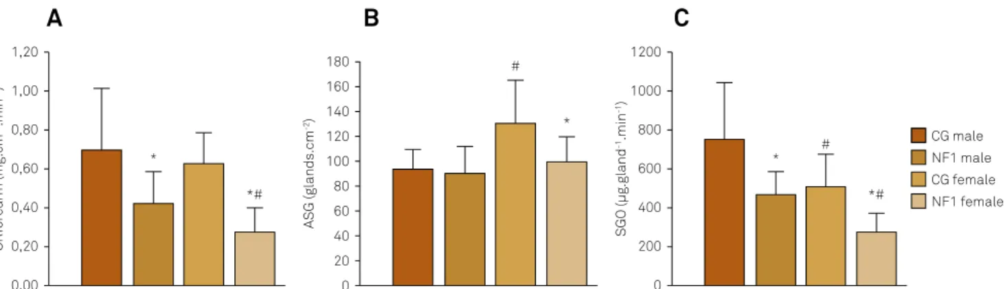
he NF1 volunteers presented with reduced sweating responses to local pharmacological stimulation (Figure 1). Both SR (males: NF1 = 0.42 ± 0.16 vs. CG = 0.71 ± 0.31; females: NF1 = 0.28 ± 0.12 vs. CG = 0.63 ± 0.16 mg.cm-2.min-1, p < 0.05)
and SGO (males: NF1 = 4.68 ± 1.20 vs. CG = 7.60 ± 2.86; females:
NF1 = 2.75 ± 0.98 vs. CG = 5.11 ± 1.62 µg.gland-1.min-1, p < 0.05)
were reduced in the NF1 group when compared to the CG, irrespective of sex. Nevertheless, the number of ASG was re -duced only among women in the NF1 group compared to the CG ( females: NF1 = 98 ± 21 vs. CG = 129 ± 33 glands.cm-2,
p < 0.05). he typical diferences in sweating between sexes was also observed: men displayed higher SGO and lower ASG than women (p < 0.05).
he PILO phase of the study was designed to prevent in -luences of core, skin and environmental temperatures on sweating. his objective was attained once there were non-signiicant variations of core (tympanic range: 37.17 ± 0.35 to 37.34 ± 0.66°C; p > 0.05), local skin (forearm range: 34.39 ± 0.25 to 34.47 ± 0.6°C; p > 0.05), and environmental temperatures (air range: 23.86 ± 0.39 C to 24.20 ± 0.58°C; p > 0.05) .
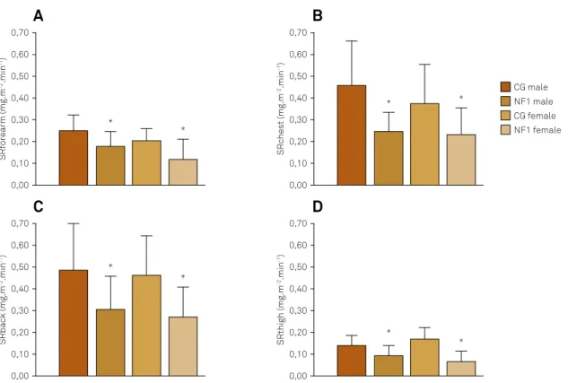
During central stimulation via passive heating (HEAT), the NF1 group sweat secretion was also reduced (Figure 2). he NF1 sub-groups showed lower SR in every region measured: (males: forearm = 0.18 ± 0.07; chest = 0.25 ± 0.09; back = 0.31 ± 0.15; thigh = 0.09 ± 0.05; and females: forearm = 0.12 ± 0.01; chest = 0.23 ± 0.13; back = 0.27 ± 0.14; thigh = 0.07 ± 0.05) when compared to the CG sub-groups (males: forearm = 0.26 ± 0.07; chest = 0.46 ± 0.21; back = 0.49 ± 0.23; thigh = 0.14 ± 0.04; and
Table 1. Physical characteristics (Mean ± SD).
Variable Age (years) Height (m) Weight (kg) BSA (m2)
GC male 39 ± 14 1.69 ± 0.06 75.89 ± 16.42 1.85 ± 0.20
NF1 male 36 ± 12 1.70 ± 0.06 74.07 ± 19.33 1.84 ± 0.21
GC female 33 ± 6 1.58 ± 0.07# 53.56 ± 3.84# 1.53 ± 0.10#
NF1 female 33 ± 9 1.55 ± 0.09# 56.10 ± 9.12# 1.54 ± 0.16#
SD: standard deviation; BSA: body surface area; CG: non-NF1 control group; NF1: neuroibromatosis type 1 individuals; # Difference between males and females.
Mean ± SD. (A) Local sweat rate (SR); (B) Number of active sweat glands (ASG); (C) sweat gland output (SGO). SD: standard deviation; CG: control group; NF1: neuroibromatosis type 1. *Difference between NF1 and GC; #Difference between males and females.
Figure 1. Sweating responses in PILO.
A
B
C
ASG (glands.cm
-2) 180 160
80 100
#
* 120
140
60 40 20 0
CG male NF1 male
NF1 female CG female
SRforearm (mg.cm
-2.min -1)
1,20
1,00
0,60
*# *
0,80
0,40
0,20
0,00
SGO (µg.gland
-1.min -1)
1200
1000
600
*#
* #
800
400
200
SRback (mg.m -2.min -1)
0,70
0,60
0,40
* *
0,50
0,30
0,10 0,20
0,00
SRforearm (mg.m
-2.min -1)
0,70
0,60
0,40
* *
0,50
0,30
0,10 0,20
0,00
SRthigh (mg.m
-2.min -1)
0,70
0,60
0,40
* *
0,50
0,30
0,10 0,20
0,00
SRchest (mg.m
-2.min -1)
0,70
0,60
0,40 * *
0,50
0,30
0,10 0,20
0,00
CG male NF1 male
NF1 female CG female
females: forearm = 0.20 ± 0.06; chest = 0.38 ± 0.18; back = 0.47 ± 0.18; thigh = 0.17 ± 0.06 mg.m-2.min-1; p < 0.05). Similarly, the SGO
was signiicantly reduced in the NF1 group when com -pared to controls in almost every region (except the back), but only in female groups (NF1: forearm = 1.01 ± 0.64; chest = 3.09 ± 1.64; back = 5.18 ± 2.87; thigh = 0.74 ± 0.63 vs. CG:
forearm = 1.89 ± 0.69; chest = 5.44 ± 3.11; back = 3.27 ± 1.81; thigh = 1.86 ± 0.68 µg.gland-1.min-1; p < 0.05). Lastly, ASG was
lower only in the back region between females NF1 (77 ± 16; 54 ± 1) and CG (70 ± 22; 98 ± 25 glands.m-2) (p < 0.05).
Heat storage rate was expressively higher in the NF1 group (males: 3212 ± 236; females: 2,065 ± 230 J.min-1.m²) than
in controls (males: 93 ± 1,171; females: 559 ± 1,198 J.min-1.m²;
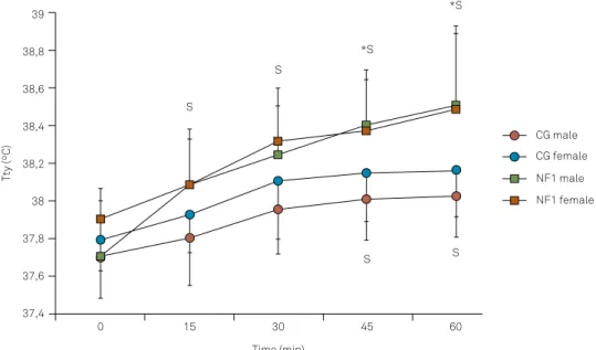
p < 0.05) and, as a consequence, NF1 patient tympanic tem -peratures showed a larger increase than controls, becom -ing higher in NF1 groups at the end of the test (Figure 3; males: NF1 = 38.5 ± 0.4°C vs. CG = 38.0 ± 0.2°C and females: NF1 = 38.5 ± 0.4 vs. CG = 38.1 ± 0.2ºC; p < 0.05). In contrast, skin temperatures decreased in a similar way during the ex -periment in all groups and in every body region measured (Tskin initial range: 33.30 ± 0.87° C to 35.01 ± 0.40°C and Tsk inal range: 32.80 ± 1.3°C to 33.72 ± 0.90°C; p > 0.05).
Table 2 shows the NF1 and control groups’ cardiovascular responses to passive heating. In both NF1 and control groups, the heart rate increased, and systolic blood pressure re -mained stable throughout the test. Nevertheless, while heart rate and systolic blood pressure behaviors were not diferent
between groups, the NF1 group’s diastolic blood pressure and mean blood pressure did not display the expected de -crease in those variables, resulting in higher end values in the NF1 group (males: -1.69 ± 4.29; females: -0.80 ± 3.77 mmHg) when compared to controls (males: -6.73 ± 4.43; females: -7.50 ± 4.64 mmHg; p < 0.05).
DISCUSSION
he present study has identiied markedly reduced thermo -regulatory capacity in NF1 patients, which could contribute to their reduced aerobic capacity and daily physical activities as well as increasing their risk of heat injury and illnesses. hese results suggest an autonomic neuropathy as the maim mecha -nism for reduced heat and exercise tolerance in NF1 patients.
Volunteers with NF1, irrespective of sex, showed lower sweat production both after local pharmacological stimu -lation and after central sweat induction by passive heating when compared to non-NF1 controls matched by age and anthropometric characteristics. he present study is the irst to describe decreased thermoregulatory responses in NF1.
Impairments in the SR to local and/or central stimuli has already been reported in other diseases afecting the neural tissues, such as diabetes11,12,16, tuberous sclerosis17,18, multiple
sclerosis19, and leprosy20.
A
C
B
D
Mean ± SD. Local sweat rate (SR) measured in forearm (A); chest (B); back (C) and thigh (D) during HEAT. SD: standard deviation; CG: control group; NF1: neuroibromatosis type 1; *Difference between NF1 and GC.
he present results showed reduction of 35% to 65% in sweat secretion (induced by PILO and HEAT protocols) among NF1 volunteers compared to non-NF1 individuals. In the PILO tests, the reduction was associated with a de -crease in sweating production (SGO = -39%) shown by NF1 males, and with a decrease in both SGO (-47%) and num -ber of active sweat glands (ASG = -34%) presented by NF1 females. However, during HEAT, only NF1 females displayed lower SGO when compared with CG females. herefore, it is not possible to conclude if the lower NF1 male SR is related to reduced ASG, SGO, or both.
Pilocarpine stimulation of eccrine sweat glands is pro -duced by direct binding of the cholinergic agonist to the mus -carinic receptors on the gland, independent of sympathetic drive21. herefore, it is possible that the NF1-reduced sweat re -sponse elicited by the present method relects disturbances in glands per se, speciically in their morphology (i.e. dystrophic or atrophic changes) and/or in their cholinergic sensitivity. Nevertheless, some degree of impairment of the central ner -vous system can also explain, at least in part, the NF1-reduced sweating. For instance, after sympathectomy, local sweating
responses to pilocarpine becomes exacerbated, but it subse -quently decreases and, in some cases, ceases completely22.
Decreased pharmacologically induced sweat production ob -served in type 1 diabetes and multiple sclerosis is an early sign of neuropathy, and it has been associated with reduced sympa -thetic nerve iber density in the sweat glands10,23,24 . In NF1, the
development of large cutaneous and subcutaneous neuroi -bromas may cause dysfunctions in peripheral nerves leading to chronic pain, sensory loss, weakness or even palsy. Additionally, a symmetrical difuse neuropathic process associated with thickening of peripheral nerves (called neuroibromatous neu -ropathy), can also cause distal sensory-motor symptoms1,14.
Drouet et al. showed that, although asymptomatic in many cas -es, NF1 peripheral neuropathy constitutes a potentially severe complication as it is associated with increased morbidity relat -ed to the neuropathy itself, to spinal cord involvement and/or degeneration of a malignant peripheral nerve sheath tumor13.
Nevertheless, until now, no studies have linked autonomic neu -ropathy to NF1 thermoregulatory capacity and heat intolerance. As has been already described, there is a 10% reduction in aerobic capacity in NF1 patients5
and, the present NF1
Table 2. Cardiovascular responses in HEAT experiments (Mean ± SD).
Group HR (bpm) SBP (mmHg) DBP (mmHg) MBP (mmHg)
Initial Final Initial Final Initial Final Initial Final
CG male 83 ± 11 93 ± 12$ 112 ± 11 112 ± 10 66 ± 6 58 ± 6$ 81 ± 7 76 ± 7$
NF1 male 82 ± 8 94 ± 13$ 120 ± 9* 121 ± 9* 71 ± 7 69 ± 8* 87 ± 8* 86 ± 8*
CG female 78 ± 11 90 ± 15$ 98 ± 6# 98 ± 6# 59 ± 5# 51 ± 5#$ 73 ± 5# 67 ± 5#$
NF1 female 79 ± 8 99 ± 12$ 107 ± 7*# 108 ± 8*# 63 ± 6# 62 ± 5*# 77 ± 6# 77 ± 6*#
SD: standard deviation; HR: heart rate; SBP: systolic blood pressure; DBP: diastolic blood pressure; MBP: mean blood pressure; NF1: neuroibromatosis type 1 individuals; CG: non-NF1 control group; *Difference between NF1 and GC; #Difference between males and females. $Difference from initial values (minute 0).
Mean ± SD. Tympanic temperature (Tty) during HEAT. SD: standard deviation; CG: control group; NF1: neuroibromatosis type 1. *Difference between NF1 and GC; $Difference from initial values (minute 0).
Figure 3. Core Temperature in HEAT.
Tty (ºC)
39
38,8
38,4 38,6
38,2
37,6 37,8 38
37,4
0 15 30
Time (min)
45 60
S
S
*S
S
*S
S
CG male
NF1 male
decrease in SR could possibly be related to the lower VO2max and/or physical training levels, as is observed in healthy indi -viduals25,26. In a previous study applying the present method,
healthy volunteers showed 20% reduced sweating response related to 20% reduced aerobic capacity9. herefore, it seems
unlikely that the 10% lower VO2max previously reported in NF1 could exclusively explain the 35% to 65% reductions in NF1 SR observed here.
he NF1 volunteers also presented with less reduction in diastolic and mean arterial blood pressure in response to heat stress. During body hyperthermia states, physiological periph -eral vasodilation mediated by neural and local (endothelial function) factors shifts part of the cardiac output to the skin surface for heat dissipation. herefore, skin blood low has a key role in normal thermoregulation and any disruption in its neural or/and local control may lead to impaired thermoregu -lation27,28,29. It is possible that some degree of endothelial dys -function observed in NF1 patients30 could contribute to the
NF1-reduced vascular thermal response in the present study. It is interesting to note that, just like sweat production/secre -tion, vascular tone is directly regulated by the sympathetic nervous system, and therefore disturbances in the vasodila -tion response could be another indirect sign of neuropathy.
References
1. Ferner RE, Huson SM, Thomas N, Moss C, Willshaw H, Evans DG et al. Guidelines for the diagnosis and management of individuals with neuroibromatosis 1. J Med Genet. 2007;44(2):81-8. doi:10.1136/jmg.2006.045906
2. Riccardi VM. Neuroibromatosis type 1 is a disorder of dysplasia: the importance of distinguishing features, consequences, and complications. Birth Defects Res A Clin Mol Teratol. 2010;88(1):9-14. doi:10.1002/bdra.20616
3. Rodrigues LOC, Batista PB, Goloni-Bertollo EM, Souza-Costa D, Eliam L, Eliam M et al. Neuroibromatoses: part 1 diagnosis and differential diagnosis. Arq Neuropsiquiatr. 2014;72(3):241-50. doi:10.1590/0004-282X20130241
4. Souza JF, Passos RL, Guedes AC, Rezende NA and Rodrigues LOC. Muscular force is reduced in neuroibromatosis type 1. J Musculoskelet Neuronal Interact. 2009;9(1):15-7.
5. Souza JF, Araújo CG, Rezende NA, Rodrigues LOC. Exercise capacity impairment in individuals with neuroibromatosis type 1. Am J Med Genet A. 2013;161A(2):393-5. doi:10.1002/ajmg.a.35729
6. Kenny GP, Yardley J, Brown C, Sigal RJ and Jay O. Heat stress in older individuals and patients with common chronic diseases. CMAJ 2010;182(10):1053-60. doi:10.1503/cmaj.081050
7. Gilsoli CV, Mora F. The hot brain: survival, temperature and the human body. London: The Massachusetts Institute of Technology; 2000.
8. Shibasaki M, Wilson TE, Crandall CG. Neural control and mechanisms of eccrine sweating during heat stress and exercise. J Appl Physiol (1805). 2006;100(5):1692-701. doi:10.1152/japplphysiol.01124.2005 9. Madeira LG, Fonseca MA, Fonseca IA, Oliveira KP, Passos RL,
Machado-Moreira CA et al. Sex-related differences in sweat gland cholinergic sensitivity exist irrespective of differences
in aerobic capacity. Eur J Appl Physiol. 2010;109(1):93-100. doi:10.1007/s00421-009-1262-8
10. Davis SL, Wilson TE, White AT, Frohman EM. Thermoregulation in multiple sclerosis. J Appl Physiol (1985). 2010;109(5):1531-7. doi:10.1152/japplphysiol.00460.2010
11. Hoeldtke RD, Bryner KD, Horvath GG, Phares RW, Broy LF, Hobbs GR et al. Redistribution of sudomotor responses is an early sign of sympathetic dysfunction in type 1 diabetes. Diabetes 2001;50(2):436-43. doi:10.2337/diabetes.50.2.436
12. Rocha CM, Madeira LG, SÁ KR, Lopes LN, Albuquerque DP, Diniz LM et al. Diabetes mellitus tipo 1 na ausência de neuropatia autonômica não altera a taxa de sudorese no exercício. Rev Bras Med Esporte. 2009;15(1):23-6. doi:10.1590/S1517-86922009000100005 13. Drouet A, Wolkenstein P, Lefaucheur JP, Pinson S, Combemale
P, Gherardi RK et al. Neuroibromatosis 1-associated neuropathies: a reappraisal. Brain. 2004;127(9):1993-2009. doi:10.1093/brain/awh234
14. Ferner RE, Hughes RAC, Hall SM, Upadhyaya M, Johnson MR. Neuroibromatous neuropathy in neuroibromatosis 1 (NF1). J Med Genet. 2004;41(11):837-41. doi:10.1136/jmg.2004.021683 15. Yerdelen D, Koc F, Durdu M, Karakas M. Electrophysiological
indings in neuroibromatosis type 1. Neurol Sci. 2011;306(1-2):42-8. doi:10.1016/j.jns.2011.03.048
16. Kihara M, Opfer-Gehrking TL, Low PA. Comparison of directly stimulated with axon-relex-mediated sudomotor responses in human subjects and in patients with diabetes. Muscle Nerve. 1993;16(6):655-60. doi:10.1002/mus.880160612
17. Chudnow RS, Wolfe GI, Sparagana SP, Delgado MR, Batchelor L, Roach ES. Abnormal sudomotor function in the hypomelanotic macules of tuberous sclerosis complex. J Child Neurol. 2000;15(8):529-32. doi:10.1177/088307380001500806
he possible mechanisms of lower responses of sweat -ing and diastolic pressure control in NF1 may include sweat gland tissue dysplasia, lower cholinergic gland sensitivity, neuropathy and impairment in the vasodilation response. herefore, the association of both lower sweat capacity and impaired vascular response is suggestive of an NF1 autonom -ic neuropathy, wh-ich should be conirmed by further histo -logical and functional studies.
Finally, during passive heating, the NF1 group’s internal temperature increased earlier and faster, reaching higher i -nal values (+ 0.6o C), leading to almost 10 times greater heat
storage rate than controls. hese indings are clear evidence of reduced thermoregulatory capacity caused by lower sweat capacity and impaired vascular response to heat stress in NF1. Nevertheless, considering the magnitude of heat stor -age observed, it should be safer to include NF1 as a predis -posing factor to the development of heat illnesses during heat stress and/or intense exercise.
18. Orozco-Covarrubias ML, Ridaura C, Tamayo-Sánchez L, Durán-Mckinster C, Ruiz-Maldonado R. [Tuberous sclerosis. Early diagnosis with autonomic nervous system responses in hypopigmented skin]. Rev Invest Clin. 1994;46(5):349-54. Spanish.
19. Davis SL, Wilson TE, Vener JM, Crandall CG, Petajan JH, White AT. Pilocarpine-induced sweat gland function in individuals with multiple sclerosis. J Appl Physiol (1985). 2005;98(5):1740-4. doi:10.1152/japplphysiol.00860.2004
20. Facer P, Mathur R, Pandya SS, Ladiwala U, Singhal BS, Anand P. Correlation of quantitative tests of nerve and target organ dysfunction with skin immunohistology in leprosy. Brain. 1998;121(12):2239-47. doi:10.1093/brain/121.12.2239
21. Randall WC, Kimura KK. The pharmacology of sweating. Pharmacol Rev. 1955;7(3):365–97.
22. Kuno Y. Human perspiration. Springield: Charles C. Thomas; 1956. 23. Maselli RA, Jaspan JB, Soliven BC, Green AJ, Spire JP, Arnason
BG. Comparison of sympathetic skin response with quantitative sudomotor axon relex test in diabetic neuropathy. Muscle Nerve. 1989;12(5):420-3. doi:10.1002/mus.880120513
24. Gibbons CH, Illigens BM, Wang N, Freeman R. Quantiication of sweat gland innervation: a clinical-pathologic correlation. Neurology. 2009;72(17):1479-86. doi:10.1212/WNL.0b013e3181a2e8b8
25. Greenhaff PL, Clough PJ.. Predictors of sweat loss in man during prolonged exercise. Eur J Appl Physiol Occup Physiol. 1989;58(4):348-52. doi:10.1007/BF00643508
26. Buono MJ, White CS, Connolly KP. Cholinergic sensitivity of the eccrine sweat gland in trained and untrained men. J Dermatol Sci. 1992;4(1):33-7. doi:10.1016/0923-1811(92)90053-E
27. Kellogg DL Jr, Crandall CG, Liu Y, Charkoudian N, Johnson JM. Nitric oxide and cutaneous active vasodilation during heat stress in humans. J Appl Physiol (1985). 1998;85(3):824-9.
28. Charkoudian N. Skin blood low in adult human thermoregulation: how it works, when it does not, and why. Mayo Clin Proc. 2003;78(5):603-12. doi:10.4065/78.5.603
29. Bruning RS, Santhanam L, Stanhewicz AE, Smith CJ, Berkowitz DE, Kenney WL et al. Endothelial nitric oxide synthase mediates cutaneous vasodilation during local heating and is attenuated in middle-aged human skin. J Appl Physiol (1985). 2012;112(12):2019-26. doi:10.1152/japplphysiol.01354.2011 30. Rodrigues LO, Rodrigues LO, Castro LL, Rezende NA, Ribeiro ALP.