Case Report
1 8 5 Arq Bras Oftalmol. 2014;77(3):185-7 http://dx.doi.org/10.5935/0004-2749.20140047
INTRODUCTION
Multiple evanescent white dot syndrome (MEWDS) is a benign self-healing disease with an unknown etiology that was first descri-bed in 1984 by Jampol et al. It is a retinal pigment epithelial (RPE) and choroidal inflammatory condition that predominantly affects young to middle-aged women, is unilateral in 80% of cases, and causes visual loss of variable degrees(1-5).
Associated clinical features include flu-like prodrome, blurred disc margins, blind spot enlargement, and temporal scotomata(2,5). Fundos-copy typically reveals multiple, 100-200 µm yellow-white outer retina dots in posterior pole and mid-periphery as well as a unique foveal granularity(1,2,4-7).
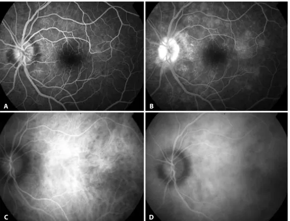
Fluorescein angiography (FA) typical aspect comprises early foveal granular hyperfluorescence, early hyperfluorescence, and late staining of the white dots in a wreath-like pattern and disc capillary leakage. Indocyanine green angiography (ICGA) shows numerous hypofluores-cent nummular lesions that are evident in the late phase and predomi-nant in the mid-periphery and around the optic disc(2,4,7).
Here, we report a case of MEWDS notable for its classic retinal appea-rance, foveal granularity, and transient disruption of the macular photoreceptor of the inner segment-outer segment (IS-OS) junction. After six months of follow up, the patient presented full visual reco-very and re-established a normal foveal aspect on Spectral Domain Optical Coherence Tomography (SD-OCT).
ABSTRACT
The purpose of this study was to describe a patient with multiple evanescent white dot syndrome (MEWDS) who presented with classic retinal findings and transient changes in outer retinal anatomy. A 20-year-old man presented with mild blurred vision in the left eye, reporting flu-like symptoms 1 week before the visual symptoms started. Fundus examination of the left eye revealed foveal granularity and multiple scattered spots deep to the retina in the posterior pole. Fluorescein angiography and indocyanine green angiography showed typical MEWDS findings. Spectral Domain Optical Coherence Tomography has shown transient changes in outer retinal anatomy with disappearance of inner segment-outer segment junction and mild attenuation of external limiting membrane. Six months later, Spectral Domain Optical Coherence Tomography has shown complete resolution with recovery of normal outer retinal aspect.
Keywords: Retinal diseases/diagnosis; Syndrome/diagnosis; Tomography, optical coherence; Case reports
RESUMO
O propósito deste estudo é descrever o caso de um paciente com síndrome dos múltiplos pontos brancos evanescentes (MEWDS), apresentando achados retinianos clássicos e alterações transitórias na anatomia retiniana externa. Paciente do sexo masculino, 20 anos de idade, apresentando embaçamento visual no olho esquerdo, relatando sintomas gripais uma semana antes do início dos sintomas visuais. Fundoscopia do olho esquerdo revelou granularidade foveal, múltiplos pontos brancos retinianos no polo posterior. A angiografia fluoresceínica e a indocianinografia verde evidenciaram achados típicos de MEWDS. A tomografia de coerência óptica de domínio espectral evidenciou alterações transitórias na anatomia retiniana externa como desapareci-mento da junção dos segdesapareci-mentos interno-externo dos fotorreceptores e leve atenuação da membrana limitante externa. Após 6 meses, a tomografia de coerência óptica mostrou completa resolução com recuperação total da anatomia retiniana externa.
Descritores: Doenças retinianas/diagnóstico; Síndrome/diagnóstico; Tomografia de coerência óptica; Relatos de casos
CASE REPORT
A 20-year-old man presented for an ophthalmologic evaluation with a 2-day history of blurred vision in his left eye and flu-like symptoms that began 1 week before the visual symptoms started. His bestcorrected visual acuity was 20/20 (OD) and 20/40 (OS); in -traocular pressure was 15 mmHg in OU. Anterior biomicroscopy was normal in OU. Fundus examination was normal in OD, and OS revea-led multiple 100-500 µm yellow-white dots deep to the retina in the posterior pole and mid-periphery, foveal granularity, and blurred disc margins (Figure 1).
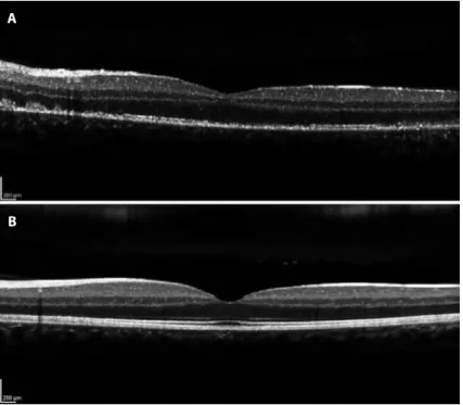
Automated perimetry showed a blind spot enlargement in the affected eye. FA had shown earlier hyperfluorescent lesions in the posterior pole and mid-periphery, with late staining of the white dots in a wreath-like pattern and late staining in the optic nerve (Figure 2). The corresponding ICGA demonstrated numerous hypofluorescent areas predominantly in the mid-periphery and around the optic disc. These hypofluorescent areas were mild and in an early angiographic phase, but became more clearly delineated in the late phase (Figure 2). SD-OCT showed mild attenuation of the external limiting mem-brane, a transient disruption of the macular photoreceptor IS-OS junction, increased RPE reflectivity and enhanced signal penetration into the underlying choroid (Figure 3). The patient was diagnosed with MEWDS and observation without treatment was suggested. The patient was lost at follow-up, but returned 24 weeks later after
Transient spectral domain optical coherence tomography findings in classic mewds:
a case report
Alterações transitórias evidenciadas na tomograia de coerência óptica de domíno espectral em
quadro clássico de mewds: relato de caso
Luciana castro Lavigne1, DaviD LeonarDo cruvineL isaac2, José osório Duarte Júnior1, Marcos PereiraDe ÁviLa2
Submitted for publication: September 17, 2013 Accepted for publication: January 27, 2014
Study conducted at Universidade Federal de Goiás, GO, Brazil.
1 Setor de Retina e Vítreo, Universidade Federal de Goiás (UFG), Goiânia, GO, Brazil. 2 Universidade Federal de Goiás (UFG), Goiânia, GO, Brazil.
Funding: No specific financial support was available for this study.
Disclosure of potential conflicts of interest: None of the authors have any potential conflicts of interest to disclose.
Transient spectral domain optical coherence tomography findings in classic mewds: a case report
1 8 6 Arq Bras Oftalmol. 2014;77(3):185-7
contacting the authors. He claimed that he had not attended the visits because he had visual recovery within one month. His BCVA was 20/20 OU. Fundus examination in the OD was normal, and the OS revealed mild foveal granularity and RPE hyperpigmentation surrounding the optic disc. Color and red-free fundus photography showed the retinal white dots disappeared. SD-OCT indicated the IS-OS disruption resolved with reestablishment of outer retina OCT anatomy (Figure 3).
DISCUSSION
MEWDS is an acute, usually unilateral retinopathy that predomi-nantly affects macular and mid-periphery regions of young women, causing mild to moderate visual loss. Characteristic symptoms inclu-de photopsia, and a viral prodrome is reported in 50% of the cases. In this case report, the patient was a young male who had unilateral visual loss. He had flu-like symptoms 1 week before the initial evalua-tion, and did not present with photopsia(1,2).
Figure 1. A) Retinography demonstrating normal foveal aspect in the right eye. B) Retinography demonstrating foveal granularity in the left eye.
A B
Figure 2. Multiple evanescent white dot syndrome lesions. A) Fluorescein angiography demonstrating early hyperluorescent lesions. B) Fluorescein angiography, in late stage, demonstrating staining of the white dots in a wreath-like pattern and disc capillary leakage. C) Indocyanine green angiography, in the early phase, showing discrete hypoluorescent nummular lesions. D) Indocyanine green angiography (late phase) showing numerous clearly delineated hypoluorescent nummular lesions.
A
C D
Lavigne LC, et al.
187
Arq Bras Oftalmol. 2014;77(3):185-7 In the present case, FA demonstrated typical early
hyperfluores-cence with late RPE staining, corresponding to hypofluorescent areas in the late phase of ICGA(1,5). These hypofluorescent areas showed moderately reflective focal lesions in the outer photoreceptor layer, where the macular photoreceptor IS-OS junction was disrupted by SD-OCT, which is associated with foveal granularity. Possibly, the foveal granularity is the most specific characteristic of MEWDS(1,4,8,9).
Considering the clinical and angiographic pattern, and through electrophysiological evidence, previous studies have suggested that the white dot lesions in MEWDS are situated in the RPE and outer re-tina. Electrophysiology analysis has revealed an electro-oculographic reduction in the light-dark ratio, and an electroretinographic altera-tion of the a-wave and early receptor potential(6,8).
Ophthalmologic studies have advocated the hypothesis that injury to the RPE is the early pathologic event in MEWDS. FA showed characteristic early hyperfluorescent lesions that were observed as window defects. It was suggested the primary site of MEWDS is on the RPE(2,6). With the clinical use of ICGA, MEWDS has become known as a choroidopathy, suggesting that the cause of the photoreceptors and RPE dysfunction is associated to decreased perfusion of the choriocapillaris(4-6,10). This supposition is based on their angiographic pattern that demonstrated hypofluorescent lesions on ICGA that may appear even in normal areas on FA and funduscopy(6).
In the case reported here, the FA pattern demonstrated characte-ristic early hyperfluorescence and late staining, which appeared to be due to RPE changes; however, the ICGA showed numerous hypofluo-rescent areas mainly in the mid-periphery. These hypofluohypofluo-rescent areas are mild in initial angiographic phases and become more evi dently delineated in the late phase, which appears to slightly outnumber those observed on FA, suggesting that MEWDS involves both RPE and choriocapillay(2,8).
In this case ICGA was not crucial to diagnosis, because of other classic findings, but it was useful to confirm diagnosis and also in diffe-rential diagnosis with other white dot syndromes. Acute posterior multifocal placoid pigment epitheliopathy (APMPPE) has been
con-sidered an important differential diagnosis. FA patterns in APMPPE show early hypofluorescence, but become hyperfluorescent later in the study. In ICGA, lesions present as early to late hypofluorescence.
MEWDS has been considered the first known disease involving electrophysiological damage of photoreceptor outer segments with complete return to normal conditions(8). We report a case of MEWDS notable for both its classic retinal appearance, confirmed by auto-mated perimetry, FA, ICGA, and SD-OCT exams. SD-OCT has showed disruption of the IS-OS junction. The patient was observed with no medical intervention, and had a favorable recovery of visual function and macular photoreceptor IS-OS junction in SD-OCT. This is consis-tent with previous report and OCT findings about the disease.
REFERENCES
1. Hua R, Chen K, Liu LM, Liu NN, Chen L, Teng WP. Multi-modality imaging on multiple evanescent white dot syndrome - A Spectralis Study. Int J Ophthalmol. 2012;5(5):644-7. 2. Kuznetcova T, Jeannin B, Herbort CP. A case of overlapping choriocapillaritis
syn-dromes: multimodal imaging appraisal. J Ophthalmic Vis Res. 2012;7(1):67-75. 3. Abu-Yaghi NE, Hartono SP, Hodge DO, Pulido JS, Bakri SJ. White dot syndromes: a
20-year study of incidence, clinical features, and outcomes. Ocul Immunol Inflamm. 2011;19(6):426-30.
4. Nguyen MH, Witkin AJ, Reichel E, Ko TH, Fujimoto JG, Schuman JS, Duker JS. Mi-crostructural abnormalities in MEWDS demonstrated by ultrahigh resolution optical coherence tomography. Retina. 2007;27(4):414-8.
5. Vianna RNG, Socci D, Nehemy MB, Deschênes J, Burnier MN Jr. The white dot syn-dromes. Arq Bras Oftalmol. 2007;70(3):554-62.
6. Penha FM, Navajas EV, Bom Aggio F, Rodrigues EB, Farah ME. Fundus autofluorescence in multiple evanescent white dot syndrome. Case Rep Ophthalmol Med. 2011;807565. 7. Slusher MM, Weaver RG. Multiple Evanescent White Dot Syndrome. Retina. 1988;
8(2):132-5.
8. Silva RA, Albini TA, Flynn HW Jr. Multiple evanescent white dot syndromes. J Ophthal-mic Inflamm Infect. 2012;2(2):109-11.
9. Saito M, Barbazetto IA, Spaide RF. Intravitreal cellular infiltrate imaged as punctate spots by spectral-domain optical coherence tomography in eyes with posterior segment in flammatory disease. Retina. 2013;33(3):559-65.
10. Papadia M, Herbort CP. Idiopathic choroidal neovascularisation as the inaugural sign of mul tiple evanescent white dot syndrome. Middle East Afr J Ophthalmol. 2010;17(3):270-4. Figure 3. A) Spectral Domain Optical Coherence Tomography (SD-OCT) demonstrating mild
attenua-tion of the external limiting membrane, disrupattenua-tion of the macular photoreceptor inner segment-outer segment (IS-OS) junction, and increased RPE relectivity. B) SD-OCT demonstrating resolution of IS-OS disruption with reestablishment of outer retina OCT anatomy.
A