Original Article
1 4 8 Arq Bras Oftalmol. 2014;77(3):148-51 http://dx.doi.org/10.5935/0004-2749.20140038
Measurement of choroid thickness in pregnant women using enhanced depth
imaging optical coherence tomography
Medição da espessura da coroide em gestantes utilizando tomograia de coerência óptica com
profundidade de imagem aprimorada
Sertan GoktaS1, ahmet BaSaran2, YaSar SakarYa1, muammer ozcimen1, zehra kucukaYdin2, raBia SakarYa1, muStafa BaSaran2, erkan erdoGan1, iSmail alpfidan1
Submitted for publication: February 12, 2014 Accepted for publication: March 24, 2014
Study conducted at Konya Training and Research Hospital, Konya, Turkey.
1 Department of Opthalmology, Konya Training and Research Hospital, Konya, Turkey.
2 Department of Obstetrics and Gynecology, Konya Training and Research Hospital, Konya, Turkey.
Funding: No specific financial support was available for this study.
Disclosure of potential conflicts of interest: None of the authors have any potential conflicts of interest to disclose.
Corresponding author: Sertan Goktas. Department of Opthalmology. Konya Research and Training Hospital, 42090 Meram - Konya, Turkey - E-mail:[email protected]
IntroductIon
Pregnancy is associated with metabolic, hormonal, and hemody-namic changes. The renin-angiotensin system regulates salt and water hemostasis in the body, and both renin and angiotensin levels increase during pregnancy. These changes lead to increasing blood volume beginning in the irst trimester(1,2). Systemic vascular
resis-tance decreases during pregnancy(3,4), and hemodynamic changes
afect blood pressure. In normal pregnancy, blood pressure initially decreases until the eighteenth to twentieth gestation week, but then increases until delivery(5,6). One study reported that total macular
vo-lume and foveal retinal thickness increase during pregnancy in the
aBStract
Purpose: To investigate choroidal thickness in healthy pregnant women during different trimesters using enhanced depth imaging optical coherence tomogra-phy (EDI-OCT ).
Methods: This prospective study included 90 healthy pregnant women in their first, second, or third trimester (groups 1, 2, and 3, respectively) and 30 non-pregnant healthy women (group 4). The age range for all groups was 18-40 years. Spectral domain optical coherence tomography scans were obtained to estimate the average choroidal thickness. Using EDI-OCT, we measured choroidal thickness manually from the outer border of the retinal pigment epithelium to the inner scleral border at the subfovea, 3 mm temporal, and 3 mm nasal to the fovea. Differences among groups were analyzed by one-way ANOVA.
results: We found a statistically significant difference between groups 2 and group 4 for subfoveal, temporal, and nasal mean choroidal thickness (p=0.007,
p<0.001, p=0.026, respectively). The mean choroidal thickness for group 2 was 395 ± 80 µm, 338 ± 74 µm, and 233 ± 61 µm at the regions subfoveal, temporal, and nasal to the fovea, respectively. In comparison, the mean choroidal thickness for group 4 was 335 ± 86 µm, 274 ± 54 µm, and 200 ± 53 μm at the regions subfoveal, tem-poral, and nasal to the fovea, respectively. No statistically significant differences were found for choroidal thickness among groups 1-4 (p=0.214, p=0.177, p=0.094, respectively) and between groups 3-4 (p=0.105, p=0.261, p=0.695, respectively) for all measured points.
conclusion: Our results suggest that choroidal thickening can occur at the regions subfoveal, temporal, and nasal to the fovea in the second trimester.
Keywords: Choroid/anatomy & pathology; Choroid/pathology; Enhanced depth imaging; Tomography, optical coherence; Diagnostic techniques, ophthalmolo-gical; Pregnancy
RESUMO
Objetivo: Investigar a espessura da coroide em gestantes saudáveis durante os di-ferentes trimestres utilizando tomografia de coerência óptica com profundidade de imagem aprimorada (EDI-OCT ).
Métodos: Este estudo prospectivo incluiu 90 gestantes saudáveis nos primeiro, segundo e terceiro trimestres da gravidez (grupos 1, 2 e 3, respectivamente) e 30 mu-lheres saudáveis não-gestantes (grupo 4) com faixa etária de 18-40 anos de idade. Foi realizada tomografia de coerência óptica espectral para estimar a espessura média da coroide. A espessura da coroide foi medida manualmente da borda externa do epitélio pigmentar da retina até o limite interno da esclera nas regiões subfoveal, 3 mm temporal e 3 mm nasal à fóvea utilizando EDI-OCT. As diferenças entre os grupos foram analisadas com o teste ANOVA unicaudal.
Resultados: Houve diferença estatística significativa na espessura média da coroide entre os grupos 2 e 4 nas regiões subfoveal, temporal e nasal à fóvea (p=0,007; p<0,001; p=0,026, respectivamente). A espessura média da coroide no grupo 2 foi: 395 ± 80 µm, 338 ± 77 µm e 233 ± 61 µm nas regiões subfoveal, temporal e nasal à fóvea, respecti-vamente. Em comparação, a espessura média da coroide no grupo 4 foi de: 335 ± 86 µm, 275 ± 54 µm e 200 ± 53 µm, nas regiões subfoveal, temporal e nasal à fóvea, respectivamente. Não foi encontrada diferença estatística significativa entre os grupos 1-4 (p=0,214, p=0,177, p=0,094, respectivamente) e os grupos 3-4 (p=0,105, p=0,261, p=0,695 respectivamente), para todas as medidas.
Conclusão: Nossos resultados sugerem que há espessamento da coroide nas regiões subfoveal, temporal e nasal à fóvea no segundo trimestre gestacional.
Descritores: Coroide/anatomia & histologia;Coroide/patologia; Tomografia de coe-rência óptica; Técnicas de diagnóstico oftalmológico; Gravidez
second and third trimesters because of luid accumulation(7). During
pregnancy, hemodynamic changes afect other parts of the body, including choroidal low.
The choroid is the vascular layer between the retina and the sclera that provides the blood supply to the eye and plays an important role in ocular nutrition. Histopathological examination showed that it is 0.22 mm thick posteriorly(8). The choroid is composed of a vascular
ne-twork that contributes to ocular nutrition through volume regulation and is extremely sensitive to blood pressure changes. The choroidal thickness is afected by blood low and perfusion pressure(9). Therefore,
Goktas S, et al.
1 4 9 Arq Bras Oftalmol. 2014;77(3):148-51
Optical coherence tomography (OCT) provides high-resolution, cross-sectional digital images of live biological tissues in vivo. With the use of enhanced depth imaging optical coherence tomography (EDI-OCT), choroid images can be obtained and the choroidal thick-ness can be measured. Using OCT, one study reported the choroid thickness as 287 μm, 261 μm, and 145 μm at subfoveal regions, 3 mm temporal to the fovea, and 3 mm nasal to the fovea, respectively, in healthy individuals(10). The change in the choroid thickness may play
a role in the pathophysiology of various ocular conditions.
In the present study, we used EDI-OCT to examine choroidal thi-ckness at each trimester in healthy pregnant women, and then com-pared these measures with those for non-pregnant healthy women.
MethodS
We examined 4 groups in the present study. Group 1 consisted of 30 eyes in 30 healthy women in the irst trimester, group 2 consisted of 30 eyes in 30 healthy women in the second trimester, and group 3 consisted of 30 eyes of 30 healthy women in the third trimester. Group 4 was the control group and consisted of 30 eyes in 30 healthy non-pregnant women. Only the right eye was assessed in each study participant. This study followed the tenets of the Declaration of Helsinki. All participants provided informed consent. The inclusion criteria for groups 1, 2, and 3 were healthy pregnant women in their irst, second, or third trimester Inclusion criteria for the control group (group 4) included an age of 18-40 years old, non-pregnant healthy regularly menstruating women. High myopic and hyperopic refrac-tive errors greater than -1.0 or +1.0 diopters, or intraocular surgical intervention were excluded from the study. Subjects with systemic diseases or conditions that might afect retinal or choroidal thickness were excluded. For example, patients with diabetes mellitus were excluded. Pregnant with high blood pressure was excluded. In addition, patients with any retinal or choroidal abnormalities detected in spectral-domain OCT scans were excluded.
All subjects underwent a thorough ocular examination, including an auto-refractometer, best-corrected visual acuity measurement, slit-lam p examination, intraocular pressure measurement, and dila ted funduscopy. Choroidal thickness was measured using a spectral-do main OCT device (Spectralis: wavelength, 870 nm; Heidelberg Engineering, Germany) with an enhanced depth-imaging mode after pupil dilation. All measurements were performed in the mor-ning. The horizontal section running through the center of the fovea was selected for further analysis. The OCT images were assessed independently by 2 ophthalmologists.
The choroidal thickness was measured from the outer portion of the hyperrelective line, corresponding to the retinal pigment
epi-thelium, to the inner surface of the sclera. Choroidal thickness was measured at the fovea and at positions 3 mm temporal, and nasal to the fovea. The values of the measurements were compared for each observer and then averaged for analysis.
Diastolic blood pressure, systolic blood pressure, and ocular per-fusion pressure were measured for each subject. Ocular perper-fusion pressure was calculated according to the following formula(11): Ocular
perfusion pressure = mean blood pressure - intraocular pressure. Statistical calculations were performed using SPSS (Statistical Package for Social Sciences version 15.0; SPSS, Inc., Chicago, IL). Cho-roidal thickness is presented as the mean ± standard deviation. The Kolmogorov-Smirnov test was used to assess correlations for data with a normal distribution. Groups were compared with an analysis of variance (ANOVA) and post hoc tests. The diferences in choroidal thickness detected by ANOVA and post hoc tests between healthy (control group) and pregnant individuals were also analyzed by the t-test. P values less than 0.05 were considered signiicant.
reSultS
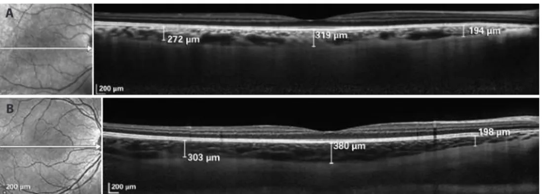
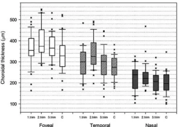
Ninety eyes in 90 healthy pregnant women and 30 eyes of 30 age-matched healthy non-pregnant women were included. The mean gestational age was 7.4 ± 2.6, 19.2 ± 2.9, and 33.1 ± 2.8 weeks in groups 1, 2, and 3, respectively. Mean age was 28.5 ± 6.4, 26.6 ± 4.2, 26.9 ± 6.2, and 29.4 ± .2 years, in groups 1, 2, 3, and 4, respecti-vely. There were no statistically signiicant diferences in age among the groups (p=0.183). Representative EDI-OCT scans for a pregnant women and the control group is presented in igure 1. Table 1 shows the mean choroidal thickness values for the groups that were mea-sured at subfoveal regions, and those 3 mm nasal to fovea, and 3 mm temporal to fovea. There were statistically signiicant diferences in subfoveal, temporal, and nasal choroidal thickness among the groups (p<0.05). The mean subfoveal, temporal, and nasal choroidal thickness was signiicantly greater in group 2 compared with the control group (p=0.007, p<0.001, p=0.026, respectively). There was no diference in mean subfoveal, nasal, and temporal choroidal thickness between group 1 and the control group (p=0.214, p=0.177, p=0.094, respectively). There was also no statistical signiicance among the 3 groups and control group for the mean subfoveal, temporal, and nasal choroidal thickness (p=0.105, p=0.261, p=0.695, respectively). Figure 2 shows the distribution of choroidal thickness according to group.
The ocular perfusion pressure was 36.3 ± 3.5 mmHg in pregnant women and 37.3 ± 2.8 mmHg in the control group. No signiicant correlations were found between the choroidal thickness and ocular perfusion pressure, gestational week.
Figure 1. A) Optical coherence tomography image from the control group demonstrating enhanced depth imaging on Spectralis (Heidelberg Engineering). The choroidal thickness was measured from the outer portion of the hyperrelective line, corresponding to the retinal pigment epithelium to the inner surface of the sclera at the subfovea, 3 mm temporal, and 3 mm nasal to the fovea. Calipers were positioned manually using computer software provided by the manufacturer. B) Optical coherence tomography image from second trimester, which depicts the increased the choroidal thickness.
a
Measurement of choroid thickness in pregnant women using enhanced depth imaging optical coherence tomography
150 Arq Bras Oftalmol. 2014;77(3):148-51 dIScuSSIon
Pregnancy can afect the eyes. Non-pathological events occurring during pregnancy includes reduced corneal sensitivity and increased corneal thickness related to the water retention. Choroidal thickness changes can be expected because of this water retention. There are some additional pathologic conditions reported to develop during pregnancy such as central serous chorioretinopathy(12).However, few
studies have investigated choroid thickness in pregnant women(13-15).
Takahashi et al. have demonstrated that there was no signiicant diference in choroidal thickness between healthy pregnant and non-pregnant women at the subfoveal and other measurement points(13). However, only the pregnant women in the third trimester
were included in that study. Similarly, we did not ind any diference in choroidal thickness measurement between pregnant women in the third trimester and the control group. Kara et al. investigated pregnant women in 15-38 weeks of gestational age(14). They reported
that subfoveal choroidal thickness increased in pregnant women but no signiicant correlation between the choroidal thickness and gestational age was found. Sayin et al. investigated pregnant women in 17-37 weeks of gestational age(15). They reported that subfoveal
choroidal thickness increased in pregnant women and found that ne-gative correlation between the choroidal thickness and gestational age. As distinct from these studies, we examined the mean choroidal thickness in pregnant subjects in each trimester via EDI-OCT. To our knowledge, the current study is the irst to investigate the choroidal thickness in three trimesters compared with non-pregnant healthy women. It can be considered as an important inding that the cho-roidal thickness signiicantly increased in the second trimester but it did not change in the irst and third trimesters.
While blood volume progressively increases, a rapid increase is ty-pically noted until mid-pregnancy, with a slower increase thereafter.
Additionally, during pregnancy, vascular resistance decreases from the ifth week of the gestation due to hormonal change(3,4). As vascular
resistance decreases, vascular compliance increases(16). The decrease
of the vascular resistance results in reduced blood pressure particu-larly in the mid-pregnancy. Thereafter, systemic pressure begins to increase again and ultimately reaches or exceeds the pre-pregnancy level(17). The reduction of blood pressure and systemic vascular
resis-tance, which is observed particularly in the middle of preg nancy, may explain the increase in choroidal thickness in the second trimester.
During pregnancy blood low increases in many organs, including the kidneys, extremities, and skin(18-20). One study reported increa sed
ocular blood low during pregnancy caused by vasodilation due to estrogen change(21). We suggest that increased choroidal thickness
may be secondary to increased blood low.
Choroidal changes during pregnancy may play a vital role in the pathophysiology of ocular diseases such as central serous choriore-tinopathy. Choroidal vasodilation and choroidal vascular hyperper-meability causes subsequent vascular leakage resulting in increased hydrostatic pressure in the choroid. Recent studies demonstrated a signiicantly increased choroidal thickness in patients with acute cen-tral serous chorioretinopathy(22,23). Central serous chorioretinopathy
may be caused by an increased hydrostatic pressure in the choroid. Pregnancy is one of the several known risk factors for central serous chorioretinopathy, which commonly develops in the third trimester(24).
We speculate the increased choroidal thickness observed in the se-cond trimester may be the causative factor underlying development of central serous chorioretinopathy in the third trimester. This may explain why central serous chorioretinopathy is more commonly observed in the third trimester.
The current study has several limitations. First, we did not measu-re ocular blood low. Color Doppler imaging can measumeasu-re the velocity of blood and vascular resistance within each vessel(25). Although this
technique is useful for determining choroidal blood low, it does not provide three-dimensional anatomical information about the cho-roidal layers. In our study, ocular blood low was not examined; the-refore, our study cannot determine the relationship between cho roidal thickness and ocular blood low. We can speculate that the thicker choroid may indicate an overall increase in choroidal blood low in pregnant women, as was previously demonstrated with a pulsatile ocular blood low pneumotonometer(21). Therefore, it is likely that
the increased choroidal thickness may be related to increased ocular blood low. Another limitation of our study was the small number of participants.
High refraction and age afect the thickness of the choroid(10,26).
Consequently, in our study we included similar groups with respect to meaningful characteristics, such as age and refraction, for both the pregnant and control groups.
In conclusion, our study showed a signiicant increase in choroi-dal thickness in the second trimester whereas there was no increase in the choroidal thickness during the irst and third trimesters. These data favor the idea that in pregnant women, increased choroidal thi ckness may lead to increased vascular permeability, which can explain the relationship between pregnancy and central serous cho-rioretinopathy. Further studies with a larger number of subjects should be performed in a pregnant population to correlate choroidal blood low with choroidal thickness.
reFerenceS
1. August P, Lenz T, Ales KL, Druzin ML, Edersheim TG, Hutson JM, et al. Longitudinal study of the renin-angiotensin-aldosterone system in hypertensive pregnant women: deviations related to the development of superimposed preeclampsia. Am J Obstet Gynecol. 1990;163(5):1612-21.
2. Pritchard JA, Rowland RC. Blood volume changes in pregnancy and the puerperium. III. Whole body and large vessel hematocrits in pregnant and nonpregnant women. Am J Obstet Gynecol. 1964;88:391-5.
3. Duvekot JJ, Peeters LL. Maternal cardiovascular hemodynamic adaptation to pregnancy. Obstet Gynecol Surv. 1994;49(12):1-14.
table 1. Mean choroidal thickness values (µm) for each group
location
Group 1 (n=30)
Group 2 (n=30)
Group 3 (n=30)
Group 4
(n=30) P* control irst
trimester
second trimester
third trimester
Subfoveal 362 ± 81 395 ± 80 368 ± 70 335 ± 86 0.037 Temporal 297 ± 73 338 ± 74 293 ± 72 274 ± 54 0.004
Nasal 225 ± 60 233 ± 61 205 ± 46 200 ± 53 0.044
Values are presented as the mean ± SD. *= ANOVA test.
Goktas S, et al.
151 Arq Bras Oftalmol. 2014;77(3):148-51
4. Gaillard R, Bakker R, Willemsen SP, Hofman A, Steegers EA, Jaddoe VW. Blood pressure tracking during pregnancy and the risk of gestational hypertensive disorders: the Generation R Study. Eur Heart J. 2011;32(24):3088-97.
5. Moutquin JM, Rainville C, Giroux L, Raynauld P, Amyot G, Bilodeau R, et al. A prospecti-ve study of blood pressure in pregnancy: prediction of preeclampsia. Am J Obstet Gynecol. 1985;151(2):191-6.
6. Macdonald-Wallis C, Tilling K, Fraser A, Nelson SM, Lawlor DA. Established pre-eclam psia risk factors are related to patterns of blood pressure change in normal term pregnan-cy: indings from the Avon Longitudinal Study of Parents and Children (ALSPAC). J Hypertens. 2011;29(9):1703-11.
7. Cankaya C, Bozkurt M, Ulutas O. Total macular volume and foveal retinal thickness alterations in healthy pregnant women. Semin Ophthalmol. 2013;28(2):103-11. 8. Ryan SJ. Retina. 4th ed. Philadelphia, PA: Elsevier Mosby; 2006. p.33-4.
9. Cioi GA, Granstam E, Alm A. Ocular circulation. In: Kaufman PL, Alm A, editors. Ader’s physiology of the eye: clinical application. 10th ed. St. Louis: Mosby; 2003. p.747-84.
10. Margolis R, Spaide RF. A pilot study of enhanced depth imaging optical coherence tomography of the choroid in normal eyes. Am J Ophthalmol. 2009;147(5):811-5. 11. Maul EA, Friedman DS, Chang DS, Boland MV, Ramulu PY, Jampel HD, et al. Choroidal
thickness measured by spectral domain optical coherence tomography: factors afecting thickness in glaucoma patients. Ophthalmology. 2011;118:1571-9. 12. Sunness JS. The pregnant woman’s eye. Surv Ophthalmol. 1988;32(4):219-38. 13. Takahashi J, Kado M, Mizumoto K, Igarashi S, Kojo T. Choroidal thickness in pregnant
women measured by enhanced depth imaging optical coherence tomography. Jpn J Ophthalmol. 2013;57(5):435-9.
14. Kara N, Sayin N, Pirhan D, Vural AD, Araz-Ersan HB, Tekirdag AI, et al. Evaluation of sub foveal choroidal thickness in pregnant women using enhanced depth imaging optical coherence tomography. Curr Eye Res. 2014:39(6):642-7.
15. Sayin N, Kara N, Pirhan D, Vural A, Araz Ersan HB, Tekirdag AI, et al. Subfoveal choroi-dal thickness in preeclampsia: comparison with normal pregnant and nonpregnant women. Semin Ophthalmol. 2014;29:11-7.
16. Spaanderman ME1, Willekes C, Hoeks AP, Ekhart TH, Peeters LL. The efect of pregnancy
on the compliance of large arteries and veins in healthy parous control subjects and women with a history of preeclampsia. Am J Obstet Gynecol. 2000;183(5):1278-86. 17. Duvekot JJ, Cheriex EC, Pieters FA, Menheere PP, Peeters LH. Early pregnancy changes
in hemodynamics and volume homeostasis are consecutive adjustments triggered by a primary fall in systemic vascular tone. Am J Obstet Gynecol. 1993;169(6):1382-92. 18. Dunlop W. Serial changes in renal hemodynamics during normal human pregnancy.
Br J Obstet Gynaecol. 1981;88(1):1-9.
19. Katz M, Sokal MM. Skin perfusion in pregnancy. Am J Obstet Gynecol. 1980;137(1):30-3. 20. Ginsburg J, Duncan SL. Peripheral blood low in normal pregnancy. Cardiovasc Res.
1967;1(2):132-7.
21. Centofanti M, Migliardi R, Bonini S, Manni G, Bucci MG, Pesavento CB, et al. Pulsatile ocular blood low during pregnancy. Eur J Ophthalmol. 2002;12(4):276-80. 22. Tan CS, Cheong KX, Sadda SR. Change in subfoveal choroidal thickness in central
serous chorioretinopathy. Eye (Lond). 2013;27(10):1221-2.
23. Imamura Y, Fujiwara T, Margolis R, Spaide RF. Enhanced depth imaging optical cohe-rence tomography of the choroid in central serous chorioretinopathy. Retina. 2009; 29(10):1469-73.
24. Gass JDM. Central serous chorioretinopathy and white subretinal exudation during pregnancy. Arch Ophthalmol. 1991;109(5):677-88.
25. Belden CJ, Abbitt PL, Beadles KA. Color Doppler US of the orbit. Radiographics. 1995; 15(3):589-608.
26. Ikuno Y, Tano Y. Retinal and choroidal biometry in highly myopic eyes with spectral-domain optical coherence tomography. Invest Ophthalmol Vis Sci. 2009;50(8):3876-80.