Arq Neuropsiquiatr 2007;65(3-B):838-840
SYNOVIAL CYST OF THE THORACIC SPINE
Case report
Helio A. Oliveira
1, Alan Chester F. de Jesus
1, Roberto César P. Prado
1,
Augusto César E. Santos
2, Paulo Marcelo S. Sobral
3, Arthur Maynart P. Oliveira
4,
Sonia Maria L. Marcena
5,Douglas Rafaele A. Silveira
6ABSTRACT - Spinal cord compressing syndrome due to synovial cyst (SC) of the thoracic spine is a rare clin-ic condition. We report a case of SC located in the thoracclin-ic spine causing spastclin-ic paraparesis in a 14 year-old female patient. The SC was removed thoroughly by laminectomy. The patient had an excellent recov-ery. The etiological and therapeutic aspects are discussed.
KEY WORDS: myelopathy, synovial cyst, spinal cord compressing, thoracic spine.
Cisto sinovial da coluna torácica: relato de caso
RESUMO - Síndrome de compressão medular causada por cisto sinovial (CS) da coluna torácica é patologia rara e pouco descrita na literatura. Descrevemos um caso de CS da coluna torácica causando paraparesia espástica em uma paciente de 14 anos de idade. O cisto foi removido através de laminectomia e a pacien-te apresentou uma excelenpacien-te recuperação. Discutimos os aspectos etiológicos e pacien-terapêuticos.
PALAVRAS-CHAVE: mielopatia, cisto sinovial, compressão medular, coluna torácica.
Hospital Universitário, Universidade Federal de Sergipe, Aracaju SE, Brazil (HC/UFS): 1
Neurologista, Departamento de Medicina, UFS; 2Neurocirurgião, HJAF, 3Médico Residente, HU/UFS; 4Médico Residente, Neurocirurgia HC/USP; 5Patologista; 6Acadêmico de Medicina, UFS..
Received 16 January 2007, received in final form 27 April 2007. Accepted 12 June 2007.
Dr. Helio Araújo Oliveira - Rua Reginaldo Passos Pina 261 - 49040-720 Aracaju SE - Brasil. E-mail: helio@infonet.com.br
Synovial cysts (SC) of the spine are cystic dilatations of the synovial sheaths commonly found in the lum-bar spine1-7, following by the cervical8-13 and rarely in
the thoracic spine14-16 usually affecting patients over
the fifth decade. These can cause myeloradiculopathy, depending on the level of occurrence due to compres-sion of the spinal cord structure or the peripherical roots2,3. These cysts have intraspinal and extradural
lo-cation and originate from the facet capsules caused by degeneration of the facet joints, being therefore known as synovial, juxtafacet, ganglion or ligamen-tum flavum cysts14. Incidentally, can be diagnosed
during pain investigation located in the spine and/or myeloradicular symptoms; now they are more easi-ly diagnosed through magnetic resonance imaging (MRI)1 and by computerized tomography (CT)18. The
SC of the thoracic spine is infrequent and the world literature shows a shortage of documented cases. This fact collaborates with the aim of this study: presentation of a case of SC in a young patient, oc-curring in the thoracic spine and developing progres-sively compressive spine symptoms.
CASE
A 14 years-old girl was admitted with a chief complain of weakness in the lower limbs which had started four months earlier. In the beginning she felt an intermittent weakness, mainly in the right, that interfered in the dance classe development. The weakness was progressive, fol-lowed by cramps, tingling and interfering in the gait. Mod-erate alteration of anal and vesical sphincter function was presented. The general examination showed good overall state. The patient was alert, lucid and guided, but with de-pressed humor. Vital signs were normal. Absence of palpa-ble ganglions was noticed. The cardio-respiratory system and the abdomen did not show any alteration during ex-am. Neurological exam presented: asymmetrical paraparesis (R>L); moderate hypertonia (R>L); increased deep tendon reflexes with clonus in the lower limbs. Bilateral Babinski sign being more evident in the right. Superficial hypoesthe-sia with sensitive level in T5 on the right and T8 at the left side. Decrease of the kinetic-postural sensibility in the low-er limbs; cranial nlow-erves altlow-erations wlow-ere not found.
T2-Arq Neuropsiquiatr 2007;65(3-B) 839
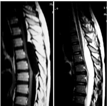
weighted sequence (Fig 1). Surgical treatment was indicat-ed and a laminectomy accomplishindicat-ed at T3-T5 level, where an extradural cystic tumor was found with poster lateral right location adherent in T4 (Fig 2).
The cyst was punctured showing a content with a lim-pid aspect, the capsule was completely removed and sent for histopathology study which showed cystic formation with thin fibrous walls covered by pavimentous cells in the internal surface; presence of thim perivascular inflamma-tory infiltrate, compatible with SC (Fig 3). In the immedi-ate postoperative the patient showed signs of clinical im-provement of the motor compromising of lower limbs. One month later the patient walked without help.
DISCUSSION
Spinal SC was initially described in 197411
. In that study the authors presented a case of SC of the cer-vical spine operated with success and confirmed by histopathological study. Other authors have reported the presence of this pathology in differents locations of the spine. The largest incidence was in the lumbar following by cervical and less frequently in the tho-racic spine. Most of the studied cases prevail in the male sex14
.The pathological substratum responsible for the development of the spinal SC includes the degeneration for arthrosis of the articulate facets, causing secondary lesion of the joint capsule and for-mation of hernia of the synovial membrane.
The presence of mixoid degeneration increases the production of hyaluronic acid with proliferation of mesenquimal cells contributing for the emergence
and size of the cysts7. Present clinical observation and
experimental date, suggest that the mechanical pres-sure in the articulate facet induces the appearance of a cascade of events as follows: upregulations and release of angiopoietin-1, interleukins-1 and 6, plate-let-derived growth factor, basic fibroblast growth fac-tor, vascular endotelial growth factor and substance P liberation, resulting in synovial hyperplasia, neo-vascularization and exudation of fluid, culminating with the formation of the cysts12
. This process could be reversible because the synovial proliferation can decrease on withdrawal of mechanical stress, what in some cases promotes the spontaneous reduction of the cysts12
.
The spondylolisthesis and the trauma can also be responsible for the emergence of these cysts. The content can be mucinous, with proteinaceous mate-rial and sometimes gaseous. They can present calci-fication focus or old intracystic hemorrhagic signs.
Fig 1. MR of the thoracic spine showed image of cystic aspect of extradural location in T4-T5, hyposignal in the weighed se-quence T1 and hypersinal in the weighed sese-quence T2.
Fig 2. Macroscopic aspect of surgical treatment showing the extradural cystic lesion.
840 Arq Neuropsiquiatr 2007;65(3-B)
Bleeding can occur by traumatism of the cyst wall due to the abundance in veined structures.
The preferential location of SC is in the great mo-bility lumbar spine segments following by the cervi-cal and less frequently in the thoracic spine. A litera-ture review shows the existence of few told cases in the thoracic location. In 2004 Cohen-Godol et al.14,
published 9 cases of SC of thoracic spine in a total of 16000 studied and submitted to the decompressive surgery of the spine due degenerative causes among others. The above mentioned cases correspond to 0.06% of the total and some literature refer that in the period from 1987 to 2001 only 10 cases have been described reaffirming the low incidence of this pathology. With the exception of a case described by Lynn et al.17 in 2000, of a patient with 24 years-old,
the average incidence of this pathology is related to patients of age above 70 years-old.
In the Brazilian literature we did not found ref-erences of SC of thoracic spine. The findings report cases of cysts of cervical and lumbar location5,10
. We report a case of a 14-year-old girl patient without significant trauma but with report of intermittent weakness during dance classes. This physical activity could be the reason for the arthritic process of articu-late facets probably exacerbated by certain reduction of the musculature facilitating a larger mobilization of the thoracic spine during the exercises. The neu-rological picture presented was of a paraparesis with pyramidal signs and sensitive deficit. The surgical and histopathological findings confirmed the diagnosis. The clinical and radiological aspects indicated the need of a differential diagnosis compatible with ex-tradural mass, tumor or herniated disc fragment14
. Aspects revealed by MRI help in the definition of the diagnosis of spinal SC.
Regarding the treatment some authors have doc-umented free remission12,19. The use of intracystic
ste-roids20 and aspiration of the same guided by CT21,22
can relieve temporarily the pain, probably due the interruption of the inflammatory cascade or the re-duction of the cyst volume12. The surgical treatment
determines the definitive cure with a minimum re-currence rate23, being the most effective treatment
in those cysts that compress the spinal cord structure
directly as in the cases of thoracic location. Common complications are referred as the intracystic hemor-rhagic or the formation of an epidural hematoma24
.
REFERENCES
1. Apostolaki E, Davies AM, Evans N, et al. MR imaging of lumbar facet joint synovial cysts. Eur Radiol 2000;10:615-623.
2. Pirotte B, Gabrovsky N, Massager N, et al. Synovial cysts of the lum-bar spine: surgery related results and outcome. J Neurosurg (Spine 1) 2003;99:14-19.
3. Shah RV, Lutz GE. Lumbar intraspinal synovial cysts: conservative man-agement and review of the world´s literature. Spine J 2003;3:479-488. 4. Khan AM, Synnat K, Cammisa FP, Girordi FP. Lumbar synovial cysts
of the spine: an evaluation of surgical outcome. J Spinal Disord Tech 2005;18:127-131.
5. Rosa ACF, Machado MM, Figueiredo MAJ, Albertoti CJ, Cerri GG. Cis-tos sinoviais lombares. Radiol Bras 2002;35:299-302.
6. Doyle AS, Menilees M. Synovial cysts of lumbar facet joints in a symp-tomatic population. Spine 2004;29:874-878.
7. Lima R, Spagamalo E, Johnston E, et al. Quistes sinoviales espinales sintomáticos. .Rev Hosp Maciel 1999;4:10-15.
8. Fonoff ET, Dias MP, Tarico MA; Myelopathic presentation of cervical juxtafacet cyst: a case report. Spine 2004;29:538-541.
9. Epstein NE, Hollingsworth R. Synovial cyst of the cervical spine. J Spi-nal Disord 1993;6:182-185.
10. Bianco AM, Madeira L, Fortini I, Elias AJR, Shibata MK. Remoção de cisto sinovial da articulação atlantoaxial pela via postero-latral trans-dural. Arq Bras Neurocir 2003;22:31-34.
11. Kao CC, Winkles SS, Turner JH. Synovial cyst of spinal facet. J Neuro-surg 1974;41:372-376.
12. Colen CB,Rengachary S. Spontaneous resolution of cervical synovial cyst: case illustration. J Neurosurg (Spine 4) 2006;4:186.
13. Tobenas-Dujardin AC, Derrey S, Proust F, Toussaint P, Laquerrierre A, Freger P. Kyste synovial atlanto-axoidien: aspects cliniques et proposi-tions chirugicalle. Neurochiorurgie 2004;50:652-656.
14. Cohen-Gadol AA, White JB, Lynch JJ, Miller GM, Krauss WE. Synovi-al cysts of the thoracic spine. J Neurosurg (Spine 1) 2004;1:52-57. 15. Graham E, Lenke LG, Hannallah D, et al. Mielopathy induced by a
tho-racic intraspinal synovial cyst: case report and review of the literature. Spine 2001;26:392-394.
16. Lopes N, Aesse F, Lopes D. Compression of thoracic nerve root by a facet joint synovial cyst: case report. Surg Neurol 1992;38:338-340. 17. Lynn B, Watkins RG, Watkins RI, et al. Acute traumatic myelopathy
sec-ondary to a thoracic cyst in a professional foot-ball player. Spine 2000; 25:1593-1595.
18. Hemninghytt S, Daniels DL, Williams AL, et al. Intraspinal synovial cyst: natural history and diagnosis by CT. Radiology 1982;145:373-376. 19. Houten J, Sanderson S, Cooper P. Spontaneous regression of synpto-matic lumbar synovial cysts. J Neurosurg (Spine 2) 2003;99:235-238. 20. Favre G, Javier-Modes RM, Hauber M, et al. Kyste symovial articulaire
lombaire revele par um syndrome pluriradiculaire: à propos de deux cas. Rev Med Interne 2004;25:230-233.
21. Abrahams JJ, Wood GW, Eames FA, et al. CT-guided needle aspiration biopsy of an intraspinal synovial cyst (ganglion): case report and re-view of the literature. AJNR 1988;9:398-400.
22. Imai K, Nakamura K, Inokuchi K, et al. Aspiration of intraspinal syno-vial cyst: recurrence after temporal improvement. Arch Orthop Trau-ma Surg 1998;20:103-105.
23. Lyons MK, Atkinson JL, Waharen RE, et al. Surgical evalution and man-agement of lumbar synovial cysts: the Mayo Clinic experience. J Neu-rosurg (Spine 1)2000;93:53-57.
