Arq Neuropsiquiatr 2004;62(3-B):879-881
Serviço de Neurologia e de Neurocirurgia do Hospital Moinhos de Vento, Porto Alegre, RS, Brazil and Faculdade de Medicina da Universidade Luterana do Brasil,Canoas, RS, Brazil: 1MD, Chief of the Service of Neurology and Neurosurgery and Professor of Neurosciences; 2Medical Student.
Received 1 December 2003, received in final form 1 April 2004. Accepted 26 May 2004.
Dr. Nilo M.M. Lopes - Rua Ramiro Barcelos 910/902 - 90035-001 Porto Alegre RS - Brasil.
PARAPLEGIA COMPLICATING PERCUTANEOUS
VERTEBROPLASTY FOR OSTEOPOROTIC
VERTEBRAL FRACTURE
Case report
Nilo M. Lopes
1, Vinicius K. Lopes
2ABSTRACT - We report a case of spinal cord and root compression during percutaneous transpedicular polyme-thylmethacrylate vertebroplasty (PTPV) for a compression fracture due to osteoporosis. Sudden onset of excruciating pain in the distribution of the right sixth intercostal nerve with hyperemia along its path, promp-ted the interruption of the procedure. Under narcotic sedation the patient was taken to the ICU and 10mg of dexamethasone was administered intravenously. Few hours later she developed paraplegia with preserva-tion of light touch and a CT scan and MRI showed epidural extravasapreserva-tion of polymethylmethacrylate with spinal cord and root compression. Surgical decompression was followed by neurological recovery. The cement could be removed after been thinned out by high speed drill, with microsurgical technique, through a wide three level laminectomy of D5 to D7. Extravasation of cement is commonly encountered in PTPV and most of the time it is asymptomatic. Root compression may require surgical intervention if nonresponsive to steroid treatment. Cord compression is less often seen and requires emergency surgery. The cement does not adhere to the duramater and it can be removed easily.
KEY WORDS: spinal fracture, polymethylmethacrylate, osteoporosis, vertebroplasty, paraplegia.
Remoção cirúrgica de polimetilmetacrilato epidural como complicação de vertebroplastia percutânea para tratamento de fratura com compressão de vértebra dorsal osteoporótica: relato de caso
RESUMO - Relatamos um caso de compressão medular e radicular durante vertebroplastia percutanea transpe-dicular com polimetilmetacrilato (VPTP) para tratamento de fratura com compressão por osteoporose. O início súbito de dor lancinante na distribuição do 6º nervo intercostal direito, com hiperemia ao longo de seu trajeto, determinou a interrupção do procedimento. Sob sedação com narcóticos, a paciente foi leva-da ao CTI, sendo administrados 10mg de dexametazona por via endovenosa. Após algumas horas, ela desenvol-veu paraplegia com preservação do tato, e a TC e a RM mostraram extravazamento epidural de polimetilmeta-crilato com compressão medular e radicular. Descompressão cirúrgica resultou em recuperação neurológi-ca. O cimento foi removido após ter sua espessura diminuída com o uso de broca de alta rotação, usando técnica microcirúrgica através de laminectomia ampla de tres níveis de T5 a T7. Extravazamento de cimen-to é comumente encontrado em VPTP, sendo assincimen-tomático na maioria das vezes. Compressão radicular pode requerer intervenção cirúrgica se não responsiva ao tratamento com corticosteróides. Compressão medular é vista com menos frequência e requer cirurgia de emergência. O cimento não adere à dura-máter e pode ser removido facilmente.
PALAVRAS-CHAVE: fratura vertebral, polimetilmetacrilato, osteoporose, vertebroplastia, paraplegia.
An increasing number of publications about percutameous transpedicular polymethylmethacry-late vertebroplasty (PTPV) can be found in the lit-terature mainly for the past six years. Although ini-tially it was done for vertebral angiomas1,2and for
metastatic vertebral disease3,4, more recently PTPV
became an alternative method to relieve pain in patients with compression fractures due to
osteopo-rosis4. Extravasation5of the cement has been
repor-ted to occur in 11 to 73% of the procedures6. In most
of these cases polymethylmethacrylate (PMMA) ends up in the soft tissues and in disc spaces and only in rare occasions it will cause symptoms7.
Intravascu-lar injection causing pulmonary embolism and epi-dural veins compromise7,8as well as cement
compression5,9should be prevented by injecting the
cement under continuos good quality fluoroscopy. We report a case of a postmenopausal woman with a symptomatic compression fracture of D6 ver-tebra due to osteoporosis who developed excrucia-ting radicular pain during the procedure and beca-me paraplegic few hours later. Neurological man-ifestations cleared after an emergency surgical de-compression was carried out.
CASE
History
This 82-year-old woman, had been continuosly under medical treatment for the past 4 months, for incapaci-tating pain due to a compression fracture of D6. Spine plain films showed deformities in at least three other vertebrae resulting in a dorsal kyphosis. She could no longer tolerate nonsteroidal antiinflammatory drugs (NSAID) and she was taking codeine several times a day. Because of severe physical limitation she was depressed and the medication made her drowsy and worsened her constipation. A bone scan and an MRI confirmed the clin-ical impression that the fractured D6 vertebral body was responsible for her pain.
Vertebroplasty
Under neuroleptoanalgesia, in the prone position, D6 was localized with fluoroscopy and the analgesia was complemented with xylocaine infiltration of all the planes from the skin to periosteum. An anesthesiologist was monitoring the patients vital signs and hemoglo-bin saturation. Fluoroscopy was switched from AP to lat-eral view once the bone biopsy needle reached the ver-tebral body in its course inside the pedicle. While inject-ing it was noticed that the cement was extrudinject-ing towards the lateral aspect of the vertebral body. The needle was withdrawn and a new needle insertion was performed through the contralateral pedicle. The lateral fluoroscop-ic view now was obscured on account of the previously extravasated cement. The injection had been uneventful up to the point when the patient began complaining of chest wall pain and the procedure was interrupted.
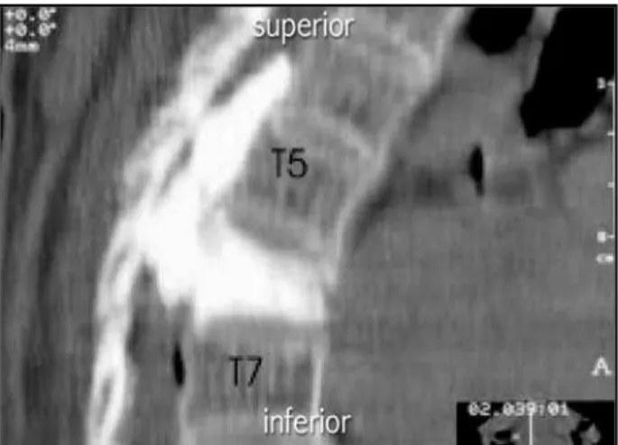
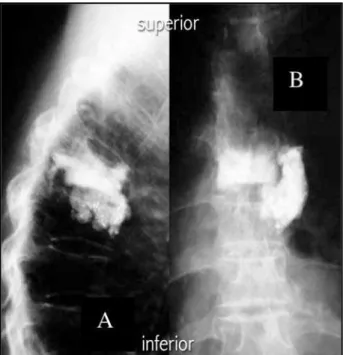
A skin rash was seen along the 6th rib. The patient was taken to ICU and had to be kept on morphine for pain relief. Dexamethasone 10mg was administered intravenously. Up to this point she was able to move both lower extremities. Repeat physical examination was im-paired because she was heavily sedated. About seven hours later she was no longer complaining of pain but she had developed paraplegia with preservation of light touch. A CT and an MRI scan revealed spinal cord and bilateral root compression from D5 to D6 (Figs 1 and 2).
Surgery
A bilateral laminectomy from D5 to D7 was carried out and the cement was drilled to the point it became
880 Arq Neuropsiquiatr 2004;62(3-B)
paper thin and easily dissected away from the dura-mater around the cord and the encased nerve roots. It was felt that the segment was stable because the ver-tebral body of D6 was filled satisfactorily with cement and the facet joint were partially preserved (Fig 3).
Postoperative course
Upon recovering from anesthesia she already had regained mobility of the lower extremities but some degree of a Brown-Sèquard syndrome remained for two weeks along with urinary retention requiring intermit-tent urethral catheterization. Within a month she was walking unaided with full bladder controll and no pain.
DISCUSSION
Percutaneous vertebroplasty with polymethyl-methacrylate proved to be a cost-effective treatment for pain relief due to compression fracture secondary
Fig 1. Sagittal CT scan of the theracic spine showing PMMA leak into the epidural space.
to osteoporosis6. It can be performed in outpatients,
with a very low morbidity rate, under local anesthe-sia, achieving lasting pain relief in more then 90% of the patients4. Spine surgeons very quickly
devel-op skills to insert a needle through the vertebral pe-dicle on account of their previous experiences with pedicular screws. Certainly there is a learning curve and one has to participate in hands-on courses before trying to perform PTPV procedure10. Complications
rates will rise as a greater number of procedures are being performed and undertrained doctors begin to undertake PTPV atracted by the good results found in the literature. PTPV has to be performed in a hos-pital with neurosurgical facilities, in order to be able to treat, without delay, complications such as cord compression.
Fluoroscopic digital images, bi-plane equip-ment and neuronavigation account for improved results due to added precision, lesser radiation ex-posure and shorter surgical time.
Even after many years in practice and more then 50 vertebrae injected without a single compli-cation, one is apt to face a spinal cord and root com-pression due to unnoticed migration of cement into the spinal canal.
Several lessons to be learned from this case and from his own experience. The radiopacity of the ce-ment has to be checked under fluoroscopy before mixing it with the monomer. Add barium as needed
Arq Neuropsiquiatr 2004;62(3-B) 881
to improve its visualization. High quality fluroscopy is a must. Keep in mind that an osteoporotic verte-brae is almost “x-ray transparent” so that the cement injection has to be monitored by fluoroscopy all the time. You donot need to fill up the vertebra. Pain relief is obtainable even with 3cc of PMMA and a unilateral pedicular injection is all you need.
It is advisible to talk to the patient and inquire him of any discomfort while injecting PMMA. The level of sedation should allow the patient to answer questions.
Check for breaches in the posterior wall of the vertebral body in the preoperative CT scan; its pre-sence is not a formal contraindication, but you must take extra care during the injection.
A vertebrogram does not insure safe PMMA in-jection. The contrast material has to be washed out with a saline flush before you start the cement in-jection.
Keeping the cement refrigerated in a bucket wi-th ice will increase your working time by a few pre-cious minutes. A thick cement lessens the chance of extravasation. Use larger gauge needle if it pro-ves to be suitable for the size of the pedicle.
Surgical removal of extravasated cement from the spinal canal and from the root foramen can be accomplished through a laminectomy using high speed drill and microsurgical techniques.
REFERENCES
1. Galibert P, Deramond H. Rosat P, Legars D. Preliminary note on the treat-ment of vertebral angioma by percutaneous acrylic vertebroplasty. Neurochirurgie 1987;233:166-168.
2. Aebli N, Krebs J, Davis G, Walton M, Williams MJA, Theis JC. Fat embolism and acute hypotension during vertebroplasty: an experimental study in sheep. Spine 2002;27:460-466.
3. Kaemmerlen P, Thiesse P, Bouvard H, et al. Percutaneous vertebroplasty in the treatment of metastases: technic and results. J Radiol 1989;70:557-562. 4. Barr JD, Barr MS, Lemley TJ, McCann RM. Percutaneous
vertebroplas-ty for pain relief and spinal stabilization. Spine 2000;25:923-928. 5. Cotten A, Dewatre F, Cortet B, et al. Percutaneous vertebroplasty for
osteolytic metastases and melanoma: effects of the percentage of lesion filling and the leakage of methylmethacrylate at clinical follow-up, Radiology 1996;200:525-530.
6. Shapiro S, Abel T, Purvines S. Surgical removal of epidural and intradur-al plymethylmetacrylate extravasation complicating percutaneous ver-tebroplasty for an osteoporotic lumbar compression fracture. J Neurosurg 2003;98(Suppl 1):S90-S92.
7. Amar AP, Larsen DW, Esnaashari N, Albuquerque FC, Lavine SD, Teitelbaum GP. Percutaneous transpedicular polymethylmethacrylate vertebroplasty for the treatment of spinal compression fractures. Neuro-surgery 2001;49:1105-1114.
8. Padovani B, Kasriel O, Brunner P et al. Pulmonary embolism caused by acrylic cement: a rare complication of percutaneous vertebroplas-ty. Am J Neuroradiol 1999;20:375-377.
9. Heini PF, Allred CD. The use of a side-opening injection cannula in ver-tebroplasty Spine 2002;27:105-109.
10. Mathis JM, Barr JD, Belkoff SM, Barr MS, Jensen ME, Deramond H. Per-cutaneous vertebroplasty: a developing standard of care for vertebral compression fractures. Am J Neuroradiol 2001;22:373-381.