379 Intradural-extramedullary cysticercosis with brain involvement
Radiol Bras 2006;39(5):379–382 Case Report
INTRADURAL-EXTRAMEDULLARY SPINAL CYSTICERCOSIS WITH
BRAIN INVOLVEMENT: A CASE REPORT AND LITERATURE REVIEW*
Luiz Antonio Rossi1, Adalberto Sestari2, Modesto Cerioni Jr.3
Report on a rare case of infestation by cysticercosis involving the cerebral subarachnoid space and the epi-dural space in the cervical and thoracic regions of a 59-year old female patient who presented nausea, signs of cerebelar ataxia and gradual loss of the sensibility in both legs. The diagnosis was based on magnetic resonance imaging of brain and cervical-thoracic spine demonstrating the presence of cysts in the subarach-noid spaces. An enzyme-linked immunosorbent assay (ELISA) performed on cerebrospinal fluid resulted positive and demonstrated a protein level of 420 mg/dl indicating the disease activity. Parasites were surgically re-moved for the necessity of thoracic spinal cord decompression. Based on findings of literature review, brief comments were made on the pathogenesis of the intradural-extramedullary spinal cysticercosis cystic form, magnetic resonance imaging findings and treatment.
Keywords: Cysticercosis; Brain; Spine; Magnetic resonance imaging.
Cisticercose intradural-extramedular cerebral e espinhal: relato de caso e revisão da literatura.
Relato de um caso raro de apresentação de infestação por cisticercose do espaço subaracnóide cerebral e intra-raquiano nas regiões cervical e torácica em mulher de 59 anos de idade, com náuseas, sinais de ataxia cerebelar e perda gradual da sensibilidade nas pernas. O diagnóstico foi feito por meio de imagens por res-sonância magnética do cérebro e da coluna cérvico-torácica, que evidenciaram a presença de cistos nos espaços subaracnóides. O exame do líquido cefalorraquiano revelou teste imunológico ELISA positivo e ele-vado nível de proteína (420 mg/dl), indicativo de atividade da doença. Os parasitos foram removidos cirur-gicamente pela necessidade de descompressão da medula espinhal torácica. Breve comentário sobre a pa-togênese da forma cística da cisticercose espinhal intradural-extramedular, aspectos das imagens de resso-nância magnética e tratamento foram feitos com base nos achados de revisão da literatura.
Unitermos: Cisticercose; Cérebro; Coluna; Ressonância magnética.
Abstract
Resumo
* Study developed at Hospital do Servidor Público Estadual “Francisco Morato de Oliveira”, São Paulo, SP, Brazil.
1. Assistant Professor of the Discipline Radiology Principles at Center of Medical and Biological Sciences, Pontifícia Universidade Católica de São Paulo.
2. Assistant Professor at Universidade da Cidade de São Paulo, Neuroradiologist at Hospital do Servidor Público Estadual “Fran-cisco Morato de Oliveira”.
3. Neurosurgeon at Hospital do Servidor Público Estadual “Francisco Morato de Oliveira”.
Mailing Address: Prof. Dr. Luiz Antonio Rossi. Rua Cornélio Pires, 284, Condomínio Campos de Santo Antonio. Itu, SP, Bra-zil, 13305-500. E-mail: [email protected]
Received June 24, 2004. Accepted after revision December 15, 2004.
INTRODUCTION
Human cysticercosis is the most com-mon parasitic disease to affect the central nervous system, caused by the larval stage of Taenia solium (cysticercus), especially in countries where the sanitary infrastruc-ture is deficient, particularly in Latin Ame-rica, Africa and Asia and yet, with relative frequency, in Portugal, Spain and European Eastern countries and endemic in devel-oped countries with high rates of immigra-tion from endemic areas(1–3).
The adult worm develops in humans after ingestion of pork, rare, infected meat
containing encysted larvae. The human cysticercosis is acquired through oral-fecal pathway after contact with member of the family or community with the adult worm, as well as by auto-infestation in patient with the intestinal adult worm. After ova ingestion, the shells of ova are digested and the larvae develops and lodge in soft-tis-sues, primarily in brain, spinal cord, heart/ extra-ocular/skeletal muscle, eyeball, fat tissue and skin(1–3).
Cerebral and spinal-medullary intrapa-renchymatous cysticercosis is caused by cysts and/or granulomatous, inflammatory nodules, whereas extraparenchymatous cys-ticercosis like those located inside perience-phalic liquoric spaces (including the sub-arachnoid space in the region of cortical sulci and encephalic cisterns), cerebral ven-tricles and intradural-extramedullary space of the vertebral column, is typically cystic. The case report includes clinical-radio-logical-pathologic aspects of cerebral and spinal intradural-extramedullary cysticer-cosis in cystic form, with a brief literature review.
CASE REPORT
A 59-year old, white woman was admit-ted for evaluation, presenting nausea, cerebelar ataxia, a gradual loss of sensibil-ity in both legs, micturition and evacuation disorders. At general physical and neuro-logical examination decreased reflexes in lower extremities and hyperesthesia at the T6 dermatome where characterized.
Magnetic resonance imaging (MRI) has revealed cystic lesions in brain, cervical-thoracic spine subarachnoid spaces with signs of compression on the thoracic spi-nal cord (Figures 1 and 2).
Laboratory tests on cerebrospinal fluid collected by means of lumbar puncture have indicated a high protein level (420 mg/dl) and the research of antibodies for cysticercus, ELISA test, was positive.
380
Rossi LA et al.
Radiol Bras 2006;39(5):379–382
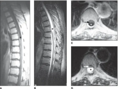
Figure 1. Dorsal column MRI. A: Sagittal plane, T1-weighted sequence showing clustered cysts (aster-isk) corresponding to an area with hyposignal. B: Sagittal plane, T2-weighted sequence showing hyperintense clustered cysts (asterisk) compressing the dorsal spinal medulla. C: Axial plane, T1-weighted sequence showing hypointense clustered cysts (asterisk). D: Axial plane, T2-weighted sequence showing hyperintense clustered cysts (asterisk) compressing the dorsal spinal medulla (arrows).
A B D
Figure 2. Axial brain MRI. A: T1-weighted showing hypointense cysts (arrows) in subarachnoid space in parietal and right frontal regions. B: T2-weighted sequence at the same level, showing hyperintense cysts (arrows).
ullary presentation being the most frequent and the intramedullary, the less frequent. Among 205 inpatients at Hospital Vera Cruz de Campinas, SP, Neurology Service, in the period between 1961 and 1987, only 18 (9.0%) had a definite diagnosis of neu-rocysticercosis, and only three (1.4%) pre-sented intradural-extramedullary spinal infestation by cysticercosis(5).
In the literature, we have found an ex-cellent review study analyzing 95 cases of spinal cysticercosis published since 1865 (Chart 1), 61 (66.0%) of them presenting intradural-extramedullary localization, and 33 (34.0%) presenting intramedullary lo-calization(4). Many studies included in this review and other more recent studies(1,3,6), likewise the present case, have shown the simultaneous involvement of liquoric, in-tradural-extramedullary spinal spaces in different segments and cerebral spaces.
The cysticerci larvae dissemination into the intradural-extramedullary spinal space has been explained by the accumulation of a great volume of blood originating from valveless epidural venous plexus, with ex-tremely thin-walled veins, which may con-duct the blood towards any direction, un-der the influence of intra-abdominal and intrathoracic pressure variations. Two sur-gically-confirmed cases of primary cys-ticerci larvae dissemination into the intra-dural-extramedullary spinal canal without cerebral involvement corroborate the hy-pothesis of retrograde flow of those larvae through the veins of the above mentioned epidural venous plexus, differently from the Isamat de La Riva’s postulate on the parasites descending migration from the cerebral subarachnoid space into the spinal space(7).
The term racemose cysticercosis ( cys-ticercus racemosus) has been utilized by several authors and characterizes the devel-opment of large translucent vesicles, with big tails, multiloculated or branched off, absent scolex, subarachnoid space-occupy-ing(1,3,5,6,8).
The intra-rachidian intradural-extra-medullary cysticercosis may be asymptom-atic or presenting few symptoms over long periods of time. Symptoms may vary, de-pending on the vertebral column segment (cervical, dorsal, lumbar or sacral) were cysts are lodged(1–3,6). When the cyst is
A B
DISCUSSION
A case of vesicles in the corpus callo-sum was first described by Paranoli in 1550. Paracelso correlated cerebral vesi-cles and convulsive attacks in 1650, while
Ridi and Malpighi characterized these para-sitic lesions in 1686. The first reference to intra-rachidian cysticercosis is attributed to Rokitansky in 1856(4).
The neurocysticercosis intra-rachidian form is quite rare, the
C
381 Intradural-extramedullary cysticercosis with brain involvement
Radiol Bras 2006;39(5):379–382
A B C
Figure 3. A: Surgical aspect following dura mater incision in the dorsal region, exposing clustered cysticercotic cysts. B: Surgical sequence showing cysts individual removal. C: Macroscopic aspect of two removed cysts.
Chart 1 Intradural-extramedullary spinal cysticercosis: published cases. . Author Westphal Richter Minor Hartman Wollenberg Sterz Vasiliu Redalie Castex Verga Morawieka Bertrand Loyo Pennybaker Fracassi De la Riva
Rocca Cabieses Cruz Calzado Canelas Staimle Absalon Alanis Trelles Castano Parker Bandres Leite Çiftçi Year 1865 1891 1899 1902 1905 1910 1921 1921 1926 1926 1927 1945 1955 1956 1956 1957 1959 1959 1961 1960 1963 1964 1965 1967 1968 1969 1988 1992 1997 1999 Country ? ? ? Germany ? ? Germany Italy France Argentina Italy ? France Mexico ? Argentina Spain Peru Peru Brazil Mexico Brazil Mexico Mexico Mexico Peru Colombia France USA Brazil, USA, Colombia USA Age ? ? ? ? ? ? ? ? ? 25-year-old ? ? ? ? ? 49-year-old ? ? (15 cases) 12-year-old 42-year-old ? 26-year-old 32-year-old 42-year-old 27-year-old 57-year-old 45-year-old 35-year-old 63-year-old 40-year-old 57-year-old 56-year-old 46--year-old 58-year-old 34-year-old 35-year-old 46-year-old 34-year-old (9 cases) 30-year-old Sex ? ? ? ? ? ? ? ? ? F ? ? ? ? ? M ? ? M M ? M F M M M M M M M F M M F F M F M F Brain* ? + + ? + + ? ? ? + + + ? ? + + + + – – ? – + + + – – – – + ? + + + – + – – + Diagnosis ? Necropsy Necropsy Lumbar puncture Necropsy Necropsy Lumbar puncture ? ? Lumbar puncture Necropsy Lumbar puncture ? ? Surgery Lumbar puncture Necropsy Lumbar puncture ? Surgery Surgery ? Surgery Surgery Surgery Surgery Surgery Surgery Surgery Surgery Surgery ? Necropsy Necropsy Necropsy Surgery Surgery Surgery Lumbar puncture Lumbar puncture, sur-gery Lumbar puncture Localization Cauda equina C4/T10 Thoracic ? Cervical-lumbar Thoracic-lumbar ? ? Cervical-thoracic ? ? Cauda equina Lumbar Cauda equina Cervical ? Lumbar Lumbar
Cervical (13), thoracic (1), lumbar (1) T3
T11/L2 e L2 L3 Thoracic T6/T9 T11/L2 T12/L2 T5/T9 Cauda equina T8/T9 C5/C7 C2/C4 L1/L2 L4/L5 C4 C4 Cauda equina Cauda equina L5/S2 L5/S1 C2/S1
Cervical-thoracic (4), cervical-thoracic-lumbar (4), cervical (2), thoracic-lumbar (1), lumbar (1) C2/C4
* Coexistence of cysts in liquoric cerebral and spinal spaces.
localized in the spine cervical-thoracic segment, the symptoms are more apparent and usually cause medullary compression syndrome, as described in this case: gradual loss of sensibility in both legs, hyperestesia at the corresponding dermatome and mic-turition and evacuation physiological dis-orders. However, in the spine lumbosacral segment, frequently the clinical picture evi-dences localized radiculopathy that may be confounded with discopathy(1–3,6).
382
Rossi LA et al.
Radiol Bras 2006;39(5):379–382
marginated by intrathecally-injected iodine contrast(2,5,6).
Presently, the imaging method of choice for intradural-extramedullary cysticercosis diagnosis is the MRI, that accurately dem-onstrates cystic lesions in subarachnoid liquoric spaces of periencephalic or spinal regions and, principally because is a non-invasive procedure(1,3,8,9).
MRI images indicate the precise local-ization and contours of these cysts whose signal intensity is similar to CSF, hyposig-nal in T1-weighted sequences and hyper-signal in T2-weighted sequences(1,3,8,9), identical to those demonstrated in this pa-tient studies.
However, these characteristics may dif-fer when cysts present inflammatory alter-ations, resulting in hyperintense images both in T1-weighted and T2-weighted sequences, as a consequence of the high protein con-tent(3). After paramagnetic contrast agent endovenous infusion, both the cyst and the meninges surrounding the adjacent medulla may be enhanced (arachnoiditis)(1–3,8), which has not occurred in this case report.
A differential diagnosis spectrum should be considered when cystic, intra-rachidian lesions are detected in MRI im-ages: congenital lesions (simple or com-plex arachnoid cysts, dermoid cysts), cysts of other parasitary etiology (hydatid), tu-berculosis, sarcoidosis or subarachnoid metastatic neoplasm(1,3).
A positive ELISA serological test and high rates of protein in CSF may be of help in the diagnosis, indicate the disease activ-ity and serve as a follow-up and control of medicamentous treatment(6). In the present case, CSF laboratory tests reveal high pro-tein levels (420 mg/dl) and positive for ELISA.
Usually, treatment is conservative, pre-senting good results with administration of cysticidal drugs like praziquantel or alben-dazole, the later demonstrating higher treat-ment efficacy(8,9). In cases where signs of medullary and/or radicular compression are detected, surgical indication becomes nec-essary, likewise in the case of our patient. An interesting aspect of the decompressive surgery in this case was the photographic register of a careful and individual surgi-cal removal of cysts, allowing the radio-logical-surgical-pathological correlation.
Concluding, cysticercosis is a severe global public health problem, including developed countries because of the high rates of immigration from endemic areas. The disease severity is determined when cystic cysticerci involve subarachnoid spaces, producing medullary symptoms when located in the cervico-thoracic seg-ment and radicular symptoms, when lo-cated in the lumbosacral segment, demand-ing surgery due to frequent unsuccessful conservative clinical treatments. The diag-nosis may be completed by a findings
com-bination: epidemiological history, classical clinical signs and symptoms, MRI images, laboratory tests and CSF serological tests.
REFERENCES
1. Leite CC, Jinkins JR, Escobar BE, et al. MR imaging of intramedullary and intradural-extra-medullary spinal cysticercosis. AJR Am J Roent-genol 1997;169:1713–1717.
2. Bandres JC, White AC Jr, Samo T, Murphy EC, Harris RL. Extraparenchymal neurocysticercosis: report of five cases and review of management. Clin Infect Dis 1992;15:799–811.
3. Çiftçi E, Diaz-Marchan PJ, Hayman LA. Intradu-ral-extramedullary spinal cysticercosis: MR im-aging findings. Comput Med Imim-aging Graph 1999;23:161–164.
4. Gallani NR, Zambelli HJ, Roth-Vargas AA, Li-moli Junior C. Spinal cord cysticercosis: report of 2 cases, review of the literature, and comments on its pathogeny. Arq Neuropsiquiatr 1992;50: 343–350.
5. Rossitti SL, Roth-Vargas AA, Moreira AR, Sper-lescu A, Araújo JF, Balbo RJ. Pure spinal leptome-ningeal cysticercosis. Arq Neuropsiquiatr 1990; 48:366–370.
6. Parker F, Hladky JP, Breton JO, Mignard C, La-porte JP, Bousquet C. Cysticercose racemeuse de la queue de cheval et arachnoidite kystique. Neu-rochirurgie 1988;34:280–285.
7. Sperlescu A, Balbo RJ, Rossitti SL. Brief com-ments on the pathogenesis of spinal cysticerco-sis. Arq Neuropsiquiatr 1989;47:105–109. 8. Martinez HR, Rangel-Guerra R,
Arredondo-Es-trada JH, Marfil A, Onofre J. Medical and surgi-cal treatment in neurocysticercosis: a magnetic resonance study of 161 cases. J Neurol Sci 1995; 130:25–34.