Arq Neuropsiquiatr 2004;62(3-B):875-878
Department of Neurosurgery, Hospital Nossa Senhora das Graças, Curitiba PR, Brazil: 1Resident in Neurosurgery; 2Neurosurgeon; 3Neurosurgeon Coordinator of the residency program.
Received 6 January 2004, received in final form 26 March 2004. Accepted 18 May 2004
Dr. Daniel Monte Serrat Prevedello - Rua Alcides Munhoz 433 - 80810-040 Curitiba PR - Brazil. E-mail: [email protected]
MANAGEMENT OF PRIMARY SPINAL
CHONDROSARCOMA
Report of two cases causing cord compression
Daniel Monte-Serrat Prevedello
1, Joacir Graciolli Cordeiro
1, Andrei Koerbel
2,
Léo Fernando da Silva Ditzel
2, João Cândido Araújo
3ABSTRACT - Chondrosarcomas are malignant tumors that rarely grow inside the spinal canal. Prognosis depends on histological features, patient’s age and surgical margins free from tumor. Response to radio and chemotherapy is poor. Ideal treatment consists of total “en-block” resection, not always achievable due to limitation of location, compromise of stability and risk of inducing neurological deficits. Two cases of spinal chondrosarcoma causing cord compression are reported, located in the cervical and thoracic spine. Microsurgical technique consisted of initial debulking followed by removal of margins until limits free from tumor were obtained. Total resection was accomplished and neurological function improved in both cas-es. Follow-up has been seven and one year respectively, with no evidence of recurrence and preserved neu-rological functions. Association between chondrosarcoma and estrogen-dependent tumor has been con-firmed in this report. Although “en-block” resection of a chondrosarcoma should be tried whenever pos-sible, tumor fragmentation should be considered in difficult cases, as in the present report, in which a long period free from recurrence with good quality of life can be obtained.
KEY WORDS: chondrosarcoma, spinal neoplasms, spinal cord compression, microsurgery.
Manejo dos condrosarcomas espinhais primários: relato de dois casos causando compressão medular
RESUMO - Os condrosarcomas são tumores malignos, raramente localizados no interior do canal espinhal, com prognóstico dependente do grau histológico do tumor, idade do paciente e margens cirúrgicas livres. Esses tumores apresentam pouca resposta à radio e quimioterapia. O tratamento ideal consiste em ressecção tumoral em bloco, condição particularmente difícil em se tratando de tumores causando compressão medular, devido à localização da lesão, comprometimento da estabilidade axial e necessidade da manutenção ou recuperação da integridade da função neurológica do paciente. Relatamos dois casos de condrossarco-mas causando compressão medular, um na coluna cervical e outro na torácica, submetidos a cirurgia com esvaziamento tumoral seguido de remoção das margens, atingindo-se ressecção total e melhora das funções neurológicas em ambos os casos. Um paciente se encontra há 7 anos e outro há 1 ano livre de recorrências, ambos com funções neurológicas preservadas. Associação entre condrosarcoma e tumor dependente de estrógeno é confirmada clinicamente pelo presente estudo. Embora uma ressecção em blo-co dos blo-condrossarblo-comas deva ser tentada quando possível, deve-se blo-considerar uma ressecção blo-com fragmen-tação tumoral em casos específicos, podendo-se atingir, como demonstrado nos presentes casos, períodos livres de recorrência e longo tempo de sobrevida.
PALAVRAS-CHAVE: condrossarcoma, neoplasias medulares, compressão da medula espinhal, microcirurgia.
Chondrosarcomas are malignant lesions, which re-present the second most common tumor of the ske-leton. However, presentation as primary tumor of the spine is extremely rare. These tumors can originate from healthy bone or develop from a cartilaginous lesion with sarcomatous degeneration1. Primary
ma-lignant tumors should be resected with wide safety margins. However, limitations to total resection may
involve risk of causing spinal instability and/or inflict-ing new deficits. Isolated cases of spinal chondrosarco-mas have been reported2-18as well as a few series with
limited number of patients19-22.
CASES
Case 1.An 80 year-old woman was admitted with pro-gressive weakness in her lower limbs, which resulted in inability to walk, one week before admission. Neurolo-gical examination showed spastic paraparesis. Hypoes-thesia with partial loss of pain and temperature below T4 level and preserved proprioception and vibration sense were also demonstrated. She was submitted to a cervical and thoracic spinal magnetic resonance study (MRI), which showed a posterior and lateral extradural tumor from C5 to T1, mainly on the left side, inside the spinal canal, causing significant cord compression. Lami-nectomies from C5 to T1 were carried out exposing an extradural mass of great proportions on the left side of the spinal canal affecting the posterior elements of C6 to T1 and causing a compressive effect on the spinal cord. The mass was completely resected in a piece-meal fash-ion until margins free of tumor were obtained. Post-oper-ative period was uneventful with gradual reversal of her neurological deficits. Pathology specimen was com-patible with chondrosarcoma and no adjuvant therapy was used.
Four years after the spinal surgery the patient under-went a histerectomy due to a endometrium carcinoma. After two years, she was submitted to another procedu-re, a radical mastectomy, due to a breast cancer.
876 Arq Neuropsiquiatr 2004;62(3-B)
Seven years have elapsed since the spinal operation with no signs of tumor recurrence.
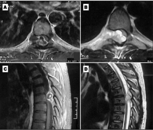
Case 2.A 25 year-old man was admitted with pro-gressive right intercostal pain irradiated from the back, at a mid thoracic level, of one-year duration. Six months prior to admission he started with gait difficulties and frequent falls, progressing to a severe paraparesis and inability to walk.
His neurological examination demonstrated bilater-al normbilater-al strength in upper extremities, and only minor proximal muscle function in his lower limbs. Partial loss of pain and temperature and diminished proprioception and vibration were seen below the T6-T7 level, in addi-tion to severe spasticity. Thoracic spine MRI demonstrat-ed a large extradural mass lesion, at T6-T7 level, 3 cm in diameter, compressing the spinal cord, growing through an enlarged right intervertebral foramen (Fig 1). The patient underwent a T6-T7 laminectomy with removal of the respective right facet, which was compromised by a whitish dense extradural tumor. The spinal cord was compressed anteriorly and shifted to the left as tumor advanced through the T6-T7 right foramen. The lesion was completely resected, in a piece-meal fashion until margins free of tumor were obtained. The T6-T7 fora-men was left wide opened and the nerve root free of
Arq Neuropsiquiatr 2004;62(3-B) 877
than subtotal resection. The tumor recurred in 64% of the patients and the median disease free interval was 16 months without adjuvant therapy20.
Shives et al. studied 20 patients with chondrosar-coma of the spine and found 100% of recurrence in patients submitted to tumor debulking only, half of the cases recurring among the six patients with inappropriate surgical margins and no recur-rences in two patients with radical resection21.
Even though rare cases with an “en-block” re-section of the lesions have been reported, some with spinal instrumentation, and the prognosis in the long run has not been clearly defined23-26.
Pa-tients submitted to radiotherapy did not demon-strate significant increase in survival in relation to those treated by surgery only19,20. There are no
studies in the literature able to demonstrate effecti-veness of radiotherapy, as well as of chemothera-py, in spinal chondrosarcoma.
Case 2 patient presented with enlargement of the intervertebral foramen, raising the suspicion of neurofibroma as first possible diagnosis27,28.
Only one case of foraminal chondrosarcoma has been reported byYünten et al., probably originat-ing from a previous osteochondroma28.
Association between chondrosarcoma and ade-nocarcinoma of breast , without previous radiother-apy, has been reported by Mummaneni and Rosen-berg29. Chondrosarcomas may suffer hormonal
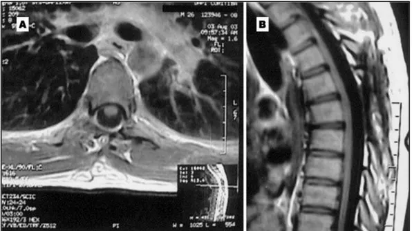
in-compression. MRI in the first post-operative day con-firmed total resection. Neurological deficits gradually im-proved. Pathology specimen was compatible with chon-drosarcoma and no adjuvant therapy was used.
One year has elapsed since the operation and the pa-tient remains asymptomatic. A new MRI was recently ob-tained confirming no signs of tumor recurrence (Fig 2).
DISCUSSION
Surgical treatment of spinal chondrosarcoma is particularly difficult. Prognosis depends mainly on tumor removal, whereas the peculiar anatomical features of the spine practically avoids an ideal “en-block” resection. When all locations are considered, survival from chondrosarcoma is relatively high, reaching more than 87% of patients at 5 years; ho-wever, it is considerably lower when only pelvic, sacral and spinal locations are considered, varying between 25 and 54%19.
There are only a few series in the literature con-cerning spinal chondrosarcomas19-22. Thoracic spine
is the most common location and the male sex is predominant20-21. According to Bergh et al.
fac-tors associated with worse prognosis are high his-tological grade, advanced age, primary surgery out of a referral center, incisional biopsy only and inadequate surgical margins19. York et al.
evaluat-ed 21 patients with spinal chondrosarcoma, dur-ing a period of 43 years, demonstratdur-ing a longer disease free interval in patients with total rather
fluence, even in the absence of detectable estro-gen receptors. High estroestro-gen levels seem to increase their growth30.
Case 1 patient developed subsequently both bre-ast and endometrial carcinoma, representing the only case reported in the literature with such an association.
Total surgical resection is the best therapeutic option for chondrosarcomas, considering its resist-ance to radiotherapy and chemotherapy. Piece-meal removal is recommended when “en-block” resection is not feasible, but assurance of margins free from tumor must be obtained31,32.
When complete resection is obtained progno-sis may be good, even in the presence of adverse factors, such as advanced age, as in case 1. Associa-tion between chondrosarcoma and estrogen-de-pendent tumors has been confirmed in this report.
REFERENCES
1. Gelabert-Gonzalez M, Reyes-Santias RM, Garcia-Pravos A. Dorsal medullary compression due to a primary vertebral chondrosarcoma. Rev Neurol 2000;30:322-324.
2. Di Lorenzo N, Palatinsky E, Artico M, Palma L. Dural mesenchymal chondrosarcoma of the lumbar spine: case report. Surg Neurol 1989;31:470-472.
3. Blaylock RL, Kempe LG. Chondrosarcoma of the cervical spine: case report. J Neurosurg 1976;44:500-503.
4. Hirsh LF, Thanki A, Spector HB. Primary spinal chondrosarcoma with eighteen-year follow-up: case report and literature rewiew. Neurosurgery 1984;14:747-749.
5. Reif J, Graf N. Intraspinal mesenchymal chondrosarcoma in a three-year-old boy. Neurosurg Rev 1987;10:311-314.
6. Lee ST, Lui TN, Tsai MD. Primary intraspinal dura mesenchymal chon-drosarcoma. Surg Neurol 1989;31:54-57.
7. Ranjan A, Chacko G, Joseph T, Chandi SM. Intraspinal mesenchymal chondrosarcoma: case report. J Neurosurg 1994;80:929-930. 8. Herman TE, McAlister WH, Dehner LP, Kaufman BA. Dedifferentiated
chondrosarcoma in childhood: report of a case. Pediatr Radiol 1995;25(Suppl 1):S140-S142.
9. Ogose A, Motoyama T, Hotta T, et al. Clear cell chondrosarcomas aris-ing from rare sites. Pathol Int 1995;45:684-690.
10. Rushing EJ, Mena H, Smirniotopoulos JG. Mesenchymal chondrosar-coma of the cauda equina. Clin Neuropathol 1995,14:150-153. 11. Ohue S, Sakaki S, Kohno K, et al. Primary spinal chondrosarcoma
local-ized in the cervical spinal canal and intervertebral foramen: case report. Neurol Med Chir 1995;35:36-39.
878 Arq Neuropsiquiatr 2004;62(3-B)
12. Vanderhooft JE, Conrad EU, Anderson PA, Richardson ML, Bruckner J. Intradural recurrence with chondrosarcoma of the spine: a case report and review of the literature. Clin Orthop 1993;294:90-95.
13. Marchi E, Antonico A Filho, Sette AA, Sette RC. Hemothorax as a pri-mary manisfestation of chondrosarcoma of the thoracic spine. Rev Assoc Med Bras 1992;38:177.
14. Tan SK, Seow KH, Chiang GS, Sim CS. Chondrosarcoma of the spine: report of two cases. Ann Acad Med Singapore 1991;20:385-388. 15. Gursel B, Yalciner G. Chondrosarcoma of the cervical vertebra: a case
report. Arch Otorhinolaryngol 1987;244:74-76.
16. Chan HS, Turner-Gomes SO, Chuang SH, et al. A rare cause of spinal cord compression in childhood from intraspinal mesenchymal chon-drosarcoma: a report of two cases and review of the literature. Neuroradiology 1984;26:323-327.
17. Ozaki T, Hillmann A, Blasius TS, Winkelmann W. Skeletal metastases of intermediate grade chondrosarcoma without pulmonary involvement: a case report. Int Orthop 1998;22:131-133.
18. Crowell RM, Wepsic JG. Thoracic cord compression due to chondrosar-coma in two cousins with hereditary multiple exostoses: report of two cases. J Neurosurg 1972;36:86-89.
19. Bergh P, Gunterberg B, Meis-Kindblom JM, Kindblom LG. Prognostic factors and outcome of pelvic, sacral and spinal chondrosarcomas: a center-based study of 69 cases. Cancer 2001;91:1201-1212.
20. York JE, Berk RH, Fuller GN, et al. Chondrosarcoma of the spine: 1954 to 1997. J Neurosurg (Spine 1) 1999;90:73-78.
21. Shives TC, McLeod RA, Unni KK, et al. Chondrosarcoma of the spine. J Bone Joint Surg 1989;71:1158-1165.
22. Camins MB, Duncan AW, Smith J, Marcove RC. Chondrosarcoma of the spine. Spine 1978;3:202-209.
23. Mandelli C, Bernucci C, Mortini P, Tartara F, Scomazzoni F, Giovanelli M. Chondrosarcoma of the thoracic spine: total en bloc sagittal resec-tion. A case report. J Neurosurg Sci 2001;45:114-119.
24. Marmor E, Rhines LD, Weinberg JS, Gokaslan ZL. Total en bloc lum-bar spondylectomy: case report. J Neurosurg 2001;95:264-269. 25. Hasegawa K, Ogose A, Kobayashi H, Morita T, Hirata Y. Simultaneous
anterior-posterior approach for excision of malignant paraspinal tumor and subsequent reconstrution: technical note. J Neurosurg 1999;91:236-240. 26. Doh JW, Halliday AL, Baldwin NG, Benzel EC. Spinal stabilization by using crossed-screw anterior-posterior fixation after multisegmental total spondylectomy for thoracic chondrosarcoma: case report. J Neurosurg 2001;94:279-283.
27. Zibis AH, Markonis A, Karantanas AH. Unusual causes of spinal foram-inal widening. Eur Radiol 2000;10:144-148.
28. Yünten N, Calli C, Zileli M, Üstün EE, Sener RN. Chondrosarcoma caus-ing cervical neural foramen widencaus-ing: case report. Eur Radiol 1997;7:1028-1030.
29. Mummaneni PV, Rosenberg WS. Spinal chondrosarcoma following ade-nocarcinoma of the breast: case report. Surg Neurol 2000;53:580-582. 30. Mackintosh D, Mason RM. Pharmacological actions of 17B-oestradiol on
articular cartilage chondrocytes and chondrosarcoma chondrocytes in the absence of oestrogen receptors. Biochim Biophys Acta 1988;964:295-302. 31. Prevedello DM, Koerbel A, Tatsui CE, et al. Prognostic factors in the treatment of the intradural extramedullary tumors: a study of 44 cas-es. Arq Neuropsiquiatr 2003;61:241-247.