Arq Neuropsiquiatr 2007;65(1):32-35
Neurology Service, Department of Internal Medicine, Medical School, Federal University of Minas Gerais, Belo Horizonte MG, Brazil:
1Resident in Neurosurgery, 2Physician, 3Assistant Professor, 4Associate Professor.
Received 29 May 2006, received in final form 30 August 2006. Accepted 23 October 2006.
Dr. Francisco E.C. Cardoso - Av Pasteur 89/1107 - 30150-290 Belo Horizonte MG - Brasil. E-mail: [email protected]
A BRAZILIAN FAMILY WITH
BROWN-VIALETTO-VAN LAERE SYNDROME WITH AUTOSOMAL
RECESSIVE INHERITANCE
José Augusto Malheiros
1, Sarah Teixeira Camargos
2,
José Teotonio de Oliveira
3, Francisco E.C. Cardoso
4ABSTRACT - We report the first Brazilian family with Brown-Vialetto-van Laere syndrome. The presence of consanguineous marriages and illness affecting three sisters and one niece support an autosomal recessive transmission. The age at onset of the illness ranged from 12 to 20 years old. The time interval between hear-ing loss and involvement of other cranial nerves varied from 3 to 12 years. MRI demonstrated bulbar atro-phy and also high intensity signal at T2 weighted and fluid attenuated inversion recovery (FLAIR) sequences.
KEY WORDS: Brown-Vialetto-van Laere syndrome, autosomal recessive inheritance, hearing impairment.
Descrição de uma família brasileira com síndrome de Brown-Vialetto-van Laere com herança autossômica recessiva
RESUMO - Descrevemos a primeira família brasileira com síndrome de Brown-Vialetto-van Laere. Os pacientes são três irmãs e uma sobrinha provenientes de casamentos consangüíneos, o que fortalece a hipótese de transmissão autossômica recessiva. A idade de aparecimento dos sintomas variou entre 12 e 20 anos. A latência entre a perda auditiva e o envolvimento de outros nervos cranianos variou de 3 a 12 anos. O estu-do de imagem por ressonância magnética demonstrou atrofia bulbar além de alteração de sinal nas seqüên-cias ponderadas em T2 e FLAIR (fluid attenuated inversion recovery).
PALAVRAS-CHAVE: síndrome de Brown-Vialetto-van Laere, herança autossômica recessiva, surdez.
Brown-Vialleto-Van Laere Syndrome – BVVL- (MIM 211530), also called “Progressive Pontobulbar Palsy with Deafness” or “Bulbar Hereditary Neuropathy type I”, is a rare entity with obscure etiologic aspects and several types of inheritance. Since its first descrip-tion at 18941 there are about 43 cases reported in the medical literature2-8. The disease is characterized by neurosensorial deafness with a variable involve-ment of cranial nerves, usually motor components of seventh, ninth to twelfth nerves; besides an upper motor neuropathy. Disease progression varies since a very slow course with motor remitting and relaps-es until fatal death. Only sporadic casrelaps-es have been described in Brazil9,10.
We report on a family with several cases of the disease in two generations of consanguineous mar-riages.
CASES
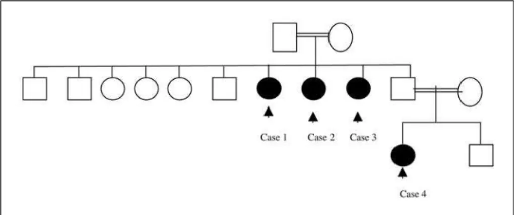
We examined four subjects of the second and third gen-eration of the kindred (Fig 1).
Case 1 – At age 20, this 55 year female developed slow-ly progressive bilateral hearing loss and mild behavioral changes, followed years later by dysarthria, dysphagia, re-duced visual acuity, muscle wasting and exercise inre-duced shortness of breath. Cognition was normal. Examination demonstrated bilateral temporal optic paleness; best cor-rected visual acuity of 20/100, absent gag reflex, tongue fasciculation and proximal muscule weakness.
Tonal audiometry demonstrated neurosensory hearing loss with absence of responses on brainstem auditory evoked potential. Needle electromyography showed den-ervation especially in sterocleidomastoideus and trapezius. Spirometry and electrocardiography were normal. Magnetic resonance imaging of the brain demonstrated bulbar atro-phy. Complete blood count and biochemistry tests were unremarkable.
Arq Neuropsiquiatr 2007;65(1) 33
reflexes were normal. Audiometry demonstrated neurosen-sorial deafness.
Case 3 – At age 12, this 48 year old female developed hearing loss. At age 15, she presented with dysarthria, dys-phagia, dysphonia and mild behavioral changes diagnosed as depression. At age 40 she also complained of exercise induced shortness of breath. Neurological examination she had tongue paralysis with widespread fasciculations, prox-imal muscle weakness more prominent in the lower limbs with normal deep tendon reflexes. There was no facial weakness. Audiometry demonstrated a severe neurosen-sorial hearing loss with no response on auditory evoked potential. Needle electromyography demonstrated dener-vation in the genioglossus muscle.
Blood chemistry, electrocardiogram, echocardiography, ergometric tests and spirometry were unremarkable. Cere-brospinal fluid cell count, protein, and glucose were nor-mal. Hematoxylin-eosin stained muscle biopsy was nornor-mal. MRI T2-weighted and fluid attenuated inversion recovery (FLAIR) sequences demonstrated a bulbar high intensity signal (Fig 2).
Case 4 – At age 18, this 23-year-old daughter of the brother of three patients described above developed pro-gressive isolated hearing loss. Neurological examination and audiometry demonstrated only bilateral neurosenso-rial hearing loss.
DISCUSSION
BVVL (Bulbar Hereditary Neuropathy type I) is a progressive pontobulbar palsy associated with a neu-rosensorial deafness. Oculomotor and trigeminal in-volvements are rare3,11. Sensorineural symptom in nearly all cases is the first symptom of the disease. There are only few cases reporting another symp-toms preceding deafness. Sathasivam et al. described one patient which the onset of symptoms was slur-ring of speech and facial weakness12. Sumners at al. described a girl with limb weakness previous on neu-rosensorial deafness13. Hearing loss has been
consis-tently described at the onset of the disease both in familiar and non- familiar cases.
The exception is Gallai’s case14with no evidence of hearing loss during the lifetime, although autop-sy showed axonal loss on the 8thnerve roots. Interval of time between hearing loss and the involvement of other cranial nerves has been variable from simul-taneous involvement (5 cases)14-16to a latency of 30 years17as shown in Table. In our series, the time inter-val between hearing loss and involvement of other cranial nerves varied from 3 to 12 years. The disease progresses from a very slow course with motor remit-ting and relapses to death8. Fazio-Londe disease (Bul-bar Hereditary Neuropathy type II) is the closest relat-ed syndrome but considerrelat-ed distinct from BVVL syn-drome because of the absence of deafness18. Bolthau-ser et al. described a similar disease, but with differ-ent aspects from the BVVL: autosomal dominant inheritance and predominant presentation of vocal alteration with dysphonia and intermittent changes on voice pitch19. Madras variant of motor neuron
dis-Fig 1. Family pedigree.
ease presents with an early onset of muscle and bul-bar involvement, with deafness occurring in two thirds of the patients9,20. Some authors consider Madras variant as clinical spectrum of the same dis-ease13,20. Three cases of the present series have prox-imal muscle weakness, supporting this hypothesis. Madras variant, however, is sporadic condition with a benign clinical course.
Dyspnea has been reported in sporadic and famil-ial cases of BVVL, especfamil-ially in younger male pati-ents18,21. This finding can be severe although there are reports of spontaneous improvement8. Cases 1 and 3 presented with dyspnea as a fluctuating symp-tom, although with mild functional impairment and normal pulmonary function tests.
Lombaert described in 197622severe neuronal changes in the brainstem reticular formation, but the reason for the fluctuating pattern is unknown. Sev-eral types of inheritance have been described in BVVL: autosomal dominant or an alternative X link-ed23,24autosomal recessive15, besides sporadic cases and even from autoimmune origin25,26. The cases described in Brazil were all sporadic9,10, and the pres-ent series is the first with a clear autosomal recessive inheritance: the pedigree showing two generations of consanguineous marriages in witch all affected
were females, strongly suggests this hypothesis. All cases described in the medical literature to date did not show any abnormality on imaging studies. We have found, however, a high intensity brainstem sig-nal in the MRI (Case 3, Fig 2) suggestive of involve-ment of the pyramidal tract.
In conclusion, we have reported a family with con-sanguineous marriages where three brothers and one niece meet diagnostic criteria of BVVL. The inher-itance of the illness is compatible with autosomal recessive transmission. One of our patients had hyper-intensity of the brainstem in the topography of the pyramidal tract. This is the first described Brazilian familial case of BVVL.
REFERENCES
1. Brown CH. Infantile amyotrofic lateral sclerosis of the family type. J Nerv Ment Dis 1894;21:707-716.
2. RamachandranNair R, Pameswaran M, Girija AS. Vialetto- Van Laere syndrome in two sisters born to consanguineous parents. Paediatr Neurol 2004;30:354-355.
3. Voudris KA, Skardoutsou A, Vagiakou EA. Infantile progressive bul-bar palsy with deafness. Brain Dev 2002;24:732-735.
4. Introini S, Sasso GM, Moioli G, Morandini WL. Case report Brown-Vialetto-van Laere syndrome. Minerva Anestesiol 2003;69:75-79. 5. Aydin OF, Ozcelikel D, Senbil N, Gurer YK. Brown-Vialetto-van Laere
syndrome: the first Turkish case. Acta Neurol Belg 2004;104:111-113. 6. Nemoto H, Konno S, Nomoto N Wakata N, Kiriraha T. A case of
Brown-Vialetto-van Laere (BVVL) syndrome in Japan. Rinsho Shinkeigaku 2005;45:356-361.
34 Arq Neuropsiquiatr 2007;65(1)
Table. Clinical summary of familiar reported cases of BVVL* (adapted from Mégarbané et al.15).
Authors Gender Age of
onset of deafness (years)
Age of onset of cranial nerves
disability (years)
Time between deafness and cranial nerves
disability
Age of death (years)
Consanguinity Inheritance
Present report 3 F 20, 18, 12 20, 27, 15 0, 9, 3 – + AR
Ramarchandran et al., 2004 2 F 7, 8 10, 11 3, 3 – + AR
Megarbane et al., 2000 3 M 2.5, 2.5, 3.5 2.5, 2.5, 3.5 0, 0, 0 –7,11 + AR
Davenport et al., 1994 1 F Childhood 18 ? – – AD
Hawkings et al., 1991 1 F 12 13 1 17 – AR
Gallai et al., 1981 1 M, 1 F 2, 1.5 14, 1.5 12, 0 –, 2 – AR
Lombaert et al., 1976 1 M, 1 F Childhood, 17
?/25 ?/8 19, 25 – AR
Boudin et al., 1971 2 F 11, 14 ?/? ?/ ? ? – AR
Van Laere, 1967 1 M 13 20 7 ? – AD? XL
Van Laere, 1966 1 F 10 10 0 ? – AR
Vialetto, 1936 1 F 0 30 30 ? – AR
Vialetto, 1936 1 F 16 35 19 ? – AR
Total 6 M,
14 F
Mean-10 Median-10
Mean-15 Median-14
Mean-6.2 Median-3
6 deaths
3 consanguineous
families
AR 10AR, 2AD, 1XL
Arq Neuropsiquiatr 2007;65(1) 35
7. Prabhu HV, Brown MJ. Brown-Vialetto-van Laere syndrome: a rare syndrome in otology. J Laryngol Otol 2005;119: 470-2.
8. Grandis D, Passadore P, Chinaglia M, Brazzo F, Ravenni R, Cudia P. Clinical features and neurophysiological follow-up in case of Brown-Vialetto-van Laere syndrome. Neuromusc Disord 2005;15:565-568. 9. Oliveira JT, Moreira PR, Cardoso F, Perpetuo FO. Brown-Vialetto-van Laere
syndrome: report of two cases. Arq Neuropsiquiatr 1995;53:789-791. 10. Rosemberg S, Lancelootti CLP, Arita F, Campos C. Progressive bulbar
paralysis of childhood with deafness: case report with clinicopatho-logic correlation. Eur Neurol 1982;21:84-89.
11. Francis DA, Ponsford JR, Wiles CM, et al. Brown-Vialetto-van Laere syndrome. Neuropathol Appl Neurobiol 1993;19:91-94.
12. Sathasivam S, O`Sullivan S, Nicolson A, et al. Brown-Vialetto-van Laere syndrome: case report and literature review. Amyotroph Lateral Scler Other Motor Neuron Disord 2000;1:277-281
13. Summers BA, Swash M, Swartz MS, Ingram DA. Juvenile onset bul-bospinal muscular atrophy with deafness: Vialetto-van Laere syndrome or Madras type motor neuron disease? J Neurol 1987;234:440-442. 14. Gallai V, Hockaday JM, Hughes JT, et al. Ponto-bulbar palsy with
deaf-ness (Brown-Vialetto-van Laere syndrome): a report of three cases. J Neurol Sci 1981;50;259-275.
15. Mégarbané AI, Desguerres I, Rizkallah E, et al. Brown-Vialetto-van Laere syndrome in a large inbred Lebanese family: confirmation of autosomal recessive inheritance? Am J Med Genet 2000;92:117-121. 16. Van Laere J. Paralysie bulbo pontine chronique progressive familiale
avec surdité: un cas de syndrome de Klippel-Trenaunay dans la meme fratrie. Rev Neurol (Paris) 1966;115:289-295.
17. Vialetto E. Contributo alla forma ereditaria della pralisi bulbare pro-gressive. Riv Sper Freniat 1936;40:1-24.
18. Voudris KA, Skardoutsou A, Vagiakou EA. Infantile progressive bul-bar palsy with deafness. Brain Dev 2002;24:732-735.
19. Bolthauser E, Lang W, Spillmann T, Holf E. Hereditary muscular atro-phy with vocal cord paralysis and sesorineural hearing loss: a domi-nant form of spinal muscular atrophy? J Med Genet 1989;26:105-108. 20. Gourie-Devi M, Suresh TG. Madras pattern of motor neuron disease
in South India. J Neurol Neurosurg Psychiatry 1988;51:773-777. 21. Dipti S, Childs AM, Livingston JH, et al. Brown-Vialetto-Van Laere
syn-drome: variability in age at onset and disease progression highlight-ing the phenotypic overlap with Fazio-Londe disease. Brain Dev 2005; 27:443–446.
22. Lombaert A, Dom R, Carton H, Brucher JM. Progressive ponto-bulbar palsy with deafness; a clinicopathological study. Acta Neurol Belg 1976; 76:309-314.
23. Van Laere J. Over een newl geval van chosnische bulbopontiene paral-ysis met doofheid. Verh Vlaan Akad Geneesk Belg 1967;30:288-308. 24. Hawkins SA, Nevin NC, Harding AE. Pontobulbar palsy and
neu-rosenssory deafness (Brown-Vialetto-van Laere syndrome) with pos-sible autosomal dominant inheritance. J Med Gen 1990;27:176-179. 25. Sztajzel R, Kohler A, Reichart M, et al. Syndrome de
Brown-Vialetto-van Laere: un cas avec anticorps anti-ganglioside GM1 et revue de la littérature. Rev Neurol (Paris) 2002;154:51-54.