1Mestranda, Laboratório de Mapeamento Cerebral e Integração Sensório-Motora, Instituto de Psiquiatria (IPUB), Universidade
Federal do Rio de Janeiro, Rio de Janeiro RJ, Brasil (UFRJ); 2Mestre em Saúde Mental, Laboratório de Mapeamento Cerebral e
Inte-gração Sensório-Motora, Instituto de Psiquiatria (IPUB), UFRJ; 3D.Sc. em Engenharia Biomédica, COPPE, UFRJ; 4Professor Adjunto III
Doutor, Coordenador do Laboratório de Mapeamento Cerebral e Integração Sensório-Motora, IPUB / UFRJ; 5Professor Adjunto II
PhD, Escola de Educação Física e Desportos (EEFD), Laboratório de Mapeamento Cerebral e Integração Sensório-Motora, IPUB / UFRJ; Professor Pesquisador, Universidade Castelo Branco (PROCIMH-UCB).
Received 13 April 2006, received in final form 17 July 2006. Accepted 20 September 2006.
Dra. Isabel Sampaio Instituto de Psiquiatria (IPUB), Laboratório de Mapeamento Cerebral e Integração SensórioMotora UFRJ -Avenida Venceslau Brás 71 / Fundos - 22290-140 Rio de Janeiro RJ - Brasil. E-mail: [email protected]
INFLUENCE OF BROMAZEPAM ON
CORTICAL INTERHEMISPHERIC COHERENCE
Isabel Sampaio
1, Fernanda Puga
1, Heloisa Veiga
2,
Maurício Cagy
3, Roberto Piedade
4, Pedro Ribeiro
5ABSTRACT - Benzodiazepines are among the most commonly prescribed medications due to their thera-peutic efficacy in reducing anxiety and inducing sleep. Consequently, they have been widely employed in the pharmacological treatment of several disorders. Nevertheless, few studies have analyzed the effects of bromazepam in electroencephalographic activity (EEG). The present study aimed at investigating the modulatory effects of this drug on brain dynamics. Specifically, the effects of bromazepam (3mg) on EEG coherence were tested in a double-blind experiment. The sample, consisting of 10 healthy subjects (5 male and 5 female), was submitted to ten minutes of EEG recording. The electrophysiological measure (coher-ence) was analyzed across three experimental conditions: bromazepam, placebo 1, and placebo 2. Results indicate that bromazepam significantly increases cortical interhemispheric coherence.
KEY WORDS: bromazepam, qEEG.
Influência do bromazepam na coerência cortical inter-hemisférica
RESUMO - Benzodiazepínicos estão entre as medicações mais comumente prescritas devido à sua eficácia terapêutica para reduzir ansiedade e induzir sono. Conseqüentemente, eles têm sido amplamente empre-gados no tratamento de diversas desordens. No entanto, poucos estudos têm analisado os efeitos do bro-mazepam na atividade eletrencefalográfica (EEG). Assim, o presente estudo teve por objetivo investigar os efeitos modulatórios desta droga na dinâmica cerebral. Especificamente, os efeitos de 3 mg de bro-mazepam na coerência eletrocortical foram analisados em um experimento duplo-cego. A amostra con-sistiu de 10 sujeitos sadios (5 homens e 5 mulheres), submetidos a dez minutos de captação do sinal de EEG. A medida eletrofisiológica (coerência) foi analisada em três condições experimentais: bromazepam, placebo 1 e placebo 2. Os resultados sugerem que o bromazepam aumenta significativamente a coerên-cia cortical inter-hemisférica.
PALAVRAS-CHAVE: bromazepam, EEGq.
It is acknowledged that the waking electroen-cephalographic activity (EEG) comprises direct rela-tions to chemical changes in the brain induced by drugs. In other words, electroenchephalography is responsive to the unique characteristics of psychoac-tive substances1. In this context, the EEG has been
wi-dely employed in the assessment of electrophysio-logical changes induced by distinct medications. Its sensibility in detecting alterations produced by a spe-cific substance may be enhanced by methods of quan-titative analyses (qEEG)2,3. Quantitative
electroen-cephalography has become, throughout the years, a tool of great clinical utility in evaluating the effects
of medications, predicting medication response, and in assessment of cognitive changes produced by such psychoactive substances. Once drugs have specific ef-fects on wave morphology, changes in qEEG variables can be used to investigate mechanisms of drug action as well as to monitor and possibly predict efficacy.
However, very few studies analyzed the effects of benzodiazepines on qEEG variables. Saletu et al.4
and lorazepam, and the exact same pattern of results, i.e., EEG profile, was observed. Specifically, studies employing bromazepam, possibly the most common-ly prescribed benzodiazepine, are practicalcommon-ly inexis-tent in the current literature. One of the few stud-ies that examined the effects of this particular drug on qEEG was conducted by Fink et al.6. The effects
of oral doses of bromazepam (9 mg) and diazepam (10 mg) in EEG data were analyzed and an analogous result was reached: increased beta and decreased alpha activity. Such changes seem to be characteris-tic for benzodiazepines.
The previously cited studies that employed qEEG, in addition to being long-standing, only analyzed power distribution. References in the current litera-ture of studies focusing on changes related to bro-mazepam intake in other qEEG variables are even scarcer. Coherence measures, for example, can be used to assess functional relationships between cor-tical areas7. Coherence is a measure of
synchroniza-tion between two areas. Hence, coherence analyses have provided the means to assess functional con-nectivity between cortical regions. The use of coher-ence analyses have gradually increased in the last two decades. Several researchers regard this meas-ure as having considerable clinical value once it can directly reflect neural network connectivity and dyna-mics8-11. Decreased coherence, for instance, is
observ-ed in major depression associatobserv-ed with organic brain disorders12, in patients with seasonal affective
disor-der13, in male depression14, and in childhood and
ado-lescent depression15. In addition, decreases in
inter-hemispheric coherence are observed in Alzheimer patients and seem to be related to other age-relat-ed decreases16,17. Furthermore, coherence analyses
have been used to characterize regional changes in neuronal couplings and information transfer relat-ed to semantic aspects of object recognition in hu-mans18, anticipation of somatosensory and
visuomo-tor events19, and central executive functions of
work-ing memory20, among others. However, none of these
studies have analyzed the effects of a specific drug on interhemispheric connectivity (i.e., coherence).
In this context, the present study aimed at inves-tigating the effects of bromazepam on the coupling of cortical areas (coherence) and the strength of inter-hemispheric connectivity.
METHOD
Subjects – The sample consisted of 10 volunteers, 5 male and 5 female, with ages varying between 21 and 38 years (27±5 years). All subjects were healthy, free of cognitive deficits and were not making use medication or any
psy-choactive or psychotropic substance at the time of the test. To assure that subjects did not present any impairment of their physical and mental health, and to identify and ex-clude from the experiment any subjects who could contam-inate future results, a questionnaire was applied. Subjects signed a consent form, where the experimental condition was thoroughly described. The experiment was submitted to the Psychiatric Institute’s ethics committee for approval.
Study design and procedures – Subjects received a cap-sule (bromazepam or glucose) on three different occasions under a randomized, double-blind, crossover study. The procedures consisted of a three-day treatment: a day of bromazepam (B) and two of placebo (P1 and P2). The pro-cedures were standardized in the following routine: 1) 10 minutes of EEG recording (5’ eyes-closed / 5’ eyes-open); 2) Administration of capsule (bromazepam or placebo); 3) The second EEG (10’), 20 minutes after drug ingestion; 4) The third EEG (10’), 60 minutes after drug ingestion.
EEG acquisition – The study design respected the Inter-national Pharmaco-EEG group guidelines. InterInter-national 10/ 20 System21for electrode placement (referred to linked
ear-lobes) was used with a 20-channel Braintech-3000 (EMSA-Medical Instruments, Brazil). The 20 monopolar electrodes were arranged in a nylon cap (ElectroCap Inc., Fairfax, VA, USA). Impedance for EEG and EOG electrodes were under 5 KΩand 20 KΩ, respectively. Visual inspection was em-ployed for detection and elimination of artifacts. The data acquired had total amplitude of less than 100 µV. The sig-nal was amplified with a gain of 22,000. Eye-movement (EOG) artifact was monitored with a bipolar electrode mon-tage using two 9-mm diameter electrodes attached above and on the external canthus of the right eye. Moreover, Independent Component Analysis (ICA) was applied to remove possible sources of artifacts. The EEG signal was analogically filtered between 0.16 Hz (high-pass) and 35 Hz (low-pass), and sampled at 200 Hz. The acquisition soft-ware, developed at the Brain Mapping and Sensorimotor Integration Lab, was employed with the following digital filters: Notch (60 Hz), high-pass of 0.3 Hz and low-pass of 25 Hz.
Data processing and analysis – At least 2 min of arti-fact-free data were extracted from the EEG record for quan-titative analysis. A Matlab 5.3®(Mathworks Inc., Naticj, MA,
USA) routine was implemented to perform a spectral analy-sis and estimate the specific parameter of interest: band-limited EEG coherence, for delta (1.0-3.5 Hz), theta (4.0-7.5 Hz), alpha (8.0-12.0 Hz), and beta (13-25 Hz) frequency bands, and between all pair combinations of electrodes. Coherence reflects the joint variation in electrical activity between homologous electrode pairs (i.e., electrodes in the same position on opposite sides of the head). In other words, coherence is an estimate of shared variance.
pari-etal electrodes were analyzed since they are representative of premotor and primary motor areas, as well as primary somatosensory and higher order somatosensory areas22.
Statistical analysis – Three-Way Anova, condition x mo-ment x electrode (3 x 3 x 3), was performed for the electro-physiological measure, i.e., coherence, in each frequency band separately (p≤0.05). A Post Hoc (Scheffé) was applied a posteriori. Experimental conditions were established as bromazepam (B), placebo 1 (P1) and placebo 2 (P2) and ex-perimental moments as 0’ (before drug administration), 20’ (twenty minutes after drug administration), and 60’ (sixty minutes after drug administration). The selected elec-trode the pairs were F3-F4, C3-C4, and P3-P4.
RESULTS
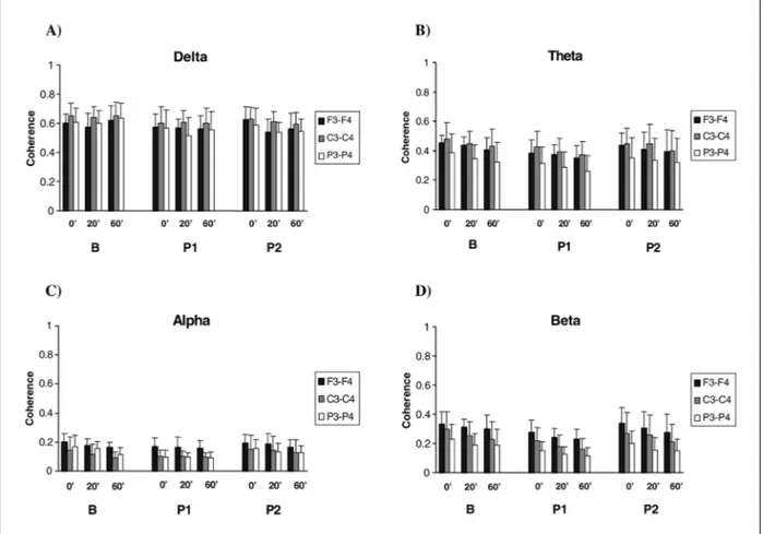
Figure illustrates coherence variations across con-ditions (B, P1, P2), moments (0’, 20’, 60’) and elec-trodes (F3-F4, C3-C4 and P3-P4) in the delta, theta, alpha, and beta frequency bands.
In delta (1-A), the three-way Anova revealed sig-nificant main effects of condition [F(2,267)=6.446; p= 0.002] and electrode site [F(2,267)=6.854; p=0.001]. No interactions were observed. The Post Hoc pointed out to the following differences: B and P1 (p=0.004),
B and P2 (p=0.024); F3-F4 and C4 (p=0.016), C3-C4 and P3-P4 (p=0.003).
In theta (1-B), the analysis revealed significant main effects of condition [F(2,267)=7.170; p=0.001], moment [F(2,267)=3.968; p=0.020], and electrode site
[F(2,267)=21.049; p=0.000]. No interactions were
observed. The Post Hoc pointed out to the following differences: B and P1 (p=0.001), P1 and P2 (p=0.038); 0’ and 60’ (p=0.020); F3-F4 and P3-P4 (p=0.000), C3-C4 and P3-P4 (p=0.000).
In alpha (1-C), main effects of condition [F(2,267)= 7.537; p=0.001], moment [F(2,267)=4.825; p=0.009],
and electrode site [F(2,267)=21.345; p=0.000] were found. No interactions were observed. The Post Hoc pointed out to the following differences: B and P1 (p=0.011), P1 and P2 (p=0.002); 0’ and 60’ (p=0.009); F3-F4 and C3-C4 (p=0.000), F3-F4 and P3-P4 (p=0.000). In beta (1-D), the three-way Anova revealed main effects of condition [F(2,267)=13.028; p=0.000], moment
[F(2,267)=6.372; p=0.002], and electrode site [F(2,267)= 36.687; p=0.000]. No interactions were observed. The Post Hoc pointed out to the following differences: B and P1 (p=0.000), P1 and P2 (p=0.001); 0’ and 60’ (p= 0.002); F3-F4 and C3-C4 (p=0.000), F3-F4 and P3-P4 (p=0.000), C3-C4 and P3-P4 (p=0.000).
DISCUSSION
The present study aimed at investigating the spe-cific effects of bromazepam on the coupling of cor-tical areas and the strength of interhemispheric con-nectivity, as expressed in terms of coherence values. Like all benzodiazepines, bromazepam facilitates the release of GABA, the major inhibitory neurotrans-mitter. Such facilitation results in inhibition of brain cognitive functions23. Thus, by using bromazepam,
one can observe the consequences of inhibition on the connectivity between different cortical areas. The present study investigated whether an inhibitory sys-tem would indeed be responsible for functional de-coupling between cortical areas, assuming that inhi-bition would weaken or even impair cortical functio-nal connections. In other words, we expected to ob-serve a decoupling between areas (i.e, lower coher-ence values) after bromazepam intake. However, the results indicated otherwise.
Specifically, coherence values increased in the bro-mazepam condition when compared to both place-bo groups in all frequency bands. It must also be stressed that a marked difference was observed between the slow bands (delta and theta) and the fast bands (alpha and beta): coherence values were significantly higher in the slower frequencies ranges. Significant differences were also observed across moments, except in the delta band. In synthesis, the drug clearly promoted an enhancement of cortical interhemispheric coherence, which is in accordance with Fingelkurts et al.24, who conducted possibly the
most expressive work related to the effects of a ben-zodiazepine on interhemispheric coherence. These authors investigated whether lorazepam would gen-erate a functional decoupling of cortical areas and concluded that, unexpectedly, the drug yielded a wi-despread increase in the inter-area functional con-nectivity. In this sense, the results of the present study indicate that the cortical inhibition produced by this benzodiazepine may be an efficient mechanism. As stated by Fingelkurts et al.24, despite the common
belief that this pharmacological class impairs brain functioning by switching off irrelevant functional connections, it seems that benzodiazepine intake in-creases the strength of functional links and promotes functional coupling between different cortical areas. These results are not so atypical. Other studies, employing coherence analysis on different experi-mental models, have reached similar outcomes. For instance, it has been shown that states of minimal cognitive processing often exhibit widespread spa-tially coherent EEG25. In the same way, an increase of
coherence values has been observed during non-REM sleep and drowsiness26, during sub-anesthetic
con-centrations27, and during meditation, which was
ac-companied by an anxiety decrease28. A possible
expla-nation for such pattern of results is that GABA seems to have an excitatory action on several brain sys-tems29. In this sense, GABA is not a single-action
neu-rotransmitter30. Therefore, the enhancement in
inter-hemispheric connectivity, expressed by an increase in coherence values, after bromazepam intake, may be the result of GABA excitatory actions.
Nevertheless, further studies, using different dos-es of bromazepam, are necdos-essary to truly understand the effects of this benzodiazepine not only on brain dynamics, but on coherence and other qEEG vari-ables. Additional studies are also necessary to thor-oughly understand how different medications affect cortical synchronicity and the strength of interhemi-spheric connectivity.
REFERENCES
1. Saletu B, Anderer P, Saletu-Zyhlarz GM. EEG topography and tomog-raphy (LORETA) in the classification and evaluation of the pharmaco-dynamics of psychotropic drugs. Clin EEG Neurosci 2006;37:66-80. 2. Anghinah R, Kanda PAM, Jorge MS, Lima EE, Pascuzzi I, Melo AC.
Estudo da coerência do eletrencefalograma para a banda de freqüên-cia alfa em indivíduos adultos normais e com provável demênfreqüên-cia do tipo Alzheimer. Arq Neuropsiquiatr 2000;58:272-275.
3. Veiga H, Deslandes A, Cagy M, Fiszman A, Piedade RA, Ribeiro P. Neurocortical electrical activity tomography in chronic schizophren-ics. Arq Neuropsiquiatr 2003;61:712-717.
4. Saletu B, Grünberger J, Linzmayer L. On the central effects of a new partial benzodiazepine agonist RO 16-6028 in man: pharmaco-EEG and psychometric studies. Int J Clin Pharmacol Ther Toxicol 1989;27:51-65. 5. Link CS, Leigh TJ, Fell GL. Effects of granisetron and lorazepam, alone and in combination, on the EEG of human volunteers. Br J Clin Pharmacol 1991;31:93-97.
6. Fink M, Irwin P, Weinfeld RE, Schwartz MA, Conney AH. Blood lev-els and encephalographic effects of diazepam and bromazepam. Clin Pharmacol Ther 1976;20:184-191.
7. Besthorn C, Förstl H, Geiger-Kabisch C, Sattel H, Gasser T. EEG coher-ence in Alzheimer disease. Electrocoher-enceph Clin Neurophysiol 1994; 90:242-245.
8. Harmony T, Marosi E, Fernandez T, Bernal J, Rodriguez M, Reyes A. EEG coherences in patients with brain lesions. Int J Neurosci 1994;74: 203-226.
9. Tatcher RW. Normative EEG databases and EEG biofeedback. J Neuro-ther 1998;2:8-39.
10. Lubar JF, White JN, Swartwood MO, Swartwood JN. Methylphenidate effects on global and complex measures of EEG. Pediatr Neurol 1999; 21:633-637.
11. Pilgreen KL. Physiologic, medical, and cognitive correlates of electroen-cephalography. In: Nunez PL (Ed). Neocortical dynamics and human EEG rhythms. New York: Oxford University Press, 1995:195-248. 12. Brassen S, Braus DF, Weber-Fahr W, Tost H, Moritz S, Adler G.
Late-onset depression with mild cognitive deficits: electrophysiological evi-dences for a preclinical dementia syndrome. Dement Geriatr Cogn Disord 2004;18:271-277.
13. Passynkova NR, Volf NV. Seasonal affective disorder: spatial organi-zation of EEG power and coherence in the depressive state and in light-induced and summer remission. Psychiatry Res 2001;108:169-185. 14. Knott V, Mahoney C, Kennedy S, Evans K. EEG power, frequency,
15. Armitage R, Hoffmann R, Emslie G, Rintelmann J, Robert J. Sleep microarchitecture in childhood and adolescent depression: temporal coherence. Clin EEG Neurosci 2006;37:1-9.
16. Wada Y, Nanbu Y, Koshino Y, Yamagushi N, Hashimoto T. Reduced inter-hemispheric EEG coherence in Alzheime disease: analysis during rest and photic stimulation. Alzheimer Dis Assoc Disord 1998;12:175-181. 17. Duff FH, McAnulty GB, Albert MS. Effects of age upon
intrehemispher-ic EEG coherence in normal adults. Neurobiol Aging 1996;17:587-599. 18. Supp GG, Schlogl A, Fiebach CJ, et al. Semantic memory retrieval: cor-tical couplings in object recognition in the N400 window. Eur J Neurosci 2005;21:1139-1143.
19. Babiloni C, Brancucci A, Vecchio F, Arendt-Nielsen L, Chen AC, Rossini PM. Anticipation of somatosensory and motor events increases cen-tro-parietal functional coupling: an EEG coherence study. Clin Neurophysiol 2006.
20. Sauseng P, Klimesch W, Schabus M, Doppelmayr M. Fronto-parietal EEG coherence in theta and upper alpha reflect central executive func-tions of working memory. Int J Psychophysiol 2005;57:97-103. 21. Jasper H. The ten-twenty electrode system of the international
feder-ation. Electroencephalogr Clin Neurophysiol 1958;10:371-375. 22. Kandel E, Schwartz S, Jessel T. Principles of neuroscience, 4.Ed. New
York: McGraw-Hill, 2000.
23. Katzung BG. Basic Clinical Pharmacology, 6.Ed. London: Pretence-Hall International, 1995.
24. Fingelkurts AA, Kivisaari R, Pekkonen E, Ilmoniemi RJ, Kahkonen S. Enhancement of GABA-related signalling is associated with increase of functional connectivity in human cortex. Hum Brain Mapp 2004;22:27-39. 25. Nunez PL. Toward a quantitative description of large-scale
neocorti-cal dynamic function and EEG. Behav Brain Sci 2000;23:371-437. 26. Bullock TH, McClune MC, Achimowicz JZ, Iragui-Madoz VG, Duckrow
RB, Spencer SS. EEG coherence has structure in the millimeter domain: subdural and hippocampal recordings from epileptic patients. Electro-encephalogr Clin Neurophysiol 1995;95:161-177.
27. Nunez PL. Neocortical dynamics and human EEG rhythms. New York: Oxford University Press, 1995.
28. Aftanas LI, Golocheikine SA. Human anterior and frontal midline theta and lower alpha reflect emotionally positive state and internalized attention: high-resolution EEG investigation of meditation. Neurosci Lett 2001;310:57-60.
29. Gulledge AT, Stuart GJ. Excitatory actions of GABA in the cortex. Neuron 2003;37:299-309.