Services of Neurosurgery1, Neurology2and Radiology3, Department of Neurology and Psychiatry, Botucatu School of Medicine,
State University of São Paulo (UNESP), Botucatu SP, Brazil.
Received 29 May 2006, received in final form 14 August 2006. Accepted 17 October 2006.
Dr. Marco A. Zanini - Depto Neurologia e Psiquiatria - Faculdade de Medicina de Botucatu - UNESP - Caixa Postal 540 - 18618-970 Botucatu SP - Brasil. E-mail: [email protected]
ABSTRACT - Thirty-four consecutive adult patients with subdural traumatic hygroma were analysed for clinical evolution, serial computed tomography scan (CT), and magnetic resonance imaging (MRI) over a period of several months. Five of the patients presented CT scan and MRI evolution data showing increas-ing density over a period of 11 days to 6 months post trauma. In these five patients, final clinical and CT scan data were benign, with complete spontaneous resolution. Descriptions in literature of evolving trau-matic subdural hygroma have presented CT scan density modifications changing into chronic subdural hematoma. Our patients show another possibility, density transformation, which sometimes show as sub-dural hematoma in CT scan and MRI, but with final evolution where clinical condition and CT scan return to normal.
KEY WORDS: traumatic subdural hygroma, evolution, head injury, CT scan .
Higroma subdural traumático: a propósito de cinco casos com modificação de densidade e re-solução espontânea
RESUMO - Analisamos 34 pacientes adultos com higroma subdural traumático quanto à evolução clínica, tomografias seriadas e ressonância magnética. Observou-se aumento da densidade do higroma subdural em cinco dos pacientes durante período que variou de 11 dias a 6 meses após o trauma. Nestes cinco pacientes, a evolução clínica foi favorável e os higromas apresentaram resolução espontânea. Há vários relatos na literatura de modificação da densidade dos higromas subdurais tramáticos, transformando-se em hematoma subdural crônico. Esta casuística apresenta outra possibilidade, ou seja, modificação da den-sidade, que pode ser apresentada como hematoma subdural pelas imagens de tomografia ou ressonância magnética, mas com resultado final das condições clínicas e de imagem retornando ao normal.
PALAVRAS-CHAVE: higroma subdural traumático, traumatismo crânio-encefálico, evolução, tomografia computadorizada.
Post-traumatic subdural hygroma is common, but its natural history is not well defined because there are few reports of clinical and computed tomogra-phy scan (CT) evolution data. These reports lack a uniform approach to method and segment time1-19.
Some authors have reported traumatic subdural hygroma presenting density modifications in the CT scan with change to chronic subdural hemato-ma4,7,9,10,12,13,15-18.
This study reports a series of South American adult patients with traumatic subdural hygroma, with em-phasis on the evolution of clinical and CT scan data.
METHOD
Thirty-four 34 consecutive adult patients with subdur-al traumatic hygroma were ansubdur-alyzed; they were ansubdur-alyzed with emphasis on patients who presented density modifi-cations over a follow-up time segment. Each patient was studied for clinical evolution, serial CT scan and magnetic resonance imaging (MRI) data for several months.
This study was approved by our University Hospital Ethics Committee for Human Research.
RESULTS
40 years. Mean evolution time was 249 days. Road traffic accidents were the main cause of head injury. The mean evolution time for subdural hygroma diag-nosis was 9 days. Twenty-one patients (61.8%) under-went conservative treatment and 13 (38.2%), surgi-cal treatment. Modifications in density on CT scan or MRI were observed in 5 patients (14.7% - mean evo-lution time 665 days).
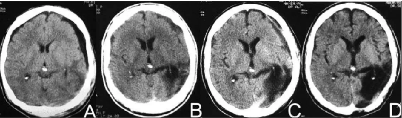
Patient 1 – A 35-year-old male patient was admit-ted with head injury after physical assault. He was confused (Glasgow Coma Scale score 10). On admis-sion, the CT scan showed left parietal hemorrhagic contusion that required surgery. The CT scan on the 9th day showed bilateral frontal subdural hygroma, mainly on the left side (Fig 1A), which presented pro-gressive enlargement and compression of the cere-bral parenchyma. On the 18thday he was submitted
to simple burr hole aspiration and irrigation of the left frontal collection. A slight xantochromic high-pressure subdural fluid was observed. On the 53rd
day, CT scan showed the hygroma with enhanced density and heterogeneous aspect (Fig 1B). The patient presented progressive clinical improvement and a conservative approach was chosen. The CT scan on the 117thday showed reduction in hygroma size,
with probable neomembrane, and without compres-sion on the underlying cerebral parenquima (Fig 1C). The CT scan on the 370thday showed cerebral
expan-sion and spontaneous resolution of the subdural col-lection (Fig 1D).
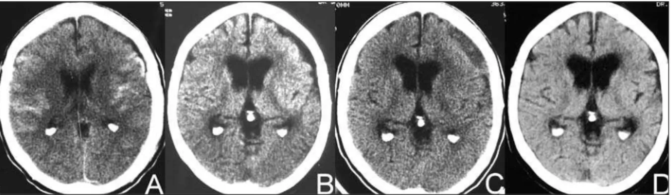
Patient 2 – A 35-year-old male patient was admit-ted with head injury after being hit by a car. He was comatose with right midriasis (Glasgow Coma Scale score 7). On admission, the CT scan showed proba-ble cerebral edema. The CT scan on the 12th day
showed bilateral frontal subdural hygroma (Fig 2A). On the following days he presented slow and pro-gressive neurological improvement. MRI on the 191st
day showed laminar subdural hematoma, without compression on the cerebral parenquima (Fig 2B). Fig 1. Case 1. (A). CT scan showing left frontal subdural hygroma (9thday). (B) Enhanced density and heterogeneous appear-ance (53rdday). (C) Reduction of the hygroma, with probable neomembrane (117thday). (D) Resolution of the subdural col-lection (730thday).
The image was enhanced after endovenous injection of paramagnetic contrast, mainly on the right side (Fig 2C). The CT scan on the 300thday showed
com-plete resolution of the subdural collection (Fig 2D). On later examination the patient was asymptomatic.
Patient 3 –A 69-year-old male patient was admit-ted with head injury after falling over. He presenadmit-ted a transient decreased conscience state and later reported headache (Glasgow Coma Scale score 14). On admission, the CT scan showed diffuse subarach-noid hemorrhage and small subdural effusion in the left frontal region (Fig 3A); on the 4thday this had
evolved into subdural hygroma, with probable com-pression of the underlying brain (Fig 3B). For the next 10 days, he presented with headache and somno-lence, with subsequent improvement. The CT scan on the 77th day showed enhanced density and
increased volume of the hygroma, with probable ipsi-lateral compression of the cortical sulci and
ventri-cles (Fig 3C). As his clinical examination was good, a conservative approach was chosen. Two years later he was asymptomatic, and the subdural collection had disappeared (Fig 3D).
Patient 4 –A 42-year-old male patient was admit-ted with polytraumas and head injury after physical assault. He was confused and agitated (Glasgow Coma Scale score 10). On admission, the CT scan showed subarachnoid hemorrhage and cerebral ede-ma (Fig 4A). The CT scan on the 26thday showed left
frontoparietal subdural hygroma with probable com-pression of the underlying brain (Fig 4B). On the 27th
day he was submitted to simple burr hole aspiration and irrigation of the left frontoparietal collection. A slight hemorrhagic clear subdural fluid was observed. He presented progressive clinical improvement. The CT scan on the 50thday showed enhanced hygroma
density (Fig 4C). Conservative treatment was chosen. The CT scan on the 240thday showed a reduction in
Fig 3. Case 3. (A) CT scan on admission showing diffuse subarachnoid hemorrhage and small subdural effusion in the left frontal region (1stday). (B) CT scan showing subdural hygroma with probable compression on the underlying brain (4thday). (C) CT scan showing enhanced density and increased volume, with probable ipsolateral compression on the underlying brain (77thday). (D) CT scan showing disappearance of the subdural collection (2 years later).
size of the subdural collection, with probable neo-membrane and septation (Fig 4D).
Patient 5 –A 36-year-old male patient was admit-ted with head injury after falling over. He was con-fused and agitated (Glasgow Coma Scale score 10). On admission, the CT scan showed laminar acute sub-dural hematoma in the right frontoparietal region (Fig 5A). He was submitted to conservative treatment. The CT scan on the 7thday showed right subdural
hy-groma adjacent to the subdural hematoma (Fig 5B); on the 11thday, it presented decreased size and
en-hanced density (Fig 5C). There was a slow and pro-gressive improvement in consciousness. Minimal cog-nitive deficits were observed on the 30thday. The CT
scan 2 years later only showed cortical atrophy (Fig 5D).
DISCUSSION
For different authors subdural hygroma is more prevalent in older patients with some degree of cere-bral atrophy4,9,14,19. In our study, 50% of our
traumat-ic subudural hygroma patients were between 16 and 40 years; this characterizes a younger and prevalent population. The mean age of 5 described cases was 43 years.
Transformations from subdural hygroma to chron-ic subdural hematoma are well documented4,7-9,13,15,18.
These transformations occurred in between 0 to 58% of cases, depending on type of study and evolution
time2,4,7,9,12,13,15,18,20-22. There were however few reports
of mean transformation times: 65.8 days7, 68 days13,
and 101 days8. In our 5 patients mean
transforma-tion time was 76.4 days. For these 5 patients with modified subdural collection after initial hygroma, enhanced density was a transitory phenomenon, and
not one of our patients needs surgery. Final clinical and imaging results presented resolution. It is not clear why our 5 patients did not develop chronic sub-dural hematoma from the enhanced density hygro-ma. This was more commonly seen in older pa-tients4,9,14,19, where some degree of cerebral atrophy
could provide the space for hematoma expansion. Four of our patients presented no cerebral atrophy.
Collection in the subdural space for more than a few weeks may induce the migration and prolifera-tion of inflammatory cells, derived from the dural border cells, originating the chronic subdural hema-toma outer membrane20,23,24. Our cases 1 and 4
pre-sented septation inside the old and transformed hy-groma delimiting areas with different densities. So, for us, the origin of these membrane cells is not clear-ly defined, but we agree that enhanced hygroma density could represent the bleeding of membranes formed in chronic phases of subdural hygroma25.
REFERENCES
1. Cornell SH, Chiu LC, Christie JH. Diagnosis of extracerebral fluid col-lections by computed tomography. Am J Roentgenol 1978;131:107-110. 2. French BN, Cob III CA, Corkill G, Youmans JR. Delayed evolution of
postraumatic subdural hygroma. Surg Neurol 1978;9:145-148. 3. Friede RL, Schachenmayr W. The origin of subdural neomembranes:
II. Fine structure of neomembranes. Am J Pathol 1978;92:69-87. 4. Hirai O, Yamakawa H, Nishikawa M, et al. Ventricular dilation during
the treatment of subdural hygromas. Neurol Med Chir 1991;31:943-947. 5. Ishibashi A, Yokokura Y, Miyagi AJ. Clinical analysis of nineteen patients
with traumatic subdural hygromas. Kurume Med J 1994;41:81-85. 6. Jaeckle KA, Allen JH. Subdural hygroma: diagnosis with computed
tomography. J Comput Tomogr 1979;3:201-206.
7. Kaufman HH, Childs TL, Wagner KA, et al. Post-traumatic subdural hygromas: observations concerning a surgical enigma. Acta Neurochir 1984;72:197-209.
8. Koizumi H, Fukamashi A, Wakao T, et al. Traumatic subdural hygro-mas in adults: on the possibility of development of chronic subdural hematoma. Neurol Med Chir 1981;21:397-406.
9. Lee KS, Bae WK, Bae HG, Yun IG. The fate of traumatic subdural hygro-ma in serial computed tomographic scans. J Korean Med Sci 2000;15:560-568.
1984;26:245-248.
16. Murata K. Chronic subdural hematoma may be preceded by persist-ent traumatic subdural effusion. Neurol Med Chir (Tokyo)1993;33:691-696.
17. Nakaguchi H, Tanishima T, Yoshimasu N. Factors in the natural histo-ry of chronic subdural hematomas that influence their postoperative recurrence. J Neurosurg 2001;95:256-262.
18. Ohno K, Suzuki R, Masaoka H, Matsushima Y, Inaba Y, Monma S.
ma. Neurosurgery 1981;8:542-550.
24. Suzuki M, Kudo A, Kitakami A, et al. Local hypercoagulative activity precedes hyperfibrinolytic activity in the subdural space during devel-opment of chronic subdural haematoma from subdural effusion. Acta Neurochir 1998;140:261-266.