Vascular Radiolesion as a Deleterious Effect of High-Dose-Rate
Intraarterial Brachytherapy with Samarium-153 in
Hypercholesterolemic Rabbits
Dalton Bertolim Précoma, Lúcia Noronha, Álvaro Vieira Moura, Airton Seiji Yamada, José Knopfholz, César Lopes
Dusilek, Rita Perussolo, Paulo Roberto Slud Brofman, Márcia Olandoski, José Cláudio Meneghetti
Pontifical Catholic University of Paraná, Radioimmunoassay and Nuclear Medicine Center of Paraná, Hospital das Clínicas Heart Institute – University of São Paulo School of Medicine (FMUSP) - Curitiba, PR - São Paulo, SP - Brazil
objective: This study was designed to evaluate vascular morphological and morphometric changes induced by brachytherapy with samarium-153 (Sm-153) at high doses in hypercholesterolemic rabbits.
Methods:Forty-three New Zealand White hypercholesterolemic rabbits were analyzed, and the total of 86 iliac arteries un-derwent balloon angioplasty injury. The rabbits were divided into three different groups: two irradiation groups (IG) assigned to 15 Gy (n=14) and 60 Gy (n=36) irradiation doses, respectively, and a control group (n = 36). Histomorphometric and qualitative histological analyses were performed for tissue evaluation.
results: Significant reductions were found in neointimal proliferation (NIP) (p< 0.0001), media area (MA) (p<0.0001) and percent stenosis (p<0.0001) in the 15-Gy IG, compared to the other groups. The 60-Gy IG had the higher rate of NIP, incre-ase in media and vessel areas (VA) and percent stenosis. The 60-Gy IG also showed the greatest number of xanthomatous cells (60-Gy IG: 86.11% and 15-Gy IG: 14.29%, p<0.0001) and the highest amount of hyaline amorphous tissue (60-Gy IG:58.33% and 15-Gy IG:0%, p=0.0001) and vascular proliferation (60-Gy IG:30.56% and 15-Gy IG:0%, p=0.0221). No statistically significant differences were found among groups concerning other tissue analyses.
conclusion: The high-dose irradiation of 60 Gy resulted in intense cell proliferation considered vascular radiolesion, unlike the 15-Gy dose, which was associated with an excellent inhibition of neointimal proliferation.
Key words: Artery injury, brachytherapy, samarium-153.
Since the advent of angioplasty, restenosis has been a great challenge in coronary intervention, and several studies have focused on examining the multiple therapeutic options. It is an attempt at tissue repair following arterial wall injury through neointimal proliferation (NIP) and remodeling. Smooth muscle cell (SMC) migration and proliferation plus extracellular matrix (ECM) deposition, associated with inflammatory and proliferative phenomena, play key roles role in this pathophysiological mechanism1-3.
Intraarterial brachyterapy was first studied in experimental models in 19944, and in 1997 study of it began in humans5.
Since then, its importance has been demonstrated in roughly 5,000 patients participating in randomized clinical trials. In the United States, more than 40,000 procedures are performed each year in 400 centers6,7. Delivery of local radiation
causes phenotypic changes that prevent cell reproduction by deoxyribonucleic acid (DNA), inhibiting smooth muscle cell (SMC) proliferation and migration and thus decreasing the degree of neointimal hyperplasia8. Our group showed that
brachytherapy with samarium-153, used for the first time, at a dose of 15 Gy effectively inhibits neointimal proliferation9.
Several studies, comparing higher doses, have demonstrated better results in NIP inhibition. However, higher doses may
have deleterious effects on the vessel wall, affecting internal vessels, as described with thoracic irradiation10. Little has
been published on the effects of intraarterial brachytherapy using higher doses.
Recently, as the effectiveness of antiproliferative drug-eluting stents was demonstrated, expectations were high for restenosis reduction11. The safety and efficacy of brachytherapy
have been established in comparative studies; however, restenosis is open field for research, because no method has thus far been able to solve this problem7,12.
This study is directed towards analyzing morphological and morphometric vascular changes induced by high dose samarium-153 (Sm-153) brachytherapy in hypercholesterolemic rabbits, compared with an optimal dose.
Methods
The animals were anesthetized intramuscularly with a mixture of xylazine (5 mg.kg-1) and Ketamine (3.5 mg.kg-1) in the thigh region. After 2 to 4 minutes, trichotomy of the lower abdominal and inguinal regions was performed, followed by a 2-3-cm longitudinal skin incision along the inguinal crease. Both the right and left femoral arteries were exposed and, through a small transverse arteriotomy, a balloon catheter 3.00 mm in diameter and 20 mm in length (balloon/artery ratio 2.5:1) was advanced over a nº 0.014 guide-wire up to the right iliac artery (RIA) and left iliac artery (LIA) lumina.
At this site, the balloon was inflated with isotonic saline solution at 5 atm for 5 minutes in the 18 rabbits (36 arteries) that comprised the control group (CG). The same procedure was performed in 7 rabbits (14 arteries), but this time the balloons were inflated with Sm-153-EDTMP solution, to deliver a dose of 15 Gy (15-Gy IG) at 1 mm from vessel surface for 13 minutes and 38 seconds. In the third group, consisting of 18 rabbits (36 arteries), the balloon was inflated with a dose calculated to reach 60 Gy at 1 mm from the vessel surface (60-Gy IG) for 6 minutes and 22 seconds, on the first day. During this phase, blood samples were collected for serum cholesterol measurements.
After 30 ± 1 days, the animals were sacrificed with a lethal dose of barbiturates, followed immediately by laparotomy to obtain access to the aorta and iliac arteries, which were flushed with a 10% formaldehyde solution for 10 minutes. The terminal aorta and iliac arteries were removed en bloc, fixed in formalin and sent to histological analysis (Fig. 1).
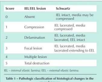
following variables were observed in every section: a) degree of vascular injury; b) degree of internal elastic lamina (IEL) and external elastic lamina (EEL) injury; c) lesional pattern of the adventitia; d) general tissue changes: d1) vascular proliferation, d2) vascular recanalization, d3) hyaline amorphous material, d4) telangiectasia, d5) endothelial characteristics, d6) granulation/ fibrous tissue, d7) cellular atypism, d8) xanthomatous cells, d9) vascular thrombosis, and d10) fibrinoid necrosis.
These variables were classified according to the scoring system shown in Table 1. (Schwartz’ modified classification 13).
Fig. 1 - Schematic representation of the aorta/iliac block and its cross-sections.
proximal medial distal
Sample preparation – After a 24-hour fixation, 3 to 6 samples were taken from each iliac artery, in 2- to 3-mm thick cross-sections, thus representing the proximal, medial and distal segments of both arteries, in all the animals. Medial segments represented the central area in most contact with the angioplasty balloon. Proximal and distal segments of the iliac artery of each side, named reference segments, corresponded to the ends relative to the central area in greatest contact with the angioplasty balloon. Randomly selected samples were also taken from aorta segments. All the material was processed according to standard technique, and 110 microscope slides stained with hematoxyline-eosine (HE) and Hardt’s elastica (orcein) were obtained, as well as 521 iliac artery cross-sections (Fig. 2).
Qualitative histological analysis - All slides were blindly analyzed using a 5-head microscope Olympus® BX 40. The
Fig. 2 - Histological sections of the three segments (proximal, medial, and distal) mounted on microscope slides.
}
}
}
Proximal segment
Medial segment
Distal segment
score iel/eel lesion schwartz
0 Absent IEL intact, media may be compressed
1 Compression IEL lacerated, media compressed 2 Delamination IEL lacerated, media
lacerated, EEL intact 3 Focal lesion IEL lacerated, media
lacerated extending to EEL 4 Multiple lesion
5 Total destruction
IEL – internal elastic lamina; EEL – external elastic lamina.
table 1 – Pathologic classification of histological changes in the several groups
Quantitative histomorphometric analysis – Morphometric measurements were performed to determine arterial lumen, IEL, EEL, neointimal area (NIA), media layer area (MA), and vessel area (VA). Slides were examined by means of Image-Pro Plus 4.5 software for Windows® coupled to an Olympus®
BX50 microscope and Sony® video camera, using the so-called
“area” morphometry application previously calibrated in µm with a 4x objective. Measurements were transferred to Excel Windows®. Morphometric measurements were performed in
all cross-sections of the microscope slide, from proximal, near slide identification, to distal. Segments with artifacts that made it impossible to obtain measurements were disregarded.
a) LA: determined by drawing a line along the luminal border perimeter; NIA: determined by drawing a line under the luminal border and the IEL perimeter line; MA: determined by IEL and EEL perimeters, i. e., EEL-IEL; d) vessel area: comprising EEL area plus NIA (EEL + NIA).
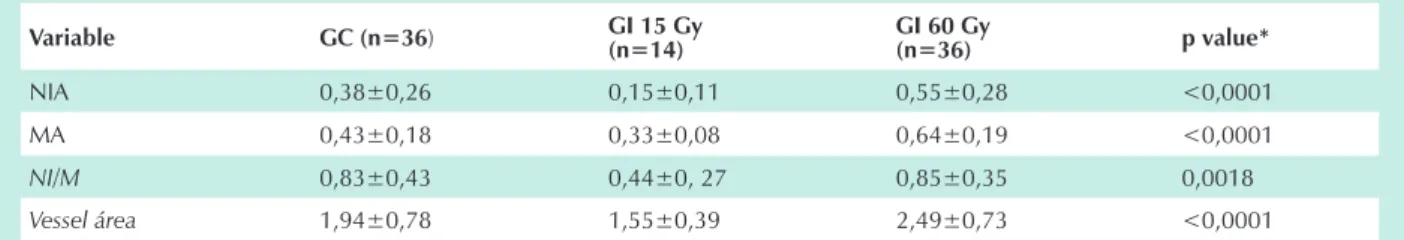
ratio (NI/M). Table 2 shows mean and standard deviation results of these variables for each group and the p values obtained from comparison of means between groups.
Neointimal area (NIA) – Reduction in neointimal area was significant in the 15-Gy IG compared with the other groups. Analysis of variance (ANOVA) showed statistically significant difference among groups (p<0.0001). When comparing two groups at a time, LDS testing pointed to statistically significant difference between the CG and 15-Gy IG (p = 0.0007), the CG and 60-Gy IG (p = 0.0121) and the 15–Gy IG and 60-Gy IG (p<0.0001). On average, the neointimal area of the 15-Gy IG was 2.5 times smaller than that of the CG and 3.6 times smaller than that of the 60-Gy IG. The CG neointimal area, in turn, was 1.4 smaller than that of the 60-Gy IG.
Media layer area (MA) – The largest MA was observed in the 60-Gy IG. When the groups were compared for MA, statistically significant differences were found between the CG and 60-Gy IG (p < 0.0001) and the 15-Gy IG and 60-Gy IG (p < 0.0001). No statistically significant difference was found between the CG and 15-Gy IG (p = 0.0516). On average, the media area of the 15-Gy IG was 1.3 times smaller than that of the CG and 1.9 times smaller than that of the 60-Gy IG. This media area was 1.5 smaller in the CG than in the 60-Gy IG.
NI/M ratio – The lowest neointima/media ratio was detected in the 15-Gy IG, with statistically significant difference among groups (p = 0.0018). A statistically significant difference was found between the CG and 15-Gy IG (p=0.0016), and the 15-Gy IG and 60-Gy IG (p=0.0010), but not between the CG and 60-Gy IG. On average, neointima/media ratio was 1.8 times lower in the 15-Gy IG compared to that of the CG and 1.9 times lower than that of the 60-Gy IG. Mean ratio between the CG and 60-Gy IG was only 1.02 fold.
Vessel area – The largest vessel area was found in the 60-Gy IG (2.49 ± 0.73mm2), the proportion of which was 38% greater than in the 15-Gy IG and 22% greater than in the CG. Analysis of variance (ANOVA) showed statistical significance, with p < 0.0001 (Tab. 2). Considering the group-to-group analysis, a statistically significant difference was found between the irradiated groups and between the 60-Gy IG and control group, with p values 0.0001 and 0.0014, respectively, using LSD analysis.
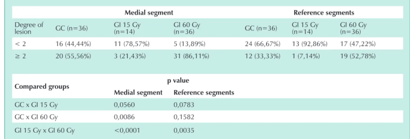
Degree of vascular lesions – The degree of vascular lesion analyzed according to Schwartz et al classification (Table1) was more frequent in the 60-Gy IG, especially in the medial segment, with a statistically significant difference between the CG (p = 0.0046) and 15-Gy IG (p = 0.0391) (Tab. 3).
Using the classification for IEL lesions (Tab.1), the greatest degree of lesion was observed in the medial segments of the three groups; however, in the group versus group analysis, no statistically significant difference was found (Tab. 4).
To analyze degree of lesion in the EEL, a classification similar to that of the IEL was used (Tab. 1). Comparing group versus
group, the greatest degree of EEL lesion was found in the 60-Gy IG medial segment, with a statistically significant difference between the CG (p=0.0086) and 15-Gy IG (p < 0.0001). There was no difference between the CG and 15-Gy IG (Tab. 5).
Qualitative histological analysis – Qualitative variables observed were xanthomatous cells, hyaline amorphous tissue,
Fig. 3 -Cross-section with morphometric measurements outlining both the lumen and the internal and external elastic laminae.
iel
eel
Degrees of vessel lesion – The pathologic classification of histological changes based on scores was adopted to assess the degree of vessel lesion (Table1). In every vessel, the medial segment was compared with reference segments (proximal and distal).
Statistical analysis was performed using Statistica/w software. Analysis of variance (ANOVA) was used to compare groups on categorical variables, and Fisher’s LSD test was used for pairwise comparison between groups. To investigate the degree of association between NIA and MA, Pearson’s correlation coefficient was estimated and the correlation between these variables tested.
Variables corresponding to cell characteristics and degree of vascular lesion were analyzed by Fisher’s exact test, comparing two groups at a time.
In regard to the degree of lesion evaluated by Schwartz’s classification, by which two groups were compared at a time, the Mann-Whitney nonparametric test was used.
Continuous variables were expressed as mean ± standard deviation, and categorical variables were expressed as frequencies and percentages. Comparisons were drawn taking into account the Bonferroni-corrected significance level (p<0.0167 for the degree of lesion and p<0.0125 for tissue changes and morphometric variables. For the other statistical tests, p-values < 0.05 were considered statistically significant.
Results
Mean total cholesterol was 1362.28 ± 273.00 mg/dL.
vascular proliferation, and granulation/fibrous tissue. The presence of xanthomatous cells in moderate/marked degree was observed in 86.11% of the arteries in the 60-Gy IG and 33.33% of the arteries in the CG, with a statistically
significant difference between these groups (p < 0.0001). When the irradiated groups were compared, the 60-Gy IG had higher rates of xanthomatous cells than the 15-Gy IG, 86.11% and 14.29%, respectively, showing a statistically
variable gc (n=36) gi 15 gy (n=14) gi 60 gy (n=36) p value*
NIA 0,38±0,26 0,15±0,11 0,55±0,28 <0,0001 MA 0,43±0,18 0,33±0,08 0,64±0,19 <0,0001
NI/M 0,83±0,43 0,44±0, 27 0,85±0,35 0,0018
Vessel área 1,94±0,78 1,55±0,39 2,49±0,73 <0,0001
CG – control group; 15-Gy IG – group irradiated with 15 Gy; 60-Gy IG: group irradiated with 60 Gy. NIA = neointimal area; MA – media area;
NI/M: neointima-to-media ratio (*) ANOVA
table 2 - Mean and standard deviation (in mm2) of the morphometric variables for each group and p values obtained from comparison of means between groups
Medial segment reference segments
Degree of
lesion GC (n=36) GI 15 Gy(n=14) GI 60 Gy(n=36) GC (n=36) GI 15 Gy(n=14) GI 60 Gy(n=36) < 2 33 (91,67%) 13 (92,86%) 22 (61,11%) 35 (97,22%) 14 (100%) 32 (88,89%)
≥ 2 3 (8,33%) 1 (7,14%) 14 (38,89%) 1 (2,78%) 0 (0%) 4 (11,11%)
compared groups p value
Medial segment reference segments
GC x GI 15 Gy 1,0000 1,0000 GC x GI 60 Gy 0,0046 0,3570 GI 15 Gy x GI 60 Gy 0,0391 0,5660
CG: control group; 15-Gy IG: group irradiated with 15Gy.; 60-Gy IG: group irradiated with 60 Gy (*) Fisher’s exact test.
table 3 – Degree of vascular lesion according to vessel segments (schwartz)13
Medial segment reference segments
Degree of
lesion GC (n=36) GI 15 Gy(n=14) GI 60 Gy(n=36) GC (n=36) GI 15 Gy(n=14) GI 60 Gy(n=36) < 2 11 (30,56%) 2 (14,29%) 8 (22,22%) 21 (58,33%) 10 (71,43%) 16 (44,44%)
≥ 2 25 (69,44%) 12 (85,71%) 28 (77,78%) 15 (41,67%) 4 (28,57%) 20 (55,56%)
compared groups p value
Medial segment reference segments
GC x GI 15 Gy 0,3030 0,5218 GC x GI 60 Gy 0,5936 0,3457 GI 15 Gy x GI 60 Gy 0,7042 0,1192
CG: control group; 15-Gy IG: group irradiated with 15Gy; 60-Gy IG: group irradiated with 60 Gy. (*) Fisher’s exact test.v
significant difference (p < 0.0001), measured by Fisher’s test. No difference was found between the CG and 15-Gy
IG. Values are shown in Table 6, and histological sections are shown in Figures 4 and 5.
Medial segment reference segments
Degree of
lesion GC (n=36) GI 15 Gy(n=14) GI 60 Gy(n=36) GC (n=36) GI 15 Gy(n=14) GI 60 Gy(n=36) < 2 16 (44,44%) 11 (78,57%) 5 (13,89%) 24 (66,67%) 13 (92,86%) 17 (47,22%)
≥ 2 20 (55,56%) 3 (21,43%) 31 (86,11%) 12 (33,33%) 1 (7,14%) 19 (52,78%)
compared groups p value
Medial segment reference segments
GC x GI 15 Gy 0,0560 0,0783 GC x GI 60 Gy 0,0086 0,1582 GI 15 Gy x GI 60 Gy <0,0001 0,0035
CG: control group; 15-Gy IG: group irradiated with 15Gy; 60-Gy IG: group irradiated with 60 Gy. (*) Fisher’s exact test.v
table 5 - Degree of lesion in the internal elastic lamina (iel) of medial and reference segments for each group
Fig. 4 - a) Aorta cross-section showing no xanthomatous cells. b) Artery cross-section from the 60-Gy IG showing slight degree of xanthomatous cells.
10x
20x
a
b
Fig. 5 - a) Artery cross-section from the CG showing moderate degree of xanthomatous cells. b) Artery cross-section from the 60-Gy IG showing marked degree of xanthomatous cells
10x
20x
Granulation/fibrous tissue was present in 27.78% in the CG; 71.43% in the 15-Gy IG, and 63.89% in the 60-Gy IG. When the groups were compared, using Fisher’s test, a statistically significant difference was found between the CG and 15-Gy IG (p = 0.0089) and the CG and 60-Gy IG (p = 0.0042). Frequencies and percentages are shown in Table 6, and histological sections are shown in Figure 6.
The presence of hyaline amorphous material was seen in 13.89% of the CG and 58.33% of the 60-Gy IG. No change was found in the 15-Gy IG. There was a statistically significant difference when the CG and 60-Gy IG (p = 0.0002) and the 15-Gy IG and 60-Gy IG were compared (p = 0.0001) by Fisher’s exact test. No statistically significant difference was found between the CG and 15-Gy IG. Frequencies and percentages are shown in Table 6.
Vascular proliferation was predominant in the 60-Gy IG, as it occurred in 30.56% of the cases. In the CG, it was detected in 11.11%. A statistically significant difference was found when the 15-Gy IG and 60-Gy IG were compared (p = 0.0221). Comparison of the other groups showed no statistically significant difference. All values are specified in Table 6.
Discussion
Using Schwartz et al’s modified classification for vascular
lesion13 the greatest degrees of lesion in the EEL were observed
in the group assigned to the highest dose of samarium-153 (60-Gy IG). The higher degrees of EEL lesion may indicate increased communication between the media and adventitia layers. These changes suggest that the effect may not be due only to the balloon trauma, since the other groups (CG and 15-Gy IG) also underwent the same pressure, but rather to the high dose of irradiation, which triggered structural modifications in the EEL.
No statistically significant difference was found among groups regarding the degree of lesion in the IEL, although this lamina was more predisposed to being compromised by the barotrauma caused by the angioplasty balloon. The role of the internal elastic lamina is to maintain vessel structure, serving as a barrier for cells and macromolecules migration between the intima and the media layers. Its thin elastic fibers have fenestrations that vary according to the species13,14.The
presence of frequent xanthomatous cells in the intimal space is typical of radiolesion associated with hypercholesterolemia. Studying IEL structural changes in hypercholesterolemic pigs, Kwon et al15 observed increased communication between the
intima and media layers. Similar findings were observed in this study. Although no significant lesion was manifest in the IEL among groups, structural changes in the IEL are associated with a considerable communication between the intima and media
variable gc (n=36) gi 15 gy (n=14) gi 60 gy (n=36) p values
Vascular proliferation 4 (0,01;0,21) 0 11 (0,16;0,46) 0,0221* Granulation/fibrous tissue 10 (0,13;0,42) 10 (0,48;0,95) 23 (0,48;0,80) 0,0089**0,0042***
Xanthomatous cell 12 (0,18;0,49) 2 (0;0,33) 31 (0,75;0,97) <0,0001*0,0001***
Hyaline amorphous material 5 (0,03;0,25) 0 21 (0,42;0,74) 0,0001*0,0002***
CG – control group; 15-Gy IG – group irradiated with 15 Gy; 60-Gy IG: group irradiated with 60 Gy. (*) Fisher’s exact test (*) 15-Gy IG x 60-Gy GI (**) CG x 15-Gy IG (***) CG x 60-Gy G.
table 6 – tissue changes found in the several groups studied – frequencies and 95% confidence intervals for the presence rates
layers, shown by the increased number of xanthomatous cells. Arteries irradiated with 60 Gy showed the highest number of these cells in moderate and marked degree, being observed in 86.11% of the arterial segments analyzed compared with only 33.33% in the control group and 14.29% in the 15-Gy irradiated group, with statistical significance among groups (p < 0.0001). The increase in media area reflects these structural changes.
Studies have demonstrated the usefulness of irradiation with beta-emitters to prevent restenosis16. Waksmanet al17
tested different doses absorbed, using 7, 14, 28 and 56 Gy of 90Sr/Y, calculated to reach 2.0 mm from the vessel wall, and achieved adequate inhibition of intimal proliferation, according to dose progression. No necrosis was observed in artery layers. In our study, doses were calculated to reach 1 mm from the vessel wall, with 15 Gy and 60 Gy, hypothesizing that the higher dose would yield better results in tissue inhibition. With the higher absorbed dose of 60 Gy, however, 63.89% of fibrosis was observed, compared with 27.78% in the CG and 71.435% in the 15-Gy IG. These findings suggest the presence of irradiation-induced lesion, probably due to an exacerbated penetration with the absorbed dose of 60 Gy of Sm-153. This is emphasized by the degree of lesion in the EEL, which was 48.5% higher in the 60-Gy IG than in the 15-Gy IG and 31% higher than in the CG. The hypothesis of radiolesion rather than lesion caused by the angioplasty balloon is thus reinforced. This finding corroborates literature data, suggesting that this fibrosis may be attributed to radiation typical of radioisotopes. Nevertheless the artery exposed to the 15 Gy dose did not show the cellular and histological abnormalities found with the 60 Gy dose, suggesting that these may be dose-response phenomena. Denham and Hauer-Jensen discussed these findings18. They described the
difference between arterial lesion caused by trauma and by radiation, the latter involving excessive production of cytokines, excessive deposition of extracellular matrix and the resulting fibrosis. Trott and Kamprad10 studied the
anti-inflammatory and proanti-inflammatory effects of radiation and point to a strong correlation with the delivered dose. This may explain the difference found between both groups of irradiated
arteries in this trial, with similar fibrosis observed with the 15 Gy dose and 60 Gy dose, but without the xanthomatous cells, hyaline amorphous material, and changes in intimal and media layers observed in the 60-Gy IG.
The primary characteristic of vascular damage following balloon angioplasty is neointimal hyperplasia, a kind of fibrocellular lesion that limits blood flow. The major characteristics are migration of media layer cells, phenotypic changes, and cellular matrix secretion19.These are some of the
main causes of restenosis in human beings undergoing balloon angioplasty, besides the elastic recoil of the arterial wall. Over recent years, intraarterial brachytherapy has been one of the main techniques for treating restenosis, through specific doses, acting as a cell growth suppressor20,21. Similar observations
were made in this study, which yield significant results in neointimal inhibition with the 15 Gy dose, unlike the arteries exposed to a 60 Gy dose, determining the deleterious effects of radiolesion using the intraarterial radiation technique.
Conclusion
Intraarterial radiation with the absorbed dose of 15 Gy (Sm-153) was adequate to reduce neointimal, media layer and vessel areas. In the absorbed dose of 60 Gy, a significant increase was found in the morphometric variables, in addition to significant abnormal cell patterns, such as xanthomatous cells and hyaline amorphous material. These data are consistent with the effects of arterial radiolesion.
Acknowledgement
Thanks to drs. Karine Dall’Oglio Tolazzi, Priscila Oliveira Silva, and Flávia Ferreira; and also to the postgraduate students Ruy Fernando Kuenzer Caetano da Silva and Manuela Oliveira, for their great help in the experimental phase.
Potencial conflict of interest
No potential conflict of interest relevant to this article was reported.
References
1. Clowes AW, Schwartz SM. Significance of quiescent smooth muscle migration in the injured rat carotid artery. Circ Res 1985;56:139-45.
2. Galis ZS, Khatri JJ. Matrix metalloproteinases in vascular remodeling and atherogenesis: the good, the bad, and the ugly.Circ Res 2002;90:251-62.
3. Schwartz SM, deBlois D, O’Brien ERM. The Intima: Soil for atherosclerosis and restenosis. Circ Res 1995;77:445-65.
4. Wiedermann JG, Marboe C, Amols H, Schwartz A, Weinberger J. Intracoronary irradiation markedly reduces restenosis after ballon angioplasty in a porcine model. J Am Coll Cardiol 1994;23:1491-8.
5. Condado JA, Waksman R, Gurdiel O, et al. Long-term angiographic and clinical outcome after percutaneous transluminal coronary angioplasty and intracoronary radiation therapy in humans. Circulation 1997;96:727-32.
6. Sapirstein W, Zuckerman B, Dillard J. FDA approval of coronary-artery brachytherapy. N Eng J Med 2001;344:297-9.
7. Teirstein PS, King S. Vascular radiation in a drug-eluting stent world it’s not over till it’s over. Circulation 2003;108:384.
8. Salame MY, Verheye S, Crocker IR, Chronos NA, Robinson KA, King SB 3rd. Intracoronary radiation therapy. Eur Heart J 2001;22:629-47.
9. Moura A, Yamada A, Hauer D, et al. Samarium-153 for intravascular irradiation therapy with liquid-filled balloons to prevent restenosis: acute and long-term results in a hypercholesterolemic rabbit restenosis model. Cardiovasc Radiat Med 2001;2:69-74.
10. Trott KR, Kamprad F. Radiobiological mechanisms of anti-inflammatory radiotherapy. Radiother Oncol 1999;51:197-203.
11. Goy JJ, Stauffer JC, Siegenthaler M, Benoit A, Seydoux C. A prospective randomized comparison between paclitaxel and sirolimus stents in the real world of interventional cardiology The TAXi trial. J Am Coll Cardiol 2005;45(2):308-11.
12. Herrmann HC. Prevention of cardiovascular events after percutaneous coronary intervention. N Engl J Med 2004;350:2708-10.
14. Wong LCY, Langille BL. Development remodeling of the internal elastic lamina of rabbit arteries. Circ Res 1996;78:799-805.
15. Kwon HM, Sangiorgi G, Spagnoli LG, et al. Experimental hypercholesterolemia induces ultrastructural changes in the internal elastic lamina of porcine coronary arteries. Atherosclerosis 1998;139:283-9.
16. Waksman R, Robinson KA, Crocker IR, et al. Intracoronary radiation before stent implantation inhibits neointima formation in stented porcine coronary arteries. Circulation 1995;92:1383-6.
17. Waksman R, Robinson KA, Crocker IR, et al. Intracoronary low-dose β -irradiation inhibits neointima formation after coronary artery balloon injury in the swine restenosis model. Circulation 1995; 92:3052-31.
18) Denham JW,Haeur-Jensen M. The radiotherapeutic injury-a complex “wound”. Radiother Oncol 2002;63:129-45.
19. Meerkin D, Tardif JC, Crocker IR, et al. Effects of intracoronary beta-radiation therapy after coronary angioplasty: an intravascular ultrasound study. Circulation 1999;99(13):1660-5.
20. Grise MA, Massullo V, Jani S, et al. Five-year clinical follow-up after intracoronary radiation: results of a randomized clinical trial. Circulation 2002;105:2737-40.