Image
Traumatic Fistula between the Right Coronary Artery and Right Atrial
Chamber
Alessandra Edna Teófilo Lemos, Antonio Luiz Júnior Araújo, Lucia de Souza Belém, Juan Alberto Cosquilo Mejia, Aluisio
Cruz Júnior, Nelson Lopes Evangelista, Luciana Santos Oliveira
Hospital de Messejana - Fortaleza, CE - Brazil
Mailing address: Alessandra Edna Teófilo Lemos •
Rua Osvaldo Cruz, 1000/1703 - 60125-150 – Fortaleza, CE - Brazil E-mail: [email protected]
Manuscript received in May 24, 2006; revised manuscript received June 12, 2006; accepted June 12, 2006.
There are two techniques to surgically repair a coronary fistula: ligation or external obliteration of the fistula, with or without distal revascularization of the coronary and closure from within the heart chamber². The second intervention was used in this case and is associated with a lower recurrence rate³.
Introduction
A 25-year old male patient, victim of attack by cutting weapon, had the side of his right hemithorax injured. In the following two weeks he developed thoracic oppressive pain, in the inframammary region. The pain was triggered by exertion, and mitigated by rest, and was followed by dyspnea. He was admitted into our facility.
Upon physical examination on admission, he presented with blood pressure at 100 x 60 mmHg, and heart frequency of 110 bpm. On auscultation, there was a continuous murmur on the left sternal border and tricuspid systolic murmur (++/+6).
He had no relevant personal or family history. He denied smoking, drinking and using illegal drugs.
The electrocardiogram on admission showed sinus rhythm with pathological Q-wave on the lower leads.
Thorax X-ray and laboratory test has normal results, including troponin I dosage.
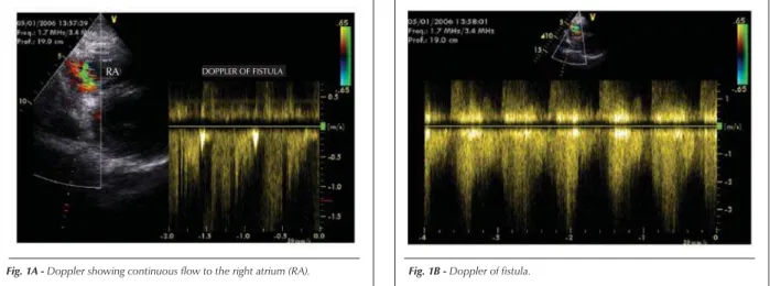
Transthoracic echocardiogram evidenced continuous flow close to the anterior leaflet of the tricuspid valve inside the right
atrium, in addition to mild to moderate tricuspid regurgitation (Fig.1A and 1B.).
Elective coronary angiography confirmed the fistula between the right coronary and the right atrium (Fig. 2A).
We performed a surgery to close the fistula from within the right atrial chamber with a saphenous vein patch (Fig. 3A and 3B)
The patient progressed without symptoms. He was normal on physical examination and underwent a second coronary investigation for control purposes, which showed a normal right coronary artery. (Fig. 2B)
Traumatic fistulae between coronary arteries and heart chambers are uncommon sequelae of thoracic trauma and require early diagnosis and intervention so as to prevent complications.
Potential complications from fistulae described in the literature include: congestive heart failure, pulmonary arterial hypertension, coronary steal syndrome with myocardial ischemia, bacterial endocarditis and formation of coronary aneurism¹.
Key words
Arterio-arterial fistula; coronary vessels; heart injuries.
Fig. 1B - Doppler of fistula.
Fig. 1A - Doppler showing continuous flow to the right atrium (RA).
RA DOPPLER OF FISTULA
Image
Lemos e cols. Fístula traumática entre coronária direita e câmara atrial direita
Arq Bras Cardiol 2007; 88(3) : e64-e65
Fig. 2A - Aortography showing the fistula (arrow) between the right coronary artery (RC); right atrium (RA); left coronary artery (LC) and Aorta (AO).
Fig. 3A - Open right atrium with fistula (arrow).
Fig. 2B - Control coronary angiography showing a normal right artery (arrow).
Referências
1. Lowe JE, Adams DH, Cumming RG, Wesly RL, Phillips HR. The natural history and recommended management of patients with traumatic coronary artery fistulas. Ann Thorac Surg. 1983; 36: 295-305.
2. Friesen CH, Howlett JG, Ross DB. Traumatic coronary artery fistula
management. Ann Thorac Surg. 2000; 69: 1973-8.
3. Trout HH, Feinberg RL.Vascular anomalies and aqcuired arteriovenous fistulas. In: Dean RH, Yao JST, Brewster DC, eds. Current diagnosis and treatment in vascular surgery. Norwalk, CT: Appleton and Lange; 1995. p. 309-24.
RC
RA
LC
Fig. 3B - Closure of fistula, from within the right atrium with a saphenous vein patch (arrow).