417
Ulisses Alexandre CROTI, Domingo Marcolino BRAILE, Sírio HASSEM SOBRINHO, Carlos Henrique DE MARCHI
Correspondência: Ulisses Alexandre Croti
Hospital de Base – FAMERP – Av. Brigadeiro Faria Lima, 5416 CEP 15090-000 – São José do Rio Preto – São Paulo
E-mail: [email protected]
Clinical-Surgical Correlation
Braz J Cardiovasc Surg 2004; 19(4): 417-418
CLINICAL DATA
A 3-year-old female Caucasian patient was referred to our department from her native city of Brasilia, Brazil. She had been asymptomatic since birth but a heart murmur was observed during a routine check-up. She was in a good general state, ruddy, hydrated with eupnaea. She had a normal heart rhythm, normal heart sounds with a continuous murmur ++/ 6+ at the left medium sternum edge. Lung auscultation was normal and the abdomen was also unchanged. Peripheral pulses were present and easily palpable.
ELECTROCARDIOGRAM
The electrocardiogram was within the normal limits with a sinusal rhythm and a heartbeat of 125 beats per minute. The electrical axis of the QRS complex was + 60º. No overload of the chambers or conduction disorders were observed.
RADIOGRAM
A cardiothoracic index of 0.53 was calculated. There was a slight increase in the thoracic aorta. The lung parenchyma was without changes.
RBCCV 44205-719
Article received in October, 2004 Article accepted in November, 2004
Case 7/2004 – Pediatric Heart Surgery Service – Hospital de Base,
Medical School, São José do Rio Preto
418
ECHOCARDIOGRAM
The patient had situs solitus and levocardia. The venoatrial, atrioventricular and ventriculoarterial connections were all concordant. The left coronary artery was dilated and presented a high-flow shunt to the left atrium.
DIFFERENTIAL DIAGNOSIS
The few clinical data may suggest patent arteriosus duct, an aneurysm of the Valsalva sinus or arterio-venous fistulae.
DIAGNOSIS
An investigation by coronary cineangiography confirmed the echocardiographic finding demonstrating an aneurysmatic dilation of the left coronary artery and a high-flow fistula to the circumflex branch of the left coronary artery to the left atrium close to the left atrial appendix.
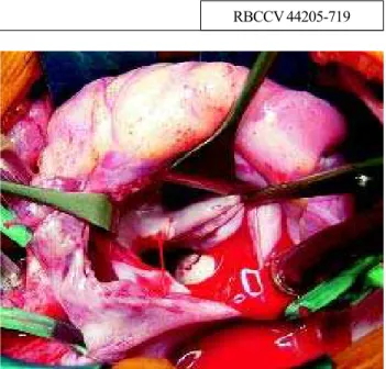
OPERATION
A transsternal median thoracotomy was performed and
CROTI, UA ET AL - Clinical-Surgical Correlation - Case 7/2004 Braz J Cardiovasc Surg 2004; 19(4): 417-418
cardiopulmonary bypass with antegrade sanguineous cardioplegia at 4ºC was established. The right atrium and interatrial septum were opened and the left atrium was exposed. The left atrial appendix was inverted and pulled inside the left atrium (Figure 1). At this time the area was washed with cardioplegia solution, enabling the identification of the exact site of the fistula. A continuous running suture using 6-0 polypropylene thread sectioning the left atrial appendix and suturing the region of the fistula was used. Thus, a second line of sutures was possible with the aim of avoiding relapse of the fistula and exclusion of the left atrial appendix. Cardiopulmonary bypass time was 55 minutes and the myocardial ischemia time was 41 minutes. In the postoperative period the patient evolved with CKMB values within the normal limits and without electrocardiographic alterations. She was released from hospital on the 5th postoperative day with a normal