Case Report
Key words
Catheters, indwelling; foreign bodies, percutaneous retrieval.
We report on the case of a patient with a long term vascular catheter embolized into the right ventricle. This case had unique characteristics since both ends of the catheter were inaccessible for snare, which made it diicult to capture them using conventional techniques. We describe a new method to retrieve the foreign body through its mid-point portion, using a single catheter with two independent snare and hook systems.
Percutaneous Retrieval of Intracardiac Foreign Body with a Novel
Technique
Estêvão Carvalho de Campos Martins and Guilherme Brenande Alves Faria
Hospital da Força Aérea do Galeão - Rio de Janeiro, RJ - Brazil
Mailing address: Estêvão Carvalho de Campos Martins •
Rua Major Rolinda da Silva, 87 - 22611-260 - Rio de Janeiro, RJ - Brazil E-mail: [email protected]
Manuscript received June 19, 2006; revised received July 14, 2006; accepted July 14, 2006.
Introduction
The endovascular pathway is frequently used for diagnostic and therapeutic purposes in clinical practice. Such use ranges from venous access for drug infusion to arterial access for endoprosthesis implantation. The use of this pathway, however, gives rise to some complications, such as the embolization of the material used in the interior of the vascular bed and heart chambers. These complications should be recognized early, and the embolized material should be removed so as to prevent a morbidity increase in the treatment of these patients.
Case Report
A 72-year old male patient, with a history of colon neoplasia, underwent resection in August 2005, and had a long term vascular catheter implanted to allow adjuvant chemotherapy. During the infusion of the chemotherapeutic agent (ifth course), the patient complained of burning near the catheter connector with concurrent local hyperemia. A thorax X-ray showed catheter embolization, with both its ends resting on the muscles of the right ventricle, and its body protruding into the right atrium (shape of a C). The patient was in stable condition, without complaints or arrhythmias. We decided to remove the foreign body percutaneously.
Procedure
We punctured the right femoral vein with local anesthesia, and introduced an 8F valved sheath using the Seldinger
technique. The position of the catheter was conirmed on angiography (fig.1). We built a single catheter with two systems: a snare and a hook system, that can be handled independently (ig. 2-A). We employed a 6F pigtail catheter, a 0.21’’ guide and a 0.014’’ x 300 cm guide wire.
The 0.014’’ guide wire was folded in its mid-point portion and each end was introduced by the pigtail through the oriices located near its distal end (ig. 2-II), and exteriorized in its proximal end (ig 2 – III), thus forming the snare system. This snare was angled so that when it was pulled it involved the whole pigtail “head” (ig. 1–E). We also used a 0.21’’ guide with the objective of controlling the distal end of the pigtail, so that when the catheter introduced this guide the catheter would be straightened and when the guide was withdrawn, the catheter reacquired the shape of a circumference, forming a hook system.
When introducing the system (0.014’’ guide wire fully pulled and 0.21’’ guide rectifying the catheter) we managed to place the pigtail snare at the level of the curvature of the foreign body. We then opened the snare system, introducing the 0.014’’ guide very carefully (fig. 2B). We withdrew the 0.21” guide slowly so that the distal end of the pigtail catheter could snare the foreign body, capturing it within its circumference (ig. 2C). We then manipulated both ends of the 0.014” guide wire to allow the snare to close progressively until the foreign body was captured, crushing it against the catheter (ig. 2D-F); we pulled the foreign body up to the tip of the 8F introducer, and removed all the set (introducer, diagnostic catheter and foreign body) at once (ig 1). We applied local compression for 10 minutes and obtained good homeostasis.
Discussion
Vascular catheters are widely used in clinical practice. Their use, and the use of other types of endovascular techniques, has led us to recognize and treat diferent complications associated with these methods, including the presence of foreign bodies in the vascular system. The irst description of embolization of a catheter fragment was made in 19541. Although it has
been reported that foreign bodies have remained in the body for up to seventeen years without major complications, the immediate removal of any embolized material is necessary due to the occurrence of well-known complications such as sepsis, endocarditis, myocardial perforation, arrhythmias and others2,3. Fisher and Ferreyro reported the incidence of
death or serious complications in up to 71% of patients in whom the foreign body is not removed4. On the other hand,
complications resulting from the removal of foreign bodies
Case Report
Martins and Faria Retrieval of foreign body with a novel technique
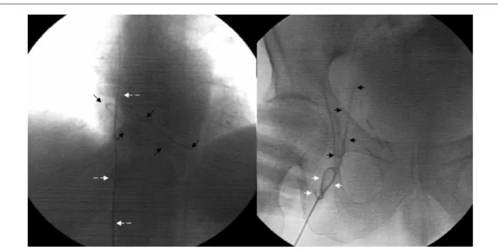
Arq Bras Cardiol 2007; 88(6) : e133-e135 Fig. 1 - To the left – Both tips of the catheter are in the right ventricle, and its body is protruding into the right atrium. To the right – removal of the whole set (catheter and foreign body) through the puncture site. Black arrow – foreign body. White arrow – pigtail catheter.
Fig. 2 - Graph showing the catheter with snare and hook. A) I – Distal portion of catheter with snare (0.014” guide) and hook (curve of catheter), II – Highlight of detail, III – Proximal portion of catheter with exteriorization of “two” 0.014” guide wires and 0.21’’ guide. B) System with snare and hook opened close to the foreign body. C) Catheter with open snare and closed hook, grasping the foreign body. D-F) Pulling of 0.014’’ guide with progressive reduction of snare and ixation of the foreign body onto the catheter.
are rare, and the success rate has been reported to reach up to 100%5.
The percutaneous removal of foreign bodies from the vascular system has been carried out since 1964, when Thomas for the first time removed a guide wire using a bronchoscope6. The snare technique is the most widely used
in the removal of foreign bodies when one of their ends is accessible with a snare7,8. The hook technique is used [to
retrieve] fragments without free ends. This technique requires venous dissection, and there are very few descriptions of it in the literature2,9.Fogarty and pigtail catheters are also used in
these procedures9,10. Combined methods are used especially
in the case of ixed or distal fragments, but here two venous accesses are required9.
One of the factors that account for unsuccessful percutaneous removal of foreign bodies is the presence of ixed fragments without a free end5. In the case reported, both
ends were inaccessible for snaring which prompted us to put together a type of catheter that worked with a double system – the snare and hook system. This enabled us to easily grasp and manipulate the mid-point portion of the foreign body with the hook while allowing us to irmly and safely pull the foreign body with the snare.
The removal of the material snared from the vascular system
Case Report
Martins and Faria
Retrieval of foreign body with a novel technique
Arq Bras Cardiol 2007; 88(6) : e133-e135
References
1. Turner DD, Sommers SC. Accidental passage of a polyethylene catheter from cubital vein to the right atrium. N Engl J Med. 1954; 251(18): 744-5. 2. Rossi P. “Hook catheter”, technique for transfemoral removal of foreign body
from the right side of the heart. Am J Roentgenol Radium Ther Nucl Med. 1970; 109(01): 101-6.
3. Ranchere JY, Thiesse P, Gordiani B, Perol M. Peripheral venous catheter embolism. Ann Fr Anesth Reanim. 1997; 16(2): 196-8.
4. Fisher RG, Ferreyro R. Evaluation of current techniques for nonsurgical removal of intravascular iatrogenic foreign bodies. Am J Roentgenol Rdium Ther Nucl Med. 1978; 130(3): 541-8.
5. Dondelinger RF, Lepoutre B, Kurdziel JC. Percutaneous vascular foreign body retrieval: experience of an 11-year period. Eur J Radiol. 1991; 12(1): 4-10.
6. Thomas J, Sinclair-Smith B, Bloomield D, Davachi A. Non-surgical retrieval of a broken segment of steel spring guide from the right atrium and the inferior vena cava. Circulation. 1964; 30: 106-8.
7. Massumi RA, Ross AM. A traumatic nonsurgical technique for removal of broken catheters from cardiac cavities. N Engl J Med. 1967; 277(4): 195-6. 8. Gabelmann A, Kramer S, Gorich J. Percutaneous retrieval of lost or misplaced
intravascular objects. Am J Roentgenol Rdium Ther Nucl Med. 2001; 176(6): 1509-13.
9. Grabenwoeger F, Dock W., Pinterits F, Apple W. Fixed intravascular foreign bodies: a new method for removal. Radiology. 1988; 167(2): 555-6. 10. Ulacker R, Lima S, Melichar AC. Intravascular foreign bodies: percutaneous
retrieval. Radiology. 1986; 160(3): 731-5.
requires special care, since the material can be a ball with three-times the caliber of the pigtail catheter plus twice the caliber of the foreign body (ig. 1F). In the case presented we used an 8F sheath that did not generate signiicant resistance to the pulling of all the block of material. However, we should highlight that interventionists should analyze the need of choosing a higher gauge introducer (9-12F) or even performing a small incision to bring the material to the exterior. This remark applies especially to those materials that have been inside the body for a longer period (with a higher possibility of deterioration and fracture),
and to more rigid foreign bodies.
This technique is described here for the irst time, and may be a useful alternative to handle more complex situations involving the removal of foreign bodies which result in higher rates of failure when the percutaneous approach is used.
Potential Conlict of Interest
No potential conlict of interest relevant to this article was reported.