In-Hospital Post-Cardiopulmonary-Cerebral
Resuscitation Survival Prognostic Factors
André Mansur de Carvalho Guanaes Gomes, Ari Timerman, Carlos Alfredo Marcílio de Souza, Carlos Maurício Cardeal Mendes, Heitor Portella Póvoas Filho, Adriano Martins de Oliveira, José Antonio de Almeida Souza Instituto do Coração do Hospital das Clínicas da FMUSP, Instituto Dante Pazzanese de Cardiologia, Hospital da Cidade - São Paulo , SP - Brazil; Escola Bahiana de Medicina e Saúde Pública, Universidade Federal da Bahia, Instituto Sócrates Guanaes - Salvador, BA - Brazil
O
BJECTIVETo assess clinical and demographic characteristics of patients who had cardiopulmonary resuscitation and identify short- and long-term survival prognostic factors.
M
ETHODSFour hundred and fifty-two (452) resuscitated patients in general hospitals from Salvador were prospectively assessed through bivariate and stratified analysis in associations between variables and survival curve for a nine-year evolution assessment.
R
ESULTSAge ranged from 14 to 93 years old, mean of 54.11 years old. Male gender patients prevailed and half of them had at least a base disease. Cardiovascular disease was the responsible etiology in 50% of cases. Cardiac arrest was observed in 77% of cases and only 69% of patients were immediately resuscitated. Initial cardiac rhythm was not diagnosed in 59% of patients. Asystole was the most frequent rhythm (42%), followed by ventricular arrhythmia (35%). Immediate survival was 24% and hospital discharge survival 5%. Cardiac arrest etiology, initial cardiac rhythm diagnosis, ventricular fibrillation or tachycardia as arrest mechanism, pre-resuscitation estimated time lower than or equal to 15 minutes and resuscitation time lower than or equal to 5 minutes were recognized as short-term prognostic factors. Non-administration of epinephrine, being resuscitated in private hospital and resuscitation time lower than or equal to 15 minutes were nine-year evolution survival prognostic factors.
Mailing Address:André Guanaes Gomes • Instituto Sócrates Guanaes - Rua Saldanha Marinho, 77-A - 40323-010 • Salvador, BA - Brazil
E-mail: andreguanaes@hospitaldacidade.com.br Received on 01/09/04 • Accepted on 03/30/05
C
ONCLUSIONData may help healthcare professionals decide when start or stop in-hospital resuscitation.
K
EY WORDSDespite huge technological advancements in the last decades, early death due to cardiovascular disease has still been a great challenge for intensive medicine all over the world. In the USA, it has been the main cause of death since 1900, except in 19181. In Brazil, 820 deaths due to
cardiovascular diseases take place every day, which represents six times acquired immunodeficiency syndrome mortality rate1. Mortality due to acute myocardial infarction
is approximately 30%, most of them suddenly and more than 50% cannot make to the hospital in time. Mortality rate decreases from 30% to 10%2 among patients who
have access to proper advanced life support treatment.
In-hospital cardiopulmonary-cerebral resuscitation (CPCR) prognosis is gloomier than outside it. Despite advanced in-hospital life support access, patients have higher disease co-morbidity and severity than those showing cardiac arrest in extra-hospital environment3.
Immediate survival perspective towards in-hospital resuscitation ranges from 30% to 50%4-6, whereas
hospital discharge survival varies from 5% to 35%5,7,
with a mean from 11% to 20%4,8-10.
This study aimed at assessing clinical and demographic characteristics of in-general hospital CPCR-submitted patients, through identification of short- and long-term survival prognostic factors in order to help healthcare professionals decide when to start or stop resuscitation.
M
ETHODS
We prospectively assessed 452 CPCR-submitted patients between July 1 and December 31, 2004, in one of the six general hospitals completing the study in the city of Salvador. Patients under 14 years old or those resuscitated before hospital admission were excluded from the study.
Cardiopulmonary-cerebral arrest (CRCA) was regarded as sudden ceasing of circulatory, respiratory, and cerebral functions, proven by the central pulse (carotid and/or femoral), ventilatory movements (apnea) absence, or agonic respiration and coma status11.
CPCR was taken into consideration whenever the patient had basic or advanced life support by means of ruling recommendations from American Heart Association,
aiming at the return of spontaneous circulation11.
Immediate survival was regarded at the time return of spontaneous circulation was achieved. It was considered for study purpose as cardiac rhythm keeping measurable pulse and/or blood pressure through palpation and/or auscultation, or when systolic pressure greater than or equal to 60 mmHg was monitored through intra-arterial cannulation, for at least an hour in the absence of external cardiac massage9,12.
Tardive survival was taken at hospital discharge and long-term survival a year after post-CPCR survival13.
Data were recorded in Utstein13-style similar
ques-tionnaire sheets. We assessed the following variables:
1) Pre-CRCA patient variables: a) demographic: sex and age; b) clinical: base disease and etiology. 2) CRCA variable: witnessed or not, estimated pre-resuscitation time, access to medications, adrenalin use and dosage, initial cardiac rhythm, defibrillation use, ventilatory support provided, interventions made. 3) Result variables: immediate survival/return of spontaneous circulation; tardive survival (hospital discharge) and long-term survival (a year or longer).
Survivors were in-hospital followed-up by assistant physician and research group and assessed through direct interviews or with relatives whenever their clinical condition did not allowed for. After discharge, besides direct interviews, patients were followed-up by telephone, mail, information from assistant physician or relatives.
The study should have been epidemiological, without any diagnostic or therapeutic intervention. The team collected data with simple and direct questions on patient health condition, his/her welfare, with no risk or damage evidence and/or physical or psychological constraint for the patient or his/her relatives.
Thus the project was submitted to and approved by Comissão de Ética para Análise de Projetos de Pesquisa (Ethics Commission for Research Project Assessment) -CAPPesq of Diretoria Clínica do Hospital das Clínicas and Faculdade de Medicina da Universidade de São Paulo, without conferring to application of reported consent in specific form.
Resuscitated patients had their features described by univariate analysis by using mean and proportion descriptive assessment. Exact binomial confidence inter-val was calculated to estimate sampling populations. Bivariate analysis was then applied to assess the asso-ciation among several patient and arrest characteristics (independent variables) with immediate survival (return of spontaneous circulation).
Association between return of spontaneous circulation and the two main prognostic independent variables from bivariate analysis (pre-resuscitation estimated time and resuscitation time) was carried out through stratified analysis and controlled through a third interest variable. Gross, combined and per stratum relative risks were calculated and homogeneity of relative risks between stratums through Mantel test statistics was tested.
Cox multivariate regression, “Proportional Hazards Regression”, was applied to estimate study variable proportional risk model, altogether on survival time and for the choice of variables included in Kaplan-Meier survival analysis. Kaplan-Meier technique was used to calculate incidence density, incidence density rate, and respective confidence intervals for pre-selected variables. Estimate of long-term survival probability was carried out from survival curves by applying Kaplan-Meier survival functions.
Association, Denmark) and STATA version 7.0 (Stata Corporation, Texas-USA) was the statistic package employed.
R
ESULTS
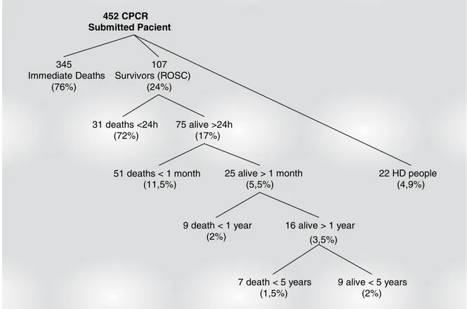
From 452 CPCR-submitted patients, 345 (76.3%) had immediate death and 107 (23.7%) achieved return of spontaneous circulation. From 107 survivors, 31 (6.9%) died in the first 24 post-resuscitation hours. Fifty-one (11.3%) patients, from 76 (16.8%) who survived for more than 24 hours, died in the first month. Only 25 (5.5%) patients survived for more than one post-CRCA month and nine (2%) of those died up to one post-CRCA year. After a year, only 16 (3.5%) initial cohort patients were alive (fig. 1).
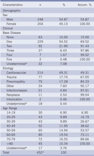
Main clinical and demographic characteristics of CPCR-submitted patients are displayed in tables 1 and 2. Concerning arrest location, ER was the most frequent with 155 (34.3%) CPCRs, followed by 138 (30.53%) in-ICU CPCRs, and 97 (21.46%) in-ward CPCRs. Despite being in-hospital, 101 (23.17%) of CRCAs were not witnessed. From 452 patients, only 106 (23.3%) were defibrillated and 60 (67.4%) of those had a total of joules lower than and/or equal to 750. Twenty-nine (32.6%) of them had a total load higher than 750 joules. Most patients, 311 (71.5%), received oxygen ventilatory prosthesis and were AMBU- (Automatic Mobile Breathing Unit) ventilated, and 91 (20.9%) of them were under
mechanical ventilation. Medications were peripheral vein administrated in 223 (57.5%) patients, with central vein used in 166 (42.8%) and orotracheal tube medication in 20 (5.1%) patients. Adrenalin was used in CPCR of 319 (87.2%) patients. In 196 (70.3%) the dose was lower than 5 mg, 65 (22.9%) received between 5 to 10 mg and 11 (3.9%), from 11 to 15 mg. In 8 (2.9%) CPCRs, adrenalin dose was higher than 16 mg.
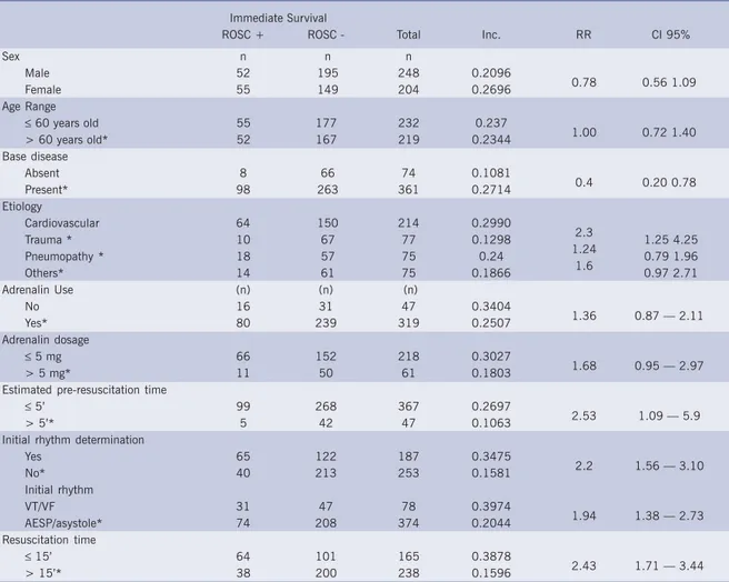
Table III shows bivariate analysis results for clinical and demographic variable assessment of CPCR-submitted patients and immediate survival (return of simultaneous circulation).
Results from association between immediate survival (return of spontaneous circulation) and estimated pre-resuscitation time as main variable, controlled by a third interest variable, are shown in table IV. Table V shows results for resuscitation time as the main immediate survival (return of spontaneous circulation) association assessment variable.
Use of adrenalin, resuscitation time higher than 15 minutes and be resuscitated in a public hospital were Cox proportional regression independent variables with statistically significant prognostic value for death risk. Patients using adrenalin had 58% higher death risk, which was statistically significant (p = 0.02). Resuscitation time higher than 15 minutes was higher death risk prognosis in 37% (relative risk 1.37; p = 0.01); Public hospital CPCR increased death risk in 37% (p = 0.007). Those
Fig. 1 - Results from 452 in-general hospital CPCR
?????? RCRC=Reanimação cardiorespiratória cerebral; RCE=Retorno a circulação espontânea; AH=Alta hospitalar ????
452 CPCR Submitted Pacient
345 Immediate Deaths
(76%)
107 Survivors (ROSC)
(24%)
31 deaths <24h (72%)
75 alive >24h (17%)
51 deaths < 1 month (11,5%)
25 alive > 1 month (5,5%)
9 death < 1 year (2%)
16 alive > 1 year (3,5%)
7 death < 5 years (1,5%)
9 alive < 5 years (2%)
Table II - CRCA and CPCR intervention characteristics
CRCA variable n % Accum. %
Witnessed CRCA
No 101 23.17 23.17
Yes 335 76.83 100.00
Undetermined* 16 3.54 Estimated pre-resuscitation
time
0 to 1' 286 68.92 68.92
1' to 5' 82 19.76 88.68
5' to 10' 26 6.27 94.94
10' to 15' 16 3.86 98.80
15' to 30' 5 1.20 100.00
Undetermined * 37 8.19 Initial Rhythm
Asystole 78 41.71 41.71
Ventricular fibrillation 65 34.76 76.47
AESP 31 16.58 93.05
Ventricular tachycardia 13 6.95 100.00 Undetermined * 265 58.63
Resuscitation time
0 to 1' 9 2.23 2.23
1 to 5' 28 6.95 9.18
5 to 10' 49 12.16 21.34
10 to 15' 79 19.60 40.94
15 to 30' 142 35.24 76.18
30 to 60' 76 18.86 95.03
>60 20 4.96 100.00
Undetermined * 49 10.84 Defibrillation
No 297 73.70
Yes* 106 23.30
Undetermined 49 10.84 Adrenalin Use
Yes 319 87.16
No 47 12.84
Undetermined 86 23.50
Total 366 100.0
* Percentage in relation to total of patients (n=452).
CRCA=cerebral arrest; CPCR= Cardiopulmonary-cerebral resuscitation; AESP=Electrical activity without pulse
Table I - Clinical and demographic characteristics of CPCR-submitted patients
Characteristics n % Accum. %
Demographic Sex
Male 248 54.87 54.87
Female 204 45.13 100.00
Clinical Base Disease
None 63 15.00 15.00
One 229 54.52 69.52
Two 92 21.90 91.43
Three 27 6.43 97.86
Four 7 1.67 99.52
Five 2 0.48 100.00
Undetermined* 32 7.08 Etiology
Cardiovascular 214 49.31 49.31
Trauma 77 17.74 67.05
Pneumopathy 75 17.28 84.33
Other 34 7.83 92.17
Infection/sepsis 21 4.84 97.01
Neoplasia 11 2.53 99.54
Intoxication 2 0.46 100.00 Undetermined* 18 0.44
Age Range
14-19 30 6.90 6.90
20-29 43 9.89 16.79
30-39 43 9.89 26.67
40-49 52 11.95 38.62
50-59 65 14.94 53.57
60-69 85 19.54 73.11
70-79 72 16.55 89.66
>80 45 10.34 100.00
Undetermined* 17 3.76
Total 452* 100
Note: univariate descriptive analysis; *percentage in relation to total of patients (n=452). CPCR= Cardiopulmonary-cerebral resuscitation
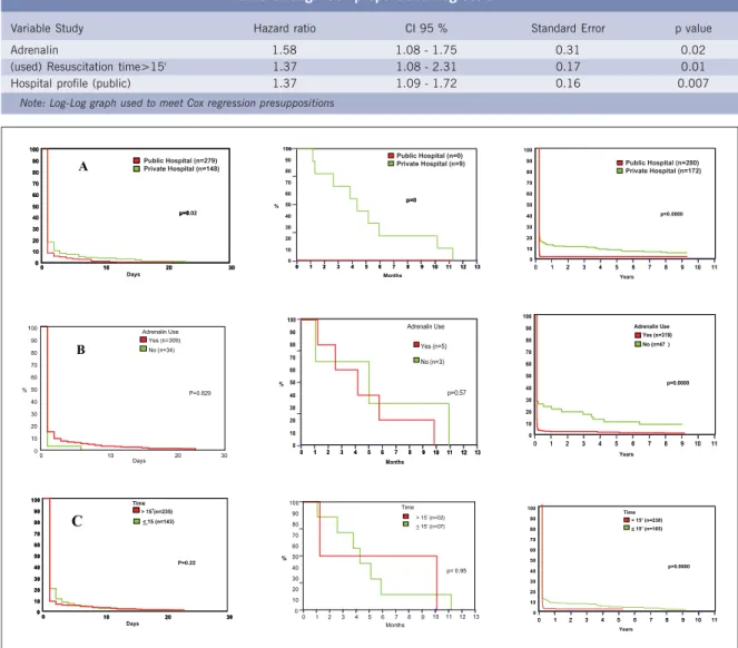
three variables were sent for survival curve assessment. The 107 CPCR surviving patients, as well as the 22 discharged ones were followed up for survival time assessment. Three periods of time were assessed: from zero to 30 post-CRCA days; from one month to a year after CRCA; and from zero to nine years, which were the base for achieving survival curves according to hospital type (fig. 2- A), adrenalin use (fig. 2-B) and resuscitation time (fig. 2-C).
D
ISCUSSION
Return of spontaneous circulation and hospital discharge survivors has been distressing and significantly unchanged in the last three decades, despite CPCR technological advancements. In-hospital CPCR needs studies to better understand its characteristics, results and peculiarities8,13,14, unlike already better studied
extra-hospital CPCRs15-22.
Situation among medical professionals is even more worrying as there are few published works on the matter
and only two national literature reports concerning results and prognosis of in-hospital resuscitated patients23, 24.
The most important and frequently analyzed variables were prospectively assessed in this study, with the aim of studying their prognostic value. There is no other study, among the most mentioned, which has simultaneously assessed so many in-hospital cardiac arrest patient characteristics. From those, only two are prospective6,8.
Such sampling represents the eighth greatest number of CRCA studied patients who were resuscitated, when compared to 33 more frequently mentioned in-hospital CPCR studies 5,6,23-26.
Table III - Clinical and demographic variable and intervention association in immediate survival CPCR-submitted patients (Return of Spontaneous Circulation)
Immediate Survival
ROSC + ROSC - Total Inc. RR CI 95%
Sex n n n
Male 52 195 248 0.2096
Female 55 149 204 0.2696
Age Range
≤ 60 years old 55 177 232 0.237
> 60 years old* 52 167 219 0.2344
Base disease
Absent 8 66 74 0.1081
Present* 98 263 361 0.2714
Etiology
Cardiovascular 64 150 214 0.2990
Trauma * 10 67 77 0.1298 1.25 4.25
Pneumopathy * 18 57 75 0.24 0.79 1.96
Others* 14 61 75 0.1866 0.97 2.71
Adrenalin Use (n) (n) (n)
No 16 31 47 0.3404
Yes* 80 239 319 0.2507
Adrenalin dosage
≤ 5 mg 66 152 218 0.3027
> 5 mg* 11 50 61 0.1803
Estimated pre-resuscitation time
≤ 5’ 99 268 367 0.2697
> 5’* 5 42 47 0.1063
Initial rhythm determination
Yes 65 122 187 0.3475
No* 40 213 253 0.1581
Initial rhythm
VT/VF 31 47 78 0.3974
AESP/asystole* 74 208 374 0.2044
Resuscitation time
≤ 15’ 64 101 165 0.3878
> 15’* 38 200 238 0.1596
*Reference stratum - which relative risk refers to (denominator) - bivariate analysis.
CPCR= Cardiopulmonary-cerebral resuscitation; ROSC=Return of spontaneous circulation; VT= Ventricular tachycardia; VF= Ventricular fibrilation; AESP=Electrical activity without pulse
0.78 0.56 1.09
1.00 0.72 1.40
0.4 0.20 0.78
2.3 1.24
1.6
1.36 0.87 — 2.11
1.68 0.95 — 2.97
2.53 1.09 — 5.9
2.2 1.56 — 3.10
1.94 1.38 — 2.73
2.43 1.71 — 3.44
Table IV - Association between immediate survival (return of spontaneous circulation) and estimated pre-resuscitation time, as main variable, controlled by a third interest variable (co-variables)
Independent Reference Favorable Stratum Combined Gross p value Variables Stratum Relative Risk (**) Relative Association
(co-variable) Relative Risk(*) Risk (***) Relative Risk (****)
Adrenalin dosage > 5mg ≤ 5mg 2.39 2.52 0.1822
5.96 1.54
Hospital profile Público Privado 2.04 2.52 0.75
2.26 1.73
Resuscitation time > 15’ ≤ 15’ 2.71 2.52 0.004
0.83 10.79
Rhythm determination No Yes 2.02 2.52 0.1094
3,68 1,05
Adrenalin used Yes No 1.6 2.52
-1.35
-Initial rhythm > 2 AESP/ASYS ≤ 2 VF/ VT 2.02 2.52 0.1094
3.68 1.05
Etiology Others CVD 2.4 2.52 0.6727
2.03 2.95
Table V - Association between immediate survival (return of spontaneous circulation) and resuscitation time, main variable, controlled by a third interest variable (co-variables)
Independent Reference Stratum Favorable Stratum Combined Gross Association p value Variables (co-variables) Relative Risk(*) Relative Risk (**) Relative Risk (***) Relative Risk (****)
Adrenalin dosage > 5mg ≥ 5mg 2.44 2.43 0.8709
2.34 2.49
Hospital Public Private 2.61 2.43 0.4972
2,24 2,87
Rhythm determination No Yes 2.80 2.43 0.6848
3.09 2.65
Adrenalin used Yes No 2.92 2.43 0.2993
2.67 7.28
Initial rhythm > 2 AESP/ ASYS ≤ 2 VF/ VT 2.80 2.43 0.6848
3.09 2.64
Etiology others CVD 2.50 2.43 0.3999
2.10 2.84
Note: Gross and combined relative and per stratum risks calculated through Mantel-Haenszel test. (*) reference stratum; (**) favorable stratum; (***) adjusted/combined; (****) gross association (bi-variate).AESP= Electrical activity without pulse; ASYS= Asystole; VF= Ventricular fibrilation; VT= Ventricular tachycardia; CVD= Cardiovascular desease
Table VI - Independent (prognostic) variable proportional risk estimate under study, concurrently on survival time through Cox proportional regression
Variable Study Hazard ratio CI 95 % Standard Error p value
Adrenalin 1.58 1.08 - 1.75 0.31 0.02
(used) Resuscitation time>15' 1.37 1.08 - 2.31 0.17 0.01
Hospital profile (public) 1.37 1.09 - 1.72 0.16 0.007
Note: Log-Log graph used to meet Cox regression presuppositions
Fig. 2 - Survival curves according to hospital type (A), adrenalin use (B) and resuscitation time (C) in the first 30 days, in the period from 1 month
to 1 year and in the period from 0 to 9 years (Kaplan-Meier technique was used)
A
0 10 20 30
100 90 80 70 60 50 40 30 20 10 0 p=0.02 Days
0 10 20 30
100 90 80 70 60 50 40 30 20 10 0 p=0 p=0 p=0 100 90 80 70 60 50 40 30 20 10 0 Months %
0 1 2 3 4 5 6 7 8 9 10 11 12 13 0 1 2 3 4 5 6 7 8 9 10 11 12 13
100 90 80 70 60 50 40 30 20 10 0 Years
0 1 2 3 4 5 6 7 8 9 10 11 0 1 2 3 4 5 6 7 8 9 10 11
p=0.0000
0 10 20 30
100 90 80 70 60 50 40 30 20 10 0 Adrenalin Use Yes (n=309) No (n=34) P=0.829 Days % B 100 90 80 70 60 50 40 30 20 10 0 Months %
0 1 2 3 4 5 6 7 8 9 10 11 12 13 0 1 2 3 4 5 6 7 8 9 10 11 12 13
p=0.57 Adrenalin Use Yes (n=5) No (n=3) 100 90 80 70 60 50 40 30 20 10 0 p=0.0000 Years
0 1 2 3 4 5 6 7 8 9 10 11 0 1 2 3 4 5 6 7 8 9 10 11
Adrenalin Use Yes (n=319) No (n=47 )
0 10 20 30
100 90 80 70 60 50 40 30 20 10 0
> 15 (n=235)’ < 15 (n=143)
P=0.22
Days
0 10 20 30
100 90 80 70 60 50 40 30 20 10 0 Time ’ C 100 90 80 70 60 50 40 30 20 10 0 Time Months %
0 1 2 3 4 5 6 7 8 9 10 11 12 13 p= 0.95 > 15’ (n=02) < 15’ (n=07)
100 90 80 70 60 50 40 30 20 10 0 p=0.0000 Years
0 1 2 3 4 5 6 7 8 9 10 11 0 1 2 3 4 5 6 7 8 9 10 11
Time > 15’ (n=238) < 15’ (n=165) Public Hospital (n=279)
Private Hospital (n=148)
Public Hospital (n=0)
Age had already been regarded as survival prognosis. Currently, most studies conclude age does not directly interfere with survival6,27. Timerman et al23 observed
unfavorable prognosis under 10 and over 70-year-old patients. However, such observation was not maintained in multivariate analysis. Likewise, in our study, age was not shown as predictor variable, as no statistically significant difference was found in bivariate analysis (relative risk: 1.0; 0.72 – 1.40; confidence interval: 95%), as well as in Cox multivariate analysis. Many authors support that base diseases may cat as confusion factor, as there is a trend for aged patients of having higher comorbidity.
By assessing CPCR-submitted patient sex, we noted that our data correspond to those from literature. That shows a slightly greater incidence among men. Such variable does not interfere with survival. In our study, women had approximately 30% more chance of immediate survival than men, which is not statistically significant7,8,23.
Cardiovascular disease is the most frequent cardiac arrest cause4,8, and also the one with the best response
to treatment, due to the highest frequency of ventricular arrhythmia in this group, which has a better prognosis.
In our study, cardiovascular disease was the main cause of cardiac arrest in almost 50% of the cases, followed by trauma (17%) and pneumopathy (17%). By taking cardiovascular diseases as survival favorable stratum, we observed an immediate survival chance almost two and a half times higher than trauma. Bedell et al8 noted in their studies that no patient who had
pneumonia or sepsis, as arrest etiology, survived hospital discharge.
In literature, base disease (subjacent) is regarded as the most prognostic powerful variable in patient survival4,23. Studies have been showing a better survival
of patients without associated, clinically diagnosed base disease compared to those who had it. Some authors noted the decrease of survival percentage according to associated disease number. Hershey and Fisher7
observed in their studies that nearly 50% of survivors did not show any associated base disease in comparison to 6.5% survivors who had three subjacent illnesses. Timerman et al23 also described base disease
as the greatest prognosis variable in sur vival determining, which was not observed in our study. Statistically significant immediate survival occurrence in non-apparent base disease patients was 10%, in comparison to 27% in those with at least one base disease (relative risk = 0.40; 0.20 to 0.78; confidence interval 95%). Such difference from literature results may be partially explained for some reasons, which have drawn attention of some researchers: great resuscitation assessment complexity, studied population diversity, varied etiologies with different severity levels, several hospital admission causes, which are often underestimated and non-notified13.
Ventilatory support type assessment for resuscitated patients showed orotracheal intubation (71.5%) as the most frequent initial approach. According to emergency care protocols11, only 26.4% had bag-valve-mask with
reservoir as first approach ventilatory support. In this study, nearly 21% of resuscitated patients were intubated and placed under mechanical ventilation. That may represent higher severity of those patients facing resuscitation procedures, as in Bedell et al8 study, mortality
of patients who needed intubation was five times higher than those without the need for it. Similar results were also observed by Robson and Hess 5.
In a 927-patient field study carried out in Seattle, in 1976, 123 (22%) from 569 witnessed CRCA patients survived, in comparison to only 14 (4%) from 358 with non-witnessed CRCA28. When CPCR starts on the street,
at CRCA place, by somebody who witnesses it and helps the victim, survival rates are higher than those without instant care by a helper18,20,28-31. Witnessed CRCA patients
showed better return of spontaneous circulation and hospital discharge rates than individuals with non-witnessed CRCA8,13,20,28,31.
Many studies have confirmed that the shorter the times, the greater the victim survival chances are. When basic life support is provided in less than 4 minutes and advanced life support in less than 8, hospital discharge survival may vary from 36% to 70%20,29,32,33.
Very important for extra-hospital CPCR studies, time variable has been currently more appraised in assessing in-hospital CPCR results. Many studies have demonstrated that in witnessed CRCAs, in which estimated pre-resuscitation time tends to be shorter, survival is greater than in non-witnessed. In BRESUS study6, 2,838 CRCAs
were assessed. Witnessed CRCA patients had 48% of return of spontaneous circulation in comparison with 32% who were found under CRCA.
very significant, both clinically and statistically. Observed and literature available data show this variable to determine time taken to start resuscitation is more efficient in predicting in-hospital survival prognosis studies than simply assess whether the arrest was witnessed, as usually analyzed in studies.
Resuscitation time is regarded as the most powerful prognostic variable on resuscitation results. In Bedell et al study8 in patients with CRCA time higher than 15 minutes,
survival decreased from 65% to 5%, and no one survived over 30 resuscitation minutes. In our study, from 165 patients with resuscitation time shorter than or equal to 15 minutes, 64 (38.78%) had return of spontaneous circulation, with a statistically and clinically significant survival chance of 2.43 times higher than the higher time stratum. Those variables kept their prognostic value when assessed in stratified multivariate model, by being concurrently analyzed with other interest co-variables.
Concerning arrest mechanism rhythm we know ventricular fibrillation is the most frequent CRCA rhythm observed in most studies and also the one with the best response to treatment, with higher survival rates8,9,34-36.
Survival chances are better if CRCA patients under ventricular fibrillation have an early approach, since as time goes by, myocardial reversibility decreases due to hypoxemia and acidemia.
In our study, almost 60% of patients did not have determined cardiac arrest rhythm. Asystole was the most frequent among those the rhythm could be determined. From 106 patients who had defibrillation, only 78 cases showed ventricular arrhythmia, which evidenced an overestimated fibrillation number. That could have been an injurious factor influencing our results. Prognostic value of whether determining CRCA initial cardiac rhythm was assessed as shown by the high indetermination percentage of 58.63% (265 CPCRs in 452 CPCRs). We noted patients who had their rhythm determined had a clinically and statistically significant 2.2 (1.55-3.1; confidence interval: 95%) times more chances to survive CRCA.
Our data showed a statistically and clinically significant return of spontaneous circulation rate (relative risk: 1.94; 1.38-2.73) almost twice as higher for patients who had ventricular arrhythmia as CRCA initial rhythm. Such observed data fully corroborate with those from literature9,13,37.
Asystole was the most frequent initial rhythm noted in our study and that is maybe a reaction from delay in initial care we noted. Such observation may have cause a decrease in the number of verified ventricular fibrillation, with a delay in initial care, evolving to asystole, which is the most unfavorable rhythm to reversion.
For a long time, epinephrine was the most important drug in CRCA approach38,39. However, its CPCR injurious
beta-adrenergic effects40 have been recently questioned.
The importance of searching for a more efficient and safe alternative than epinephrine in classic dosage of 0.1 mg/
kg, recommended in American Heart Association protocols11,41,42, has already been understood. Such trend
has been recently achieved with the last recommendations from American Heart Association: epinephrine went from class I to undetermined class32, pointing at the need for
better controlled clinical assays in order to determine the real role of that drug and its indications. We analyzed survival association with who used epinephrine or not. A favorable survival chance of 36% for who did not use epinephrine was noted (relative risk: 1.36; 0.87-2.11; confidence interval: 95%). However, it maybe did not represent statistically significant due to the sample. When comparing who used epinephrine considering administrated dosage, we observed a clinically and statistically significant favorable immediate survival incidence for those who used dosage lower than or equal to 5 mg of the referred drug, nearly 70 %,.
Kyff et al4 found the worst survival results among
ward-resuscitated patients (4.5%). Many studies also showed that ward CRCA, besides more frequent, had worse results. Another study, assessing 79 in-general hospital patients, noted survival among ward-resuscitated patients was lower, only 3%7, when compared to other in-hospital
locations. Those data confront ours, contributing to explain hospital discharge results os 5%.
This group was 9-year evolution followed, with no loss of segment among those 22 hospital discharged resuscitated patients, which increases analysis power23,43
very much. Such fact is important because there has not been many prospective in-hospital CPCR studies6,8. Thus,
variables with more survival analysis power – resuscitation time, type of hospital and use and non-use of epinephrine, assessed in short- and medium-term (from one month to one year) and long-term (nine years) - from Cox multivariate regression analysis, were selected.
By assessing short- and long-term patient survival, we note: 1) private hospital mortality rate (0.5%) was 15 times lower when compared to public hospital rate (7.5%), showing a statistically significant difference (p = 0.0000). There was also a favorable statistically significant difference for a lower mortality in approximately 20% in private hospital in the first 30 days; 2) Patients who did not use epinephrine had a statistically significant difference of 12.5 times more chance of survival, comparing with those who used it (p=0.0000); 3) Nine-year assessment for those who had resuscitation time shorter than or equal to 15 minutes showed almost 20 times higher survival chance.
R
EFERENCES
Information exposed may provide assistance to frontline healthcare professionals, involved with potentially severe and/or sudden death risk patients, who are therefore
involved with the possibility of having to resuscitate and better decide when starting and when stopping resuscitation efforts.
1. Zheng ZJ, Croft JB, Giles WH, Mensah GA. Sudden cardiac death in the United States, 1989 to 1998. Circulation 2001; 104: 2158-63.
2. Lown B, Wolf M. Approaches to sudden death from coronary heart disease. Circulation 1971; 44: 130-142.
3. Chamberlain D, Cummins RO. Recommended guidelines for uniform reporting of data from out-hospital cardiuac arrest: the “Utstein Style”. The European Resuscitation Council. American Heart Association, Heart and Stroke Foundation of Canada and Australian Resuscitation Council. Eur.J. Anesthesiol.,v 9,p.245-56,1992
4. Kyff J, Puri VK, Raheja R, Ireland T. Cardiopulmonary resuscitation in hospitalized patients: continuing problems of decision-making. Crit Care Med 1987; 15: 41-3.8
5. Robinson Gr, Hess D. Postdischarge Survival And Functional Status Following In-Hospital Cardiopulmonary Resuscitation. Chest 1994; 105: 991-996.
6. Tunstall-Pedoe H, Bailey L, Chamberlain DA, Marsden AK, Ward ME, Zideman DA. Survey of 3,765 cardiopulmonary resuscitations in British Hospitals (The Bresus Study): Methods and overall results. Br Med J 1992; 304: 1347-51.
7. Hershey CO, Fisher L. Why Outcome of cardiopulmonar y resuscitation in general wards is poor. Lancet 1982; 1: 31-4.
8. Bedell SE, Delbanco TL, Cook EF, Epstein FH. Sur vival after cardiopulmonary resuscitation in the hospital. N Engl J Med 1983; 309: 569-76.
9. Castagna J, Weil MH, Shubin H. Factors determining survival in patients with cardiac arrest. Chest 1974; 65: 527-9.
10. Lemire JG, Johnson AL. Is cardiac resuscitation worthwhile? A decade of experience. N Engl J Med 1972; 286: 970-2.
11. Guidelines For Cardiopulmonary Resuscitation And Emergency Cardiac Care. JAMA 1992; 268: 2199-2241.
12. Stiell IG, Hebert PC, Weitzman BN et al. High-dose epinephrine in adult cardiac arrest. N Engl J Med 1992; 327: 1045-50.
13. Cummins RO, Chamberlain D, Hazinski MF et al. Recommended Guidelines For Reviewing, Reporting, and Conducting Research on In-Hospital Resuscitation: The In-Hospital ‘Utstein Style’. Resuscitation 1997; 34: 151-83.
14. Cummins RO, Chamberlain DA, Abramson NS et al. Recommended Guidelines for Uniform Reporting of Data from out-of-Hospital Cardiac Arrest: The Utstein Style. A Statement for Health Professionals from a Task Force of The American Heart Association, The European Resuscitation Council, The Hear t and Stroke Foundation of Canada, and The Australian Resuscitation Council. Circulation 1991; 84: 960-75.
15. Becker L, Eisenberg M, Fahrenbruch C, Cobb L. Public locations of cardiac arrest. Implications for public access defibrillation. Circulation 1998; 97: 2106-109.
16. Kudenchuk PJ, Cobb L A, Copass MK et al. Amiodarone for resuscitation after out-of-hospital cardiac arrest due to ventricular fibrillation. N Engl J Med 1999; 341: 871-8.
17. Stults KR, Brown DD, Schug VL, Bean JA. Prehospital defibrillation performed by emergency medical technicians in rural communities. N Engl J Med 1984; 310: 219-223.
18. Tweed WA, Bristow G, Donen N. Resuscitation from cardiac arrest: assessment of a system providing only basic life support outside of hospital. Can Med Assoc J 1980; 122: 297-300.
19. Valenzuela TD, Roe DJ, Nichol G, Clark LI, Spaite DW, Hardman RG. Outcomes of rapid defibrillation by security officers after cardiac arrest in casinos. N Engl J Med 2000; 343: 1206-09.
20. Weaver WD, Copass MK, Bufi D, Ray R, Hallstrom AP, Cobb LA. Improved neurologic recovery and survival after early defibrillation. Circulation 1984; 69: 943-8.
21. Weaver WD. Resuscitation outside the hospital-what’s lacking? N Engl J Med 1991; 325: 1437-9.
22. White RD, Hankins DG, Bugliosi TF. Seven years’ experience with early defibrillation by police and paramedics in an emergency medical services system. Resuscitation 1998; 39: 145-51.
23. Timerman A, Piegas LS, Sousa JE. Results of cardiopulmonary resuscitation in a cardiology hospital. Resuscitation 1989; 18: 75-84.
24. Timerman A, Sauaia N, Piegas LS et al. Prognostic factors of the results of cardiopulmonary resuscitation in a cardiology hospital. Arq Bras Cardiol 2001; 77:152-60
25. Goldberg RJ, Gore JM, Haffajee CI, Alpert JS, Dalen JE. Outcome after cardiac arrest during acute myocardial infarction. Am J Cardiol 1987; 59: 251-5.
26. Tor tolani AJ, Risucci DA, Powell SR, Dixon R. In-hospital cardiopulmonary resuscitation during asystole. Therapeutic factors associated with 24-hour survival. Chest 1989; 96: 622-6.
27. Burns R, Graney MJ, Nichols LO. Prediction of in-hospital cardiopulmonary arrest outcome. Arch Intern Med 1989; 149: 1318-21.
28. Eisenberg MS, Bergner L, Hallstrom A. Cardiac resuscitation in the community. importance of rapid provision and implications for program planning. JAMA 1979; 241: 1905-07.
29. Cummins RO, Eisenberg M, Bergner L, Murray JA. Sensitivity, accuracy, and safety of an automatic external defibrillator. Lancet 1984; 2: 318-20.
30. Cummins RO, Eisenberg MS. Prehospital cardiopulmonar y resuscitation. Is it effective? JAMA 1985; 253: 2408-12.
31. Eisenberg MS, Copass MK, Hallstrom AP et al. Treatment of out-of-hospital cardiac arrests with rapid defibrillation by emergency medical technicians. N Engl J Med 1980; 302: 1379-83.
32. Guidelines 2000 for cardiopulmonary resuscitation and emergency cardiovascular care. Circulation 2000; 102: I86-9.
33. Lund I, Skulberg A. Cardiopulmonary resuscitation by lay people. Lancet 1976; 2: 702-04.
34. Landry FJ, Parker JM, Phillips YY. Outcome of cardiopulmonary resuscitation in the intensive care setting. Arch Intern Med 1992; 152: 2305-08.
36. Peatfield RC, Sillett RW, Taylor D, Mcnicol MW. Survival After Cardiac Arrest In Hospital. Lancet 1977; 1: 1223-5.
37. Hollingsworth JH. The Results Of Cardiopulmonary Resuscitation. A 3-year University Hospital Experience. Ann Intern Med 1969; 71: 459-66.
38. Barton C, Callaham M. High-dose epinephrine improves the return of spontaneous circulation rates in human victims of cardiac arrest. Ann Emerg Med 1991; 20: 722-5.
39. Safar P. History of cardiopulmonary resuscitation. Acute Care 1986; 12: 61-2.
40. Tang W, Weil MH, Sun S, Noc M, Yang L, Gazmuri RJ. Epinephrine increases the severity of postresuscitation myocardial dysfunction. Circulation 1995; 92: 3089-93.
41. Standards and Guidelines for Cardiopulmonary Resuscitation (Cpr) and Emergency Cardiac Care (Ecc). JAMA 1980; 244: 453-509.
42. Standards and Guidelines for Cardiopulmonary Resuscitation (Cpr) and Emergency Cardiac Care (ECC). JAMA 1986; 255: 2989-2905.