J. Braz. Chem. Soc., Vol. 15, No. 2, 324-326, 2004. Printed in Brazil - ©2004 Sociedade Brasileira de Química 0103 - 5053 $6.00+0.00
Short Report
* e-mail: [email protected]
Hot-Filament Metal Oxide Deposition (HFMOD): A Novel Method for Depositing Thin
Films of Metallic Oxides
F. Paulo Rouxinol, B. Cláudio Trasferetti, Richard Landers and Mário A. Bica de Moraes*
Instituto de Física “Gleb Wataghin”, Universidade Estadual de Campinas, CP 6165, 13.087-970 Campinas - SP, Brazil
O presente artigo descreve um novo método para a síntese de filmes finos de óxidos metálicos. Um filamento de metal, que pode ser aquecido por uma fonte de corrente alternada é instalado numa câmara de vácuo. Por meio de um fluxômetro de massa, oxigênio pode ser admitido no interior da câmara. Da reação entre o oxigênio e o metal do filamento aquecido, espécies de óxidos voláteis MexOy, onde Me é o metal, são formadas, se condensando num substrato colocado próximo ao
filamento, formando o filme. Foi observado que os filmes finos de WxOy e MoxOy podem ser
depositados de forma satisfatória por este novo método. Embora várias outras técnicas de análise tenham sido usadas para caracterizar os óxidos, esta nota enfatiza os resultados obtidos por espectroscopia de fotoelétrons de raio-X (XPS).
This short report describes a novel method for the synthesis of metal oxide thin films. The experimental setup consists of a metal filament installed inside a vacuum chamber. The filament can be heated by an ac power supply while oxygen is admitted into the chamber using a mass flowmeter. From reactions between oxygen and the heated metal filament, volatile oxide species, MexOy, where
Me is the metal, can be formed, condensing on a nearby substrate. We have observed that thin films of WxOy and MoxOy can be satisfactorily deposited by this novel method. Although several techniques were used to characterize the oxides, this note emphasizes the results obtained by X-ray Photoelectron Spectroscopy (XPS).
Keywords: WO3, MoO3, thin films, XPS, CVD
Introduction
Metallic oxides are important materials from the standpoint of both fundamental and applied science. Particularly, tungsten and molybdenum oxide films have been the focus of extensive scientific investigations due to their prospective technological applications in (i) electrochromic devices,1,2 (ii) gasochromic sensors,3,4 and
(iii) electrocatalysis.5,6 However, their most intensively
investigated property so far is the electrochromism. WO3 films are considered one of the most viable options in emerging electrochromic technology,1 being applicable
for regulating the throughput of radiant energy in smart windows and antidazzling mirrors. These oxides have also been used as “templates” in the synthesis of composites of the type transition metal oxide/conducting polymer, generating promising results in the area of cathodic materials in secondary lithium batteries.7
Several deposition techniques such as sputtering,8-10
thermal evaporation,3,11 plasma-enhanced chemical vapor
deposition12 and sol-gel13,14 have been used to obtain
325 Hot-Filament Metal Oxide Deposition (HFMOD)
Vol. 15, No. 2, 2004
properties carried out on samples of WO3 show that their optical efficiency is higher than those of WO3 films obtained by the above-mentioned techniques. It is also important to remark that this technique differs from a deposition technique called hot filament chemical vapor deposition (HFCVD), which have been used to deposit siloxane15 and diamond-like films,16 because the filament
used here is not just a “catalyst” used to activate chemical species; it is also a reactant in the reaction.
Experimental
Figure 1 depicts the experimental deposition setup that was designed in our laboratory for the deposition of transition metal oxides. The filament (F), which is made of the metal of the oxide to be deposited, is resistively heated by an ac current supply. Oxygen is admitted to the chamber via an electronic mass flowmeter. Pressure measurements are made using a capacitance manometer. During the depositions the chamber is continuously pumped by a Roots pump and the oxygen pressure is adjusted using the flowmeter. The chamber base pressure is about 2.0 x 10⁻² Pa.
Substrate temperatures are measured using a chromel-alumel thermocouple and filament temperatures are determined with an optical pyrometer through a glass viewpoint in the wall of the deposition chamber.
In order to characterize the structure of the deposited films, several techniques were used such as Infrared Reflection-Absorption Spectroscopy (IRRAS), Rutherford Backscattering (RBS), X-ray Photoelectron Spectroscopy (XPS), UV-vis Absorption Spectroscopy, Single Wavelength Ellipsometry (SWE) and Raman Spectroscopy. The electrochromic properties of the films were also investigated by means of spectroelectrochemical measure-ments in the visible range. Manuscripts describing all these
results are currently being prepared and will be submitted to scientific journals.
For the purposes of this report, only XPS results will be presented and discussed. For the XPS analyses, a hemispherical spectrometer using the unmonochromatized Kα X-ray line of aluminum was employed. To investigate the possible tungsten or molybdenum valence states, the 4f-doublet peak or the 3d-doublet peak, respectively, were fitted with Gaussian peaks corresponding to known bonding states of tungsten and oxygen.
Results and Discussion
The XPS data were gathered for a WxOy and a MoxOy sample deposited with the conditions given in Table 1.
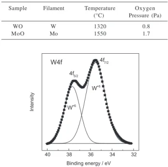
The tungsten 4f XPS spectrum (squares) for sample WO is shown in Figure 2. As can be seen from that figure, the 4f profile can be fit by two Gaussian peaks centered at 37.7 and 35.5 eV which are, respectively, the binding energy of electrons in the 4f5/2 and 4f7/2 levels of tungsten in the W+6 valence state,17 indicating that the film is
composed of stoichiometric WO3.
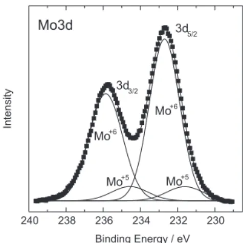
Figure 3 presents the XPS spectrum of the Mo3d core levels for sample MoO. As can be seen from that figure, the 3d profile can be fit by two pairs of Gaussian peaks. The most intense pair is centered at 235.8 and 232.7 eV which
Table 1. Deposition parameters of the WxOy and MoxOy films pre-sented in this work
Sample Filament Temperature Oxygen (°C) Pressure (Pa)
WO W 1320 0.8
MoO Mo 1550 1.7
Figure 1. Schematic representation of the experimental arrange-ment inside the chamber. F- filaarrange-ment of 0.4 mm (tungsten) and 0.6 mm (molybdenum) diameter metal wire, shaped into a seven-turn coil of 8 mm diameter and 35 mm length; S - substrate; SH - copper substrate holder of 50 X 30 x 7 mm3; TC - thermocouple;
filament-to-substrate separation: 30 mm.
326 Rouxinol et al. J. Braz. Chem. Soc.
are, respectively, the binding energy of electrons in the 3d3/2 and 3d5/2 levels of molybdenum in the Mo+6 valence state.
The other pair is centered at 234.6 and 231.6 eV corresponding to the binding energy of electrons in the same 3d levels, but in MoO+5 valence state. For both valence
states, these binding energies are in close agreement with literature values. 18,19 Therefore, it was concluded that the
Mo atoms were in mixed valence states, Mo+6 and Mo+5,
with a high predominance of the former over the latter. Thus the film was in the overall MoOx stoichiometry, with x smaller but close to 3. The observation that the film stoichiometry is close to that of MoO3, is consistent with the expectation that the film is formed from MoO3 and MoO2 species desorbed from the Mo filament and that the desorption rate of the former is greater than that of the latter.
The spectra of Figures 2 and 3 are representative of other spectra of samples prepared with oxygen flow rates in the interval between 6.0 and 21 sccm and the same filament temperatures. Thus the chemical composition of the films does not significantly change in this oxygen flow rate range.
Conclusions
By means of the unexpensive and simple deposition technique described in this note, it was possible to deposit WxOy and MoxOy films with x close to 3. Several details about the role played by the deposition parameters on the overall structure and properties of the films are underway in our laboratory.
Acknowledgements
The authors thank the Fundação de Amparo à Pesquisa do Estado de S. Paulo-FAPESP (grants number 02/07482-6 and 98/10979-2), the Conselho Nacional de
Desenvol-vimento Científico e Tecnológico (CNPq), and Coorde-nação de Aperfeiçoamento de Pessoal de Nível Superior for financial support. Thanks are also due to R. C. G. Vinhas who provided excellent technical assistance with XPS analysis.
References
1. Granqvist, C. G. In The CRC Handbook of Solid State
Electro-chemistry, Gellings, P. J.; Bouwmesster, H. J. M. eds.; CRC Press, Inc.: Cleveland Ohio, 1997, ch. 16.
2. Bange, K.; Sol. Energy Mater. Sol. Cells1999, 58, 1. 3. Lee, S. –H.; Cheong, H. M.; Liu, P.; Smith, D.; Tracy, C. E.;
Mascarenhas, A.; Pitts, J. R.; Deb, S. K.; J. Appl. Phys.2000,
88, 3076.
4. Solis, J. L.; Saukko, S.; Kish, L.; Granqvist, C. G.; Lantto, V.;
Thin Solid Films2001, 391, 255.
5. Roland, J. F.; Anson, F. C. ; J. Electroanal. Chem.1992, 336, 245.
6. Habazaki, H.; Hayashi, Y.; Konno, H.; Electrochim. Acta2002,
47, 4181.
7. Varela, H.; Huguenin, F.; Malta, M.; Torresi, R. M.; Quim. Nova2002, 25, 287.
8. Witham, H. S.; Chindaudom, P.; An, I.; Collins, R. W.; Messier, R.; Vedam, K.; J. Vac. Sci. Technol. A1993, 11, 1881. 9. Taylor, T. A.; Patterson, H. H.; Appl. Spectrosc.1994, 48, 674. 10. Cazzanelli, E.; Papalino, L.; Pennisi, A.; Simone, F.;
Electrochim. Acta2001, 46, 1937.
11. Bohnke, O.; Frand, G.; Fromm, M.; Weber, J.; Greim, O. ;
Appl. Surf. Sci.1996, 93, 45.
12. Henley, W. B.; Sachs, G. J.; J. Electrochem. Soc.1997, 144, 1045.
13. Santato, C.; Odziemkowski, M.; Ulmann, M.; Augustynski, J.;
J. Am. Chem. Soc.2001, 123, 10639.
14. Sharma, N.; Deepa, M.; Varshney, P.; Agnihotry, S. A.; Thin Solid Films2001, 401, 45.
15. Pryce Lewis, H. G.; Casserly, T. B.; Gleason, K. K.; J. Electrochem. Soc.2001, 148, F212.
16. Li, C.; Feng, K. C.; Yuan, H. T.; Xiong, Y.; Feng, K.; Appl.
Surf. Sci.2003, 207, 169.
17. Wagner, C. D.; Riggs, W. M.; Davis, L. E.; Moulder, J. F. In
Handbook of X-ray Photoelectron Spectroscopy; Muilenberg, G. E.; ed., Perkin Elmer Corp.: Eden Praire, MN, 1979, p. 146, and references therein.
18. Fleisch, T. H.; Mains, G. J.; J. Chem. Phys.1982, 76, 780. 19. Colton, R. J.; Guzman, A. M.; Rabalais, J. W.; J. Appl. Phys.
1978, 49, 409.
Received: July 29, 2003
Published on the web: February 13, 2004
FAPESP helped in meeting the publication costs of this article. Figure 3. XPS spectrum of the Mo3d core levels for sample MoO