#
From the Psychiatric Institute, Hospital dasClínicas, Faculty of Medicine, University of São Paulo.
CASE REPORTS
DEPRESSION AND DEMENTIA OF
CEREBROVASCULAR ORIGIN
Paulo Germano Marmorato, Ricardo Alberto Moreno, Silvia Belk Keila and Doris Hupfeld Moreno
RHCFAP/3065
MARMORATO PG et al. - Depression and dementia of cerebrovascular origin. Rev. Hosp. Clín. Fac. Med. S. Paulo 57(1): 25-30, 2002.
We report the case of a patient who presented various psychiatric syndromes at the time of evaluation – partial complex epileptic seizures, personality change, and severe depression, which eventually progressed to dementia – resulting from multiple cerebral infarctions of probable neuro-angiopathic origin, of unknown etiology. Aspects related to depression following cerebrovascular accidents, as well as how cerebrovascular accidents can result in different disorders depending on the variables, are discussed based on the data from current literature.
DESCRIPTORS: Depression. Vascular dementia. Epilepsy. Personality change. Stroke.
Cerebrovascular disorders contrib-ute greatly to the sum of psychiatric disability, chiefly in the elderly popu-lation and mainly as a result of stroke. Depression is the most prevalent psy-chiatric disorder to follow stroke; the rates are between 30% and 60%, de-pending on the population studied1-4.
However, mood disorders are not rec-ognized in many stroke patients, who remain untreated, a fact that results in a poorer outcome for the patients be-cause of its significant impact on func-tional recovery and rehabilitation. Vas-cular accidents are the second most fre-quent cause of dementia, accounting for 15% to 40% of all dementia cases5.
Depression or other mental disorder is often the first symptom in cases of de-mentia of vascular origin5. Although
most of the studies of the psychiatric consequences of stroke have concen-trated on depression, evidence suggests that other psychiatric disorders such as
schizophreniform and paranoid psy-chosis, manic states, and personality changes are also important18, and
should be considered in a complete evaluation.
CASE REPORT
J. A. C., a previously healthy 72-year-old retired stonemason master, began to present at the age of 66 years acute (lasting a few minutes) changes of behavior: automatic and stereotyped movements of the face and upper limbs, grimacing, purposeless walking around his bed, and stepping non-ex-istent stairways. He had no recollection of these episodes. No medical advice was sought at that time. He was a very
$
parietal lobe was diagnosed. After dis-charge, a mild paresis remained, and there was intensification of the depres-sion with psychotic symptoms—visual and auditory hallucinations and perse-cutory delusions. He also attempted suicide on several occasions. He was admitted to the in-patient ward and be-gan treatment with mirtazapine, 45 mg/ day and, after partial remission, was discharged to outpatient follow-up. He came back after a week asking to be admitted again because of the burden he represented to his family and the fear of committing suicide. Electrocon-vulsive therapy was indicated, but it could not be done because of the risk of new hemorrhagic episodes. An epi-leptic focus at the right temporal lobe was found at electroencephalogram (frequent acute waves projecting to right anterior temporal region—F8 and Z2, diffusing to T4), and there was re-mission of the fits with the use of carbamazepine, 800 mg/day (plasma level, 9.7 µg/dL). Imipramine was tried at doses up to 225 mg/day (plasma level, 15.1 ng/dL) because of the nonresponse to lower doses and the se-verity of the disorder. After 15 days, delirium took place suddenly with os-cillation between somnolence and in-tense agitation, remarkable cognitive deficits, and visual hallucinations.
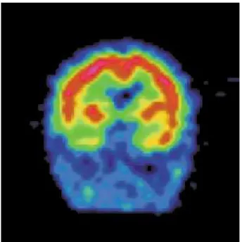
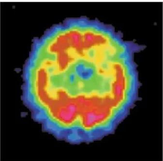
Magnetic resonance showed recent frontal hemorrhage at the left frontal lobe, as well as multiple subacute and chronic lesions widespread throughout the white matter (Fig. 1). The SPECT scans (Figs. 2 and 3) showed focal hypoperfusion of the right temporal and left frontal lobes, which is evidence of functional damage.
Delirium cleared after 1 week of taking risperidone, 2 mg/day, but psy-chotic depression persisted. In consid-eration of the fact that the anticholin-ergic effects of imipramine could be contributing to the delirium, treatment was changed to sertraline, up to 300 mg/day, combined with nortriptyline,
Figure 2 - SPECT scan – coronal section – right temporal hypoperfusion.
Figure 1 - Nuclear Magnetic Resonance – frontal hemorrhage at the left frontal lobe as well as multiple subacute and chronic lesions widespread through the white matter.
up to 100 mg/day, with the result of a subsequent remission of the depressive symptoms. The combination of ser-traline and nortriptyline was justified because of the linear pharmacokinetics of sertraline23, the resistant depression,
and the better tolerability to side effects by the patient. Important deficits per-sisted in the cognitive areas of atten-tion, memory, calculaatten-tion, abstracatten-tion,
and praxis, as did the slowing of psy-chomotor activity. There was evolution to a severe dementia with symptoms of frontal lobe syndrome, such as thought perseveration, emotional incontinence, and lack of initiative, planning, and in-sight.
%
neuro-angiopathy, possibly the result ofvasculitis, an investigation into the etiology based on inflammatory func-tion tests, liquor, and cerebral biopsy was carried out, but results were incon-clusive.
DISCUSSION
Magnetic resonance showed multi-ple subcortical hemorrhagic small le-sions of the whole brain, such as in the frontal and temporal lobes, subacute and chronic, that indicate the occurrence of ischemic lesions over the course of the past few years and months. This fact seems to have contributed to the pro-gressive psychopathological manifesta-tions, initially expressed by a focal and intermittent manifestation, progressing to a personality change, then to psy-chotic depression, and eventually to a severe dementia.
The first reported mental change occurred 6 years before admission as partial complex seizures. Such epilepsy of late onset was possibly the manifes-tation of an ischemic lesion of the right temporal lobe, a fact that can be in-ferred by the ancient lesion found in
this region. Epileptic phenomena, es-pecially those of temporal lobe epi-lepsy, are clear examples of acute psy-chological disturbances that result from focal brain dysfunction, as are some of the disturbances seen after small acute cerebrovascular accidents5.
After four years, a gross change in the patient’s personality traits occurred. Behavioral tendencies that had previ-ously been enduring characteristics of the individual were altered. Brain dam-age often results in changes of tem-perament, or changed patterns of reac-tion to events and to other people, as well as a difficulty in adjusting to new circumstances. Such personality changes of organic origin have been attributed to widespread vascular al-terations within the damaged brain and usually predict a progressive dement-ing illness5.
A depressive disorder of progressive severity with many similarities to major functional depression took place after 6 months of personality change. Since there was evidence of cerebral damage that could be assumed as the etiology for that syndrome, the diagnosis was or-ganic depressive disorder (F06.32), ac-cording to the ICD-106.
The previous history raises the hy-pothesis of a possible dysthymic disor-der and/or a personality with remark-able anancastic traits. Previous affec-tive disorders have been found to be important risk factors for developing post-stroke depression7. Due to the
paucity of reliable data, since the pa-tient lived most of his life apart from his family, there are difficulties in clari-fying this issue.
Some studies have shown that post-stoke depression cannot be attributed just to a psychological reaction to physical and social disability8. The
present case illustrates this fact, since depression was initiated before devel-opment of a paresis that imposed little functional impairment. Several studies suggest that left frontal lobe lesions are more frequently associated with further course of depression, even in the pres-ence of bilateral injury, and a signifi-cant correlation has been found be-tween severity of depression and prox-imity of the lesion to the frontal pole 8-11. However, a recent systematic review
showed no evidence of a relationship between lesion location and depression after stroke, so this hypothesis remains a subject of controversy3,22. Lesions in
this case report showed this pattern of distribution. Depression secondary to lesions in the left frontal lobe would be associated with greater severity in com-parison with that caused by lesions in other areas12, as would be a
predomi-nantly dysphoric mood, anxiety, in-somnia, poorer memory performance13,
and pathologic emotionalism14.
Sub-cortical atrophy was another finding present in this case that has been asso-ciated with higher incidence of depres-sion and its clinical variability7,19.
&
should not be related to the frontal lobe stroke that happened during in-patient treatment, since postural hypotension is associated with ischemic, not hemo-rrhagic strokes. It has been suggested that post-stroke depression may be the consequence of severe depletions of norepinephrine and/or serotonin pro-duced by frontal or basal ganglia le-sions18.
The depressive disorder was com-plicated by delusions and hallucina-tions that added additional distress to the patient. Some authors have associ-ated lesions in the right hemisphere, especially in temporo-parieto-occipital areas, as well as subcortical atrophy, with risk factors for further manifesta-tion of psychosis16,17.
Such findings support the sugges-tion that neurophysiological (mainly caused by damage to frontal/subcorti-cal brain circuitry22) processes may
play an important role—together with other variables such as social support and pre-existing personal characteris-tics—in the etiology of post-stroke de-pression.
A vascular pattern of dementia was found in the present case: cognitive losses progressing in a step-like fashion, initial manifestation of emotional changes, and fluctuation of level of con-sciousness after hemorrhagic episodes. The dementia syndrome consists of a constellation of symptoms that suggests widespread brain dysfunction. Some forms of dementia are best regarded as the end result of multiple focal patho-logies that coalesce and combine to im-pair functions globally, as in the vascu-lar dementias5. The pattern of lesions in
the white matter for the vascular pattern
is different from that of hypertensive origin, in which there are preponderant lesions of the basal ganglia, which were spared in the present case. The inci-dence of predominantly subcortical lit-tle lesions associated with perforating arteries found at cerebral arteriography strongly suggests a neuro-angiopathy that would cause a major susceptibility to bleeding. A cause for the neuro-an-giopathy was searched for through in-flammatory activity exams and cerebral biopsy. The fact that no significant changes were found does not rule out the hypothesis of some kind of vasculi-tis as the etiology.
Two important functional changes were seen at the SPECT scan: focal hypoperfusion of right temporal and left frontal lobes. Changes in these cerebral regions are shown in the literature to be related to memory deficits, personality changes, depressive symptoms, and frontal lobe syndrome17, 18—all clinical
changes that were presented by the pa-tient along the course of his illness.
Concerning treatment, the use of nortriptyline was found to produce bet-ter results than the placebo in one of the few post-stroke depression treat-ment studies performed20,23. Treatment
with nortriptyline had a significant ef-fect, but complete remission of depres-sive symptoms was not achieved. This result is not surprising, since wide-spread lesions and the consequent overall brain dysfunction have been suggested to be associated with poor remission of symptoms1.
This case illustrates the various psychopathological manifestations caused by progressive damage to the brain. In 6 years, more severe
symp-toms were added to preexisting ones that eventually culminated in severe dementia. Inferences can be made re-lating specific symptoms and syn-dromes to dysfunctions in localized brain areas that agree with data re-ported in the literature. However, many of these studies need to be replicated. Since the relationships between brain damage and mental symptoms are quite complex, other possible etiolo-gical factors such as disability, lesion size, cognitive impairment, and social functioning should be taken into ac-count18,22.
The clinician ought to remain aware that occasionally mental illness present-ing as personality change, affective dis-turbance, and psychotic symptoms may be related to the early stages of cerebral disease and that such cases are not unu-sual, especially in elderly patients. The importance of recognizing post-stroke depression should be emphasized be-cause of its high prevalence and bebe-cause when it is not correctly diagnosed and effectively treated, the course of disease will worsen due to lack of cooperation with rehabilitation and the deficiencies caused by the stroke. The study of such patients provides us with the opportu-nity not only to examine those factors that produce psychiatric disorder but also factors that exert protective effects against it or that cannot ameliorate this disorder.
ACKNOWLEDGEMENT
'
REFERENCES
1. STARKSTEIN ES, ROBINSON RG & PRICE TR - Comparison of recovered versus nonrecovered patients with poststroke depression. Stroke 1988;19:1491-1496.
2. ROBINSON RG, MORRIS PL & FEDOROF JP - Depression and cerebrovascular disease. J Clin Psychiatry 1990; 51(suppl. July): 26-31.
3. PRIMEAU F - Post-stroke depression: A critical review of the literature. Can J Psychiatry 1998; 33(8):757-764.
4. OPPENHEIMER S & HACHINSKI V - Complications of acute stroke. Lancet 1992; 339:721-724.
5. LISHMAN WA - Organic Psychiatry. 3th ed. Oxford, Blackwell
Science, 1998.
6. OMS - Classificação de transtornos mentais e de comportamento da CID-10. S. Paulo, Artes Médicas, 1993.
7. ANDERSEN G, VESTERGAARD K, INGEMANN-NIELSEN M et al. - Risk factors for post-stroke depression. Acta Psychiatrica Scand 1995; 92:193-198.
8. ROBINSON RG, KUBOS KL, STARR LB et al. - Mood disorders in stroke patients: importance of location of lesion. Brain 1984; 107,81-93.
9. LIPSEY JR, ROBINSON RG, PEARLSON GD et al. - Mood change following bilateral brain injury. Brit J Psychiatry 1983; 143:266-273.
10. EASTWOOD MR, RIFAT SL, NOBBS H et al. - Mood disorder following cerebrovascular accident. Brit J Psychiatry 1989; 154:195-200.
11. CARSON JA, MACHALE K, ALLEN K et al. – Depression after stroke and lesion location: a systematic review. Lancet 2000; 356:122-126.
12. BOLLA-WILSON K, ROBINSON RG, STARKSTEIN SE, et al. – Lateralization of dementia of depression in stroke patients. Am J Psychiatry 1989; 146:627-634.
13. STERN AR & BACHMAN DL - Depressive symptoms following stroke. Am J Psychiatry 1991; 148:351-356.
RESUMO RHCFAP/3065
MARMORATO PG e col. - Depressão e demência de origem cerebrovas-cular. Rev. Hosp. Clín. Fac. Med.
S. Paulo 57(1):25-30, 2002.
Relatamos o caso de um paciente que apresentou diversas síndromes psi-quiátricas ao longo do tempo - crises epilépticas parciais complexas,
altera-ção de personalidade e depressão gra-ve que finalmente progrediu para de-mência - resultantes de múltiplos infartos cerebrais de provável origem neuroangiopática cuja etiologia perma-neceu indeterminada. São discutidos aspectos relacionados a depressões se-cundárias a acidentes cerebrovascu-lares, assim como estes podem
resul-tar em diferentes transtornos de acor-do com algumas variáveis que são dis-cutidas com base nos dados da litera-tura atual.
DESCRITORES: Depressão.
!
14. HOUSE A, DENNIS M, MOLINEUX A et al. - Emotionalism after stroke. Br Med J 1989; 298:991-994.
15. RABINS PV, STARKSTEIN SE & ROBINSON RG - Risk factors for developing atypical (schizophreniform) psychosis following stroke. J Neuropsychiatry Clin Neurosci 1991; 3:6-9.
16. LEVINE DN & FINKLESTEIN S - Delayed psychosis after right temporo-parietal stroke or trauma: relation to epilepsy. Neurology 1982; 32:267-273.
17. ROBINSON RG & STARKSTEIN SE - Neuropsychiatric aspects of cerebrovascular disorders. In: SADOCK BJ & SADOCK VA -Comprehensive Textbook of Psychiatry. 7thed. Philadelphia,
Lippincott, 2000,
18. BARBER R, SHELTENS P, GHOLKAR A et al. – White matter lesions on magnetic resonance imaging in dementia with Lewy bodies, Alzheimer’s disease, vascular dementia and normal aging. J Neurol Neurosurg Psychiatry 1999;67:66-72.
19. LIPSEY JR, ROBINSON RG, PEARLSON GD et al. - Nortriptyline treatment of post-stroke depression: a double blind study. Lancet 1984; 1:297.
20. JOHNSON GA - Research into psychiatric disorder after stroke: the need for further studies. Aust N Z J Psychiatry 1991; 25:358-370. 21. RAO R - Cerebrovascular disease and late life depression: an age old association revisited. Int J Geriatric Psychiatry 2000; 15,419– 433.
22. KIMURA M, ROBINSON RG & KOSIER JT - Treatment of cognitive impairment after post-stroke depression: a double-blind treatment trial. Stroke 2000; 31:1482-1486.
23. THE UNITED STATES Pharmacopeial Dispensing Information. Sertralina systemic. In: Drug Information for the Health Care Professional. USP-DI, Massachusetts, Micromedex, 2000. p. 2566-2570.