Evaluation of the neonatal sepsis
diagnosis: use of clinical and laboratory
parameters as diagnosis factors
AVALIAÇÃO DO DIAGNÓSTICO DA SEPSE NEONATAL: USO DE PARÂMETROS LABORATORIAIS E CLÍNICOS COMO FATORES DIAGNÓSTICOS
EVALUACIÓN DEL DIAGNÓSTICO DE SEPSIS NEONATAL: USO DE PARÁMETROS LABORATORIALES Y CLÍNICOS COMO FACTORES DIAGNÓSTICOS
RESUMO
Objetivou-se descrever e comparar as carac-terísticas clínicas, laboratoriais e assistenci-ais de RN que apresentaram sepse compro-vada tardia e de RN que apresentaram sepse não comprovada tardia. Em seguida, avaliar se houve diferença entre os grupos, além de descrever os germes prevalentes na unida-de neonatal estudada. Estudo unida-descritivo, envolvendo 168 casos. Observou-se que 33,3% tiveram sepse tardia provada. A ida-de no momento da sepse, o tempo total ida-de internação, a quantidade total de neutró-filos, a quantidade de neutrófilos imaturos e o valor da PC-r mostraram bons parâme-tros na diferenciação entre os dois grupos quando analisados de forma isolada. A
Klebisiella pneumoniae, o Staphylococcus coagulase negativo e o S. aureus foram as bactérias mais comumente isoladas.
DESCRITORES
Sepse. Recém-nascido. Diagnóstico.
Enfermagem neonatal.
1 Neonatology Nurse, Universidade Federal do Rio de Janeiro. Graduate Student in Hospital Infection Control, Universidade Federal Fluminense. Trainee,
Hospital Infection Control Commission, Hospital Universitário Antônio Pedro, Universidade Federal Fluminense. Resident Nurse in Neonatology at Clínica Perinatal Laranjeiras, Rio de Janeiro. Niterói, RJ, [email protected] 2 Adjunct Professor, Maternal-Infant Nursing Department, Medical
School, Universidade Federal Fluminense. Clinical Head, Neonatal Unit, Hospital Universitário Antônio Pedro, Universidade Federal Fluminense. Clinical Head, Perinatal Unit, Hospital de Clínicas de São Gonçalo, Rio de Janeiro. Niterói, RJ, Brazil. [email protected] 3 Nursing Student, Universidade Federal
Fluminense. Trainee, Hospital Infection Control Commission, Hospital Universitário Antônio Pedro, Universidade Federal Fluminense. Niterói, RJ, Brazil.
O
RIGINAL
A
R
TICLE
Luciano de Assis Meireles1, Alan Araújo Vieira2, Carolina Roella Costa3
ABSTRACT
The purpose of this study was to describe and compare the clinical, laboratory and health care characteristics of newborns (NBs) with confirmed late onset sepsis and NBs with unconfirmed late sepsis, verify if there were any differences between the groups, and describe the germs prevalent in the studied neonatal unit. This is a de-scriptive study, involving 168 cases. It was observer that 33.3% had a confirmed diag-nosis for late onset sepsis. The age at the time of sepsis onset, the length of stay, the total number of neutrophils, the number of immature neutrophils and the value of PC-r proved good parameters to differenti-ate between the two groups when analyzed separately. The most common isolated bac-teria were: Klebsiella pneumoniae, Staphy-lococcus coagulase negative and S. aureus.
KEY WORDS
Sepsis.
Infant, newborn. Diagnosis. Neonatal nursing.
RESUMEN
Se objetivó describir y comparar las carac-terísticas clínicas, laboratoriales y asistenciales de RN que presentaron sepsis comprobada tardía y de RN que presenta-ron sepsis no comprobada tardía para, en-tonces, evaluar si hubo diferencia entre los grupos, además de describir los gérmenes prevalentes en la unidad neonatal estudia-da. Estudio descriptivo, involucrando 168 casos, 33,3% tuvieron sepsis tardía proba-da. La edad al momento de la sepsis, el tiempo total de internación, la cantidad total de neutrófilos, la cantidad de neutró-filos inmaduros y el valor de la PC-r mos-traron buenos parámetros en la diferencia-ción entre los dos grupos cuando fueron analizados en forma aislada. La Klebsiella pneumoniae, el Staphylococcus coagulase
negativo y el S. aureus fueron las bacterias aisladas con mayor prevalencia.
DESCRIPTORES
INTRODUCTION
Late-onset sepsis is significantly associated with pro-longed hospitalization and mortality in newborn infants (NI) hospitalized at the intensive care unit (ICU). The incidence of sepsis is higher in low-weight NI and can reach approxi-mately 25%(1). In developing countries, sepsis is responsible
for 30 - 40% of neonatal deaths(2).
Sepsis should be adequately diagnosed at the start of the problem as, if not, actually affected NI can rapidly evolve to septic shock, disseminated intravascular coagulation and death(3).
As they spend more time with the NI, nurses and their team can detect clinical alterations compatible with the emergence of sepsis faster, provided that they know the parameters and factors that should be overseen, signaliz-ing a rapid intervention when necessary.
At the neonatal ICU, empirical antibiotics therapy is commonly started for infants with suspected late-onset sepsis, although many of these NI are not
de-veloping sepsis, but a non-specific manifes-tation that can confound professionals. When antibiotics are administered but not actually necessary, the number of multi-resistant germs increases, as well as hospital cost and the chance of related adverse effects(4).
Many studies try to correlate clinical and laboratory findings with the presence of proven sepsis. Until date, none of them has managed to define the most adequate pa-rameters to diagnose neonatal sepsis with certainty(5). In addition, there is the
aggravat-ing factor that no laboratory tests and
clini-cal signs exist with sufficiently high sensitivity and negative predictive values for a diagnosis with certainty.
This study aims to describe and compare the clinical, laboratory and care characteristics of NI with late-onset primary bloodstream infection (proven sepsis) and NI with late-onset unproven sepsis (unproven sepsis), and then assess whether differences existed between both groups, besides describing the prevalent germs at the neonatal unit under analysis.
METHOD
A descriptive study was carried out between April 1st
2004 and December 31st 2008, based on prospective data
collected by the Hospital Infection Control Commission (HICC).
The study started with data collection from the NI epi-demiological surveillance forms that contained the diag-nosis of late-onset hospital infection, based on an active
search by the HICC at Hospital Universitário Antônio Pedro. Then, these NI's patient files were systematically analyzed for clinical alterations, care characteristics and laboratory results that supported the suspected diagnosis of late-on-set neonatal sepsis.
The definition of sepsis published by the Brazilian Na-tional Health Surveillance Agency (ANVISA) was consid-ered, in which it is defined as a systemic response, with-out any other recognized cause than infection, associ-ated with at least two or more of the following signs and symptoms: thermal instability, bradycardia, apnea, food intolerance, worsening of respiratory discomfort, glucose intolerance, hemodynamic instability, hypoactivity and lethargy(6-7).
Infants diagnosed with proven or unproven hospital in-fection were included in the research, who were obligato-rily hospitalized at the ICU for more than 72 hours of life, independently of their Gestational Age or weight range at birth.
The NI with late-onset sepsis, presence of two clinical signs and at least one altered labo-ratory result, associated with the obligatory use of antibiotics, were divided and studies in two different groups, which were:
Group 1: Unproven sepsis - NI with com-patible clinical and laboratory situation, as-sociated with negative blood culture (BC), obligatorily treated with antibiotics for at least seven days.
Group 2: Proven sepsis - RN with clinical and laboratory situation suggesting sepsis, associated with positive BC and who obliga-torily received antimicrobial treatment for at least seven days.
The collected data were analyzed in Statistical Package for the Social Sciences (SPSS) software for Windows 16.0. Continuous variables were described using central tendency measures (mean, median, standard deviation and variance), while categorical variables were described through relative frequencies. Parametric tests were used for comparisons (t-test for variables with normal distribution), as well as non-parametric tests (when the variable showed no normal dis-tribution), chi-square test and Fisher's exact test (when necessary), with significance set at 95%. Approval for this research was obtained from the Institutional Review Board at Hospital Universitário Antônio Pedro (HUAP).
RESULTS
During this period, 168 cases of late-onset neonatal sep-sis occurred at HUAP, but no cases of meningitis and uri-nary tract infections, which are cases of proven sepsis.
As they spend more time with the NI, nurses and their team
can detect clinical alterations compatible with the emergence of sepsis faster, provided
that they know the parameters and factors
The study cases were joined in two groups:
Group 1- Cases of unproven sepsis: with positive clini-cal signs and negative blood culture (66.7% - 112 cases).
Group 2- Cases of proven sepsis - PBSI: with positive clinical signs and positive blood culture (33.3% - 56 cases).
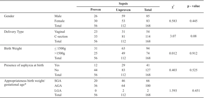
As for the clinical characteristics, the groups differed in terms of the NI's age at the moment the sepsis was diagnosed and the total hospitalization time, which were higher in the group of NI with PBSI. No significant differ-ences were found for the other characteristics (Tables 1 and 2).
Table 1 - Comparison between clinical characteristics of infants under analysis - categorical variables - Niterói - 2008
Sepsis
χ2 p - value
Proven Unproven Total
Male 26 59 85
Female 30 53 83 0.583 0.445
Gender
Total 56 112 168
Vaginal 23 31 54
C-section 33 81 114 3.07 0.08
Delivery Type
Total 56 112 168
< 1500g 31 63 94
>1500g 25 49 74 0.012 0.912
Birth Weight
Total 56 112 168
Yes 12 29 41
No 44 83 127 0.403 0.525
Presence of asphyxia at birth
Total 56 112 168
SGA 20 46 66
AGA 36 64 100
LGA 0 2 2
Appropriateness birth weight/ gestational age*
Total 56 112 168
1.593 0.451
Obs.: *Weight for gestational age curve proposed by Alexander GR et al.. A United States national reference for fetal growth. Obstet Gynecol. 1996;87(2):163-8.
Table 2 - Comparison between clinical characteristics of infants under analysis (56 infants with proven sepsis and 112 with unproven sepsis) - continuous variables - Niterói - 2008
Sepsis Mean SD Median Vm - VM p - value
Birth weight (grams) Proven 1612.86 821.31 1442.5 530-3505
Unproven 1614.73 851.06 1335.0 530-4430 0.989
Gestational age (weeks) Proven 31.95 3.85 32.0 26-41
Unproven 32.24 4.27 32.0 24-42 0.664
Age of infant at sepsis (day) Proven 30.93 41.10 13.5 3-213
Unproven 18.15 25.93 11.5 3-150 0.015
Age at the start of enteral diet (day)* Proven 9.98 19.51 3.0 0-70
Unproven 5.95 10.32 3.0 0-70 0.921
Days to reach Total Oral Diet Proven 14.09 10.61 11.0 0-42
Unproven 12.34 9.48 11.0 0-47 0.260
Hospitalization time (day) Proven 62.70 60.90 37.5 6-225
Unproven 44.14 42.82 30.0 3-225 0.023
Obs.: PBSI: Primary bloodstream infection; N: quantity; SD: Standard deviation; Vm: minimum value; VM: maximum value; *Kruskal-Wallis non-parametric statistical comparison method - t-test for others; (+)/(-) Positive blood culture /Negative blood culture, respectively.
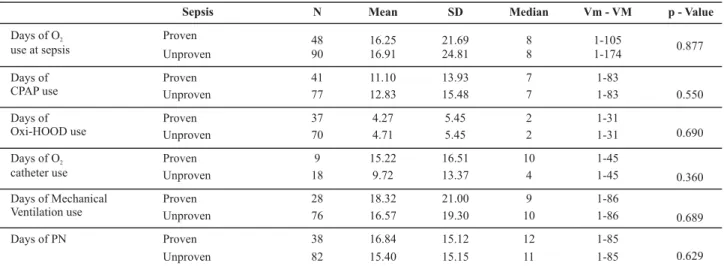
As for the presence and duration of invasive support, no significant differences between the groups appeared (Table 3). No difference was found either for usage time of peripherally inserted central catheter (PICC) (N=37 - mean
Table 3 - Comparison between usage time of invasive support in infants who used this support in the study groups - Niterói - 2008
Sepsis N Mean SD Median Vm - VM p - Value
Proven 48 16.25 21.69 8 1-105
Days of O2
use at sepsis Unproven 90 16.91 24.81 8 1-174 0.877
Proven 41 11.10 13.93 7 1-83
Days of
CPAP use Unproven 77 12.83 15.48 7 1-83 0.550
Proven 37 4.27 5.45 2 1-31
Days of
Oxi-HOOD use Unproven 70 4.71 5.45 2 1-31 0.690
Proven 9 15.22 16.51 10 1-45
Days of O catheter use
2
Unproven 18 9.72 13.37 4 1-45 0.360
Proven 28 18.32 21.00 9 1-86
Days of Mechanical
Ventilation use Unproven 76 16.57 19.30 10 1-86 0.689
Proven 38 16.84 15.12 12 1-85
Days of PN
Unproven 82 15.40 15.15 11 1-85 0.629
Obs.: PBSI: Primary bloodstream infection; N: quantity; SD: Standard deviation; Vm: minimum value; VM: maximum value; (+)/(-) Positive blood culture / Negative blood culture, respectively. PN: parenteral nutrition; O2: oxygen; CPAP: continuous positive airway pressure; Oxi- HOOD: oxygen therapy hood.
None of the clinical manifestations showed differences between the study groups, as shown in Table 4.
Table 4 - Comparison between presence of clinical alterations in study groups -Niterói - 2008
Blood culture result
χ2
p - value
Positive Negative Total
Yes 28 49 77
No 28 63 91
Respiratory Problems
Total 56 112 168
0.587 0.443
Yes 9 30 39
No 47 82 129
Total 56 112 168
Intestinal Tract Manifestations
2.404 0.121
Yes 24 65 89
No 32 47 79
Neurological Manifestations
Total 56 112 168
3.453 0.063
Hypothermia 4 8 12
Normothermia 36 83 119
Hyperthermia 16 21 37
Temperature
Total 56 112 168
2.144 0.342
Yes 1 5 6
No 55 107 162
Total 56 112 168
Coagulation manifestations
0.778 0.378
Yes 15 32 47
No 41 80 121
Cardio-Respiratory manifestations
Total 56 112 168
0.059 0.808
Yes 4 7 11
No 52 105 157
Metabolic Alterations
Total 56 112 168
0.049 0.825
As for the laboratory tests, differences were found in the total quantity of neutrophils and the quantity of
Table 5 - Comparison between hematologic parameters in study groups - Niterói - 2008
Sepsis N Mean SD Median Vm - VM p- value
Proven 56 13,814 9,069 11200.00 1,600-56,600
White cells
(cells/mm3) Unproven 112 13,580 9,446 11000.00 2,300-70,000 0.831
Proven 56 61 15 61 27-90
Total
Neutrophils(%) Unproven 112 54 15 52 16-92 0.004
Proven 56 12 11 10 0-55
Immature
Neutrophils* (%) Unproven 112 8 7 7 0-41 0.010
Proven 56 0.20 0.17 0.19 0-0.9
Ratio of Immature on
Total Neutrophils Unproven 112 0.15 0.13 0.13 0-0.63 0.350
Proven 56 192,898 153,647 148,000 1,000-590,000
Platelets
(units/mm3) Unproven 112 240,042 158,695 223,000 890-913,000 0.700
Proven 23 5,50 4,89 3,7 0,04-17,01
Serum value of C-reactive protein
(mg%)** Unproven 35 1,17 2,18 1,0 0,02-10,16 0.000
Obs: PBSI: Primary bloodstream infection; N: quantity; SD: Standard deviation; Vm: minimum value; VM: maximum value; *Kruskal-Wallis non-parametric statistical comparison method - t-test for others. ** Values related to 2007 and 2008.
A significant difference was found for C-reactive protein (PC-r) levels between the study groups. These data, how-ever, only refer to NI hospitalized between 2007 and 2008 (N=65), when this test was routinely collected (Table 5).
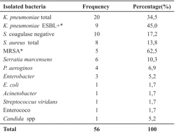
The most commonly isolated bacteria were Klebsiella pneumoniae, Staphylococcus coagulase-negative and S. aureus. Out of the 50 germs isolated in the cultured blood samples, 14 bacteria were multi-resistant, nine of which were Klebsiella pneumoniae extended-spectrum beta-lactamases (ESLB) positive and five S. aureus methicillin-resistant (MRSA) (Table 6).
Table 6 - Frequency of isolated bacteria in blood cultures of infants with proven sepsis - Niterói - 2008
Isolated bacteria Frequency Percentage(%)
K. pneumoniaetotal 20 34,5
K. pneumoniae ESBL+* 9 45,0
S.coagulase negative 10 17,2
S. aureus total 8 13,8
MRSA* 5 62,5
Serratia marcensens 6 10,3
P. aeroginos 4 6,9
Enterobacter 3 5,2
E. coli 1 1,7
Acinetobacter 1 1,7
Streptococcus viridans 1 1,7
Enterococo 1 1,7
Candida spp 1 5,2
Total 56 100
* Multiresistant bacteria; ESBL:xxx ; MRSA:xxx
The clinical characteristics observed at the start of late-onset sepsis (24 hours before and after the start of the con-dition) in the NI under analysis did not show precision to distinguish between the two study groups, except for age at the moment of sepsis and total hospitalization time, which were both higher in infants with proven sepsis.
The NI's higher age at the moment they presented proven sepsis reflects greater exposure to risk factors and invasive procedures occurred during the hospitalization period, an important fact to increase the risk of proven sep-sis. On the other hand, the fact of presenting nosocomial sepsis generates an often prolonged stay and, thus, in-creased hospitalization time. The duration of hospitaliza-tion is significantly longer in children who develop late-onset sepsis in comparison with those who do not develop sepsis(1). The time of hospital stay and use of invasive
de-vices make the NI more vulnerable to hospital sepsis(8). In
this study, it was verified that longer hospitalization is re-lated with higher incidence of proven sepsis.
It is important to highlight that no difference occurred in the use of invasive devices between the two groups (PICC, vesical catheter and umbilical catheter). As reported in lit-erature(9-10), the prolonged use of invasive support devices
put the NI at risk of systemic and local infectious complica-tions by potentially pathogenic germs like Staphylococcus coagulase negative spp, Enterococcus, Staphylococcus aureus, Enterobacter spp, Candida albicans, Pseudomonas areuginosa and Klebsiella pneumoniae.
The presence of certain clinical manifestations did not distinguish between the NI in both study groups. Clinical alterations are described as predictive signs with low diag-nostic value for sepsis, needing other associated diagnos-tic proof to confirm the condition(11). Even if the clinical
al-terations associated with neonatal sepsis are not specific, but can be attributed to another problems or diseases than neonatal sepsis, authors(12) report that, besides laboratory
alterations, the patient's clinical situation should be valued,
DISCUSSION
as the risk of bacterial infections in asymptomatic infants is very low.
Negative blood culture results do not imply the inexis-tence of bacterial sepsis in NI, as the sensitivity of blood cul-ture is low(13), which justifies the difficulty to isolate the germ
even when present in the bloodstream. The number of po-sitive blood culture results varies, ranging between 33 and 53% in neonatal sepsis cases(4). In this study, it
correspon-ded to 33.3% among cases treated as late-onset sepsis. The total quantities of neutrophils and immature neutro-phils were higher in the NI group with proven sepsis. A study(14)
involving patients with late-onset sepsis showed that increased quantities of total neutrophils showed low sensitivity (65%) and low positive predictive value (18%) for the sepsis diagno-sis, but an excellent negative predictive value (98%).
Thus, normal values could exclude the occurrence of sepsis and calculating the relation between the absolute number of total and immature leukocytes can help to diag-nose neonatal sepsis, showing the best sensitivity among all hematological parameters(15).
This study did not intend to calculate sensitivity, specific-ity, positive and negative predictive values of the clinical, labo-ratory and care parameters under analysis, as no proven non-infected group was included for the sake of comparison.
In this study, the PC-r showed to be an excellent labora-tory method to distinguish between the NI in both study groups (Table 5). Levels were higher for NI with proven sep-sis, suggesting that this dosage can support the initial diag-nosis of proven neonatal sepsis. Nowadays, in function of its high negative predictive value, PC-r dosage is used, when negative, to discard the sepsis diagnosis, and also guides the treatment time with antibiotics therapy(16).
Bacteria are the main responsible for infectious com-plications in neonates. The epidemiological profile of these germs apparently changes at each neonatal ICU. Today, a larger number of cases caused by gram-negative bacteria is observed in late-onset sepsis(8), but the proportion of
late-onset sepsis cases associated with gram-positive bacteria has progressively increased over the last two decades and, today, S. aureus, S. coagulase negative and Enterococos are responsible for 30 to 50% of cases(8). In this study, these
bacteria represented 34% of all isolated germs.
As for the bacteria isolated in blood samples of NI with late-onset sepsis, the most important bacteria were K. pneumoniae, S. coagulase negativo, S. aureus, in line with current medical literature(5).
The main infectious agent of late-onset infections is Klebsiela(17). In this study, these bacteria were responsible
for 34.5% of sepsis cases. The K. pneumoniae that produce positive ESBL are increasingly common in hospital sepsis in neonates and are associated with increased morbidity due to the difficult and prolonged treatment(18). In this research,
45% of isolated K. pneumoniae were ESBL positive. Another germ constantly related with the severity of late-onset nosocomial infection and increased morbidity is methicillin-resistant S. aureus (MRSA)(8,1 ). This germ
repre-sented 62.5% of S. aureus isolated in blood cultures in this research. In a study(19) of NI with proven late-onset sepsis
caused by S. aureus, only 8% were due to MRSA.
CONCLUSION
Clinical manifestations were insufficient to distinguish be-tween NI with proven sepsis and NI with unproven sepsis. Alert-ness to these manifestations can be very important though, which can often represent early signs of proven late-onset sep-sis. Neonatology Nurses need to rapidly identify these suspi-cious signs, besides other factors, supporting a fast diagnosis. Care characteristics have shown that age at the moment the sepsis is diagnosed and total hospitalization time can contribute to an early suspicion of sepsis, as they distin-guished between both study groups.
In the laboratory tests, the PC-r serum level found was different in the two groups. Medical literature, however, only indicates its use to discard sepsis cases in case of nega-tive results, due to its high neganega-tive predicnega-tive value.
Total and immature neutrophils efficiently distinguished the study groups and, hence, can help to diagnose sepsis. They should not be analyzed separately though, due to their low positive and negative predictive value.
The search for more efficient methods to identify proven sepsis should be a constant focus in research, as this disease is one of the main causes responsible for infant mortality at neo-natal ICUs and, consequently, for high social and financial costs.
REFERENCES
1. Stoll BJ, Hansen N, Fanaroff AA, Wright L, Carlo WA, Ehrenkranz RA, et al. Late-onset sepsis in very low birth weight neonates: the experience of the NICHD Neonatal Research Network. Pediatrics. 2002;110(2 Pt 1):285-91.
2. World Health Organization (WHO). Young Infants Study Group. Bacterial etiology of serious infections in young infants in devel-oping countries. Pediatr Infect Dis J. 1999;18(10 Suppl):S17-22.
3. Chiesa C, Panero A, Osborn JF, Simonetti AF, Pacifico L. Diagnosis of neonatal sepsis: a clinical and laboratory challenge. Clin Chem. 2004;50(2):279-87.
4. Hudome SM, Fisher MC. Nosocomial infections in the Neona-tal Intensive Care Unit. Curr Opin Infect Dis. 2001;14(3):303-7.
6. Brasil. Ministério da Saúde. Agencia Nacional de Vigilância Sa-nitária. Neonatologia: Critérios Nacionais de Infecção Relacio-nadas à Assistência à Saúde. Brasília; 2008.
7. Bone RC. The patoghenesis of sepsis. Am Intern Med. 1991;115(6):457-69.
8. Sundaram V, Kumar P, Dutta S, Mukhopadhyay K, Ray P, Gautam V, et al. Blood culture confirmed bacterial sepsis in neonates in a North Indian tertiary care center: changes over the last decade. Jpn J Infect. Dis. 2009;62(1):46-50.
9. Perlman SE, Saiman L, Larson EL. Risk factors for late-onset health care-associated bloodstream infections in patients in neonatal intensive care units. Am J Infect Control. 2007;35(3):177-82.
10. Ottolini MC, Lundgren K, Mirkinson LJ, Cason S, Ottolini MG. Utility of complete blood count and blood culture screening to diagnose neonatal sepsis in the asymptomatic at risk newborn. Pediatr Infect Dis J. 2003;22(5):430-4.
11. Weber MW, Carlin JB, Gatchalian S, Lehmann D, Muhe L, Mulholland EK, et al. Predictors of neonatal sepsis in developing countries. Pediatr Infect Dis J. 2003;22(8):711-7.
12. Escobar GJ, Li DK, Armstrong MA, Gardner MN, Folck BF, Verdi JE, et al. Neonatal sepsis workups in infants >/=2000 grams at birth: a population-based study. Pediatrics. 2000;106(2 Pt 1):256-63.
13. Gerdes JS. Clinicopathologic approach to the diagnosis of neonatal sepsis. Clin Perinatol. 1991;18(2):361-81.
14. Berger C, Uehlinger J, Ghelfi D, Blau N, Fanconi S. Comparison of C-reactive protein and white blood cell count with differential in neonates at risk for septicaemia. Eur J Pediatr. 1995;154(2):138-44.
15. Manroe BL, Weinberg AG, Rosenfeld CR, Browne R. The neo-natal blood count in health and disease. I. Reference values for neutrophilic cells. J Pediatr. 1979;95(1):89-98.
16. Rezende Junior DC, Moraes JMMF, Lucca MG, Orrico SRP, Spegiorin MA, Christiano Junior AC, et al. O rápido declínio da concentração sérica de proteína C-Reativa na fase inicial da sepse é preditivo de boa evolução. Rev Bras Terapia Inten-siva. 2005;17(2):104-7.
17. Gaynes RP, Edwards JR, Jarvis WR, Culver DH, Tolson JS, Martone WJ. Nosocomial infections among neonates in high-risk nurseries in the United States. Pediatrics. 1996;98(3 Pt 1):357-61.
18. Sirot D. Extended-spectrum plasmid-mediated beta-lactamases. J Antimicrob Chemother. 1995;36 Suppl A:19-34.