Major Article
Corresponding author: Dra Sara M. Robledo.
e-mail: [email protected]
Received 31 October 2016
Accepted 17 January 2017
In vitro and in vivo antileishmanial activity of
Artemisia annua L. leaf powder and its potential
usefulness in the treatment of uncomplicated
cutaneous leishmaniasis in humans
Luz Estella Mesa
[1], Daniel Vasquez
[1], Pierre Lutgen
[2],
Iván Darío Vélez
[1],
Adriana María Restrepo
[1], Isabel Ortiz
[3]and Sara María Robledo
[1][1]. Programa de Estudio y Control de Enfermedades Tropicales-PECET, Instituto de Investigaciones Médicas, Facultad de Medicina, Universidad de Antioquia, Medellín, Colombia. [2]. Iwerliewen Fir Bedreete Volleker-IFBV- Réseau belgo-luxembourgeois de valorisation des herbes médicinales-BELHERB, Niederanven,
Luxembourg. [3]. Grupo de Investigación Biología de Sistemas, Universidad Pontiicia Bolivariana. Medellín, Colombia.
Abstract
Introduction: Cutaneous leishmaniasis (CL) is a tropical disease that affects millions of individuals worldwide. The current drugs for CL may be effective but have serious side effects; hence, alternatives are urgently needed. Although plant-derived materials are used for the treatment of various diseases in 80% of the global population, the validation of these products is essential. Gelatin capsules containing dried Artemisia annua L. leaf powder were recently developed as a new herbal formulation (totum) for the oral treatment of malaria and other parasitic diseases. Here, we aimed to determine the usefulness of A. annua gel capsules in CL. Methods: The antileishmanial activity and cytotoxicity of A. annua L. capsules was determined via in vitro and in vivo
studies. Moreover, a preliminary evaluation of its therapeutic potential as antileishmanial treatment in humans was conducted in 2 patients with uncomplicated CL. Results:Artemisia annua L. capsules showed moderate in vitro activity in amastigotes of
Leishmania (Viannia) panamensis; no cytotoxicity in U-937 macrophages or genotoxicity in human lymphocytes was observed. Five of 6 (83.3%) hamsters treated with A. annua capsules (500mg/kg/day) for 30 days were cured, and the 2 examined patients were cured 45 days after initiation of treatment with 30g of A. annua L. capsules, without any adverse reactions. Both patients remained disease-free 26 and 24 months after treatment completion. Conclusion: Capsules of A. annua L. represent an effective
treatment for uncomplicated CL, although further randomized controlled trials are needed to validate its eficacy and safety.
Keywords: Cutaneous leishmaniasis. Artemisia annua. Leishmania panamensis. Anti-leishmanial activity. Therapeutic response.
INTRODUCTION
Leishmaniasis is a tropical disease that is caused by >20 species of Leishmania, an intracellular protozoa transmitted by
sandlies of the Phlebotomus and Lutzomyia spp. The disease is clinically manifested as cutaneous leishmaniasis (CL), mucosal leishmaniasis (ML), and visceral leishmaniasis (VL); these clinical forms differ in terms of immunopathology and the degree of morbidity and mortality1. The disease is endemic in 98 countries in
5 continents, primarily including poorly developed or developing nations. The World Health Organization (WHO) estimates that approximately 12 million individuals are affected and 350 million
people are at risk ο infection. CL is the most prevalent clinical
manifestation with 0.7-1.2 million new cases per year, whereas
VL affects 0.2-0.4 million individuals; however, the number of such cases in endemic areas is known to be underreported2. The
standard treatment for CL includes a pentavalent antimonial [meglumine antimoniate (MA) or sodium stibogluconate], miltefosine, pentamidine isethionate, or amphotericin B. Although these drugs are still effective, they have serious side effects3;
hence, there is an urgent need for research and development to produce safer and effective, but also inexpensive drugs. Plants represent a natural source of medications to treat human diseases, and thousands of potent biological metabolites
have been identiied thus far from plants. Moreover, several
such metabolites have been used to discover and develop drugs or have been used as scaffolds for designing better pharmacologically active compounds4. Approximately 80% of
the global population use phytomedicine for treating infectious and non-infectious diseases; hence, herbal medicine has received
marked scientiic interest, particularly for the validation of the
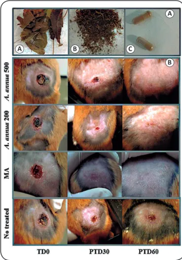
A
B C
A
B
FIGURE 1 - Treatment with Artemisia annua L. leaf powder and the evolution of lesions in hamsters. In panel (A),the image shows the process of obtaining capsules from the leaves of Artemisia annua: the leaves and thin stems are selected from the whole plant (a), are pulverized (b), and are then powdered and packaged in gelatin capsules (c). In panel (B), the image shows the clinical appearance of the lesion in a representative hamster before (pre-treatment) and 30 and 60 days after treatment (PTD30 and PTD60, respectively) with 500 or 200mg/kg/day (oral) of Artemisia annua L. leaf powder or meglumine antimoniate (intramuscular injection). MA: meglumine antimoniate;
TD0: treatment day 0; PTD30: post-treatment day 30; PTD60: post-treatment day 60.
recently updated the Traditional Medicine Strategy for 2014-2023 to prioritize traditional and complementary medicine products5.
An overwhelming number of scientiic reports have focused
on compounds from plants with validated, both in vitro and in vivo, antileishmanial activity; however, very few plant products have advanced to the clinical phase of evaluation in humans.
Artemisia annua L. (Asteraceae), commonly named as sweet wormwood or Qing Hao, is one such plant. It is a Chinese medicinal plant traditionally used for the treatment of fever and chills, as well as malaria6,7. The main metabolites present
in this plant, including artemisinin, coumarins, lavonoids,
saponins, and terpenoids, have demonstrated a wide range of pharmacological activities against metabolic diseases such as cancer8 and diabetes9, as well as infectious agents such as
viruses10,11 and Trypanosoma and Leishmania protozoa12-16.
Pulverized entire leaves of A. annua L. (80% leaves and 20% thin branches), known as totum, seem to represent a novel botanical drug for the treatment of infectious diseases, including malaria, with good results and without side effects17.
The phytochemical analysis of this A. annua L. powder indicated the presence of 0.1% artemisinin, 0.1% scopoletin, 0.2% alpha-pinene, 0.5% limonene, 0.6% eucalyptol, 1.6% artemisia ketone, 0.2% camphor, 0.8% caryophyllene, and 0.8% polyphenols (Laboratoire National de Sante, 3583 Dudelange, Luxemburg). Moreover, an unexpectedly high level of transfer of artemisinin from the plant material into the bloodstream, as compared to the pure drug, had already been observed18.
Considering the potential usefulness of A. annua L. and its botanical products, in the present study, we assessed the
in vitro and in vivo antileishmanial activity of the A. annua L. leaf powder in L. (V) panamensis. Furthermore, the potential usefulness of this herbal product was evaluated in 2 CL patients with a short evolution history. To our knowledge, this is the
irst study to report the in vitro and in vivo activity of A. annua L. in L. (V) panamensis, and the potential usefulness of a herbal product comprising the entire leaves of A. annua L. in the treatment of uncomplicated CL.
METHODS
Artemisia annua L. leaf powder
Artemisia annua L. material was collected in Walferdange-Luxembourg in September 2012, from plants cultivated in
the Colabor Garden. Certiied organic material, including 1
specimen registered as MNHNL17732, was deposited in the Herbarium LUX, at the Musée National d'Histoire Naturelle, Luxembourg. The material was analyzed in the Laboratoire National de la Santé de Luxembourg, which is ISO 17025 certified. The leaves and thin branches were harvested, dried for 3 days at 35°C, and pulverized to obtain a powder that was dispensed in gelatin capsules (Figure 1A). All the processes were conducted using Good Manufacturing Practice
(Phytosanitary Certiicate # EC/LU/11773) at the semi-industrial
facilities of Téi vum Séi in Winseler-Luxembourg.
For the in vitro assays, the powder of 3 capsules was solubilized in a phosphate-buffered saline (PBS) solution
with 0.05% dimethyl sulfoxide (DMSO; Sigma); the concentration was adjusted to 200, 100, 50, 25, 12.5, and 6.25µL/mL in a complete medium containing Roswell Park Memorial Institute (RPMI-1640) medium (Sigma-Aldrich, St Louis MO, USA) supplemented with 10% fetal bovine serum (FBS; Gibco, Life technologies Gaithersburg MD, USA) and a 1% penicillin (10,000 units/mL)-streptomycin (10mg/mL) solution (Sigma) to determine cytotoxicity or at 100, 25, 6.25, and 1.56µL/mL to determine antileishmanial activity.
For the in vivo assays, the powder of 10 capsules was solubilized in ultrapure water with 0.05% DMSO via sonication and homogenization.
Cell line
incubated at 37°C under 5% CO2. The medium was changed every 3 days until use.
Parasites
Leishmania (Viannia) panamensis transfected with the green
luorescent protein (GFP), MHOM/CO/87/UA140-pIR-eGFP,
were maintained as promastigotes via culture in a biphasic Novy-MacNeal-Nicolle (NNN) medium, and incubated at 26°C.
In vitro cytotoxicity assay
The cytotoxicity of the A. annua L. leaf powder was determined in mammalian U-937 cells based on its effect on cell growth, as determined by the [3-(4,5-Dimethylthiazol-2-yl)-2,5-Diphenyltetrazolium Bromide]-MTT microenzymatic assay described previously19.
Genotoxicity test
The effect of A. annua L. powder on chromosome alteration was determined in vitro using the Organisation for Economic Co-operation and Development (OECD) guideline No. 473 chromosome aberration test20.
In vitro antileishmanial activity of Artemisia annua capsules
The antileishmanial activity was tested in intracellular amastigotes obtained after the infection of U937 cells with promastigotes of L. (V) panamensis UA140-pIR-eGFP, as described previously19.
Leishmanicidal response in hamsters treated with Artemisia annua L. leafpowder
The therapeutic response to A. annua L. leaf powder was evaluated in hamsters (Mesocricetus auratus) experimentally infected with L. (V) panamensis, as described previously21. Five
experimental groups (n = 6 each) consisting of males (n = 3) and females (n = 3), were established. Two groups were treated with 100µL A. annua L. leaf powder at 500 and 200mg/kg/day, which was administered orally, once daily, for 30 days. The third group was treated with 100µL MA at 120mg/kg/day via an intramuscular injection, once daily, for 10 days. The fourth
and ifth groups corresponded to non-infected hamsters and
hamsters that were infected but not treated.
Treatments were initiated immediately after the development of a typical ulcer. The ulcer areas and body weight were measured at 2-week intervals from the start of treatments to the end of the study which occurred 2 months after completion of treatment. The overall time points of evaluation were as follows: pre-treatment day (TD0), end of treatment (TD14), and post-treatment days (PTD) 30 and 60.
At PTD60 (end of the study), hamsters were humanely
sacriiced, and after necropsy, liver and kidney biopsies were
conducted for histopathological studies, and sample of the ulcer was also used to determine the number of living L. (V) panamensis parasites through the ampliication of a single copy
gene of Leishmania using quantitative reverse transcription polymerase chain reaction (qRT-PCR)22.
The effectiveness of each treatment was assessed by comparing the lesion sizes before and after treatments. The treatment outcome at the end of study was recorded as cured (healing of 100% of the ulcer area and complete disappearance of the lesion), improvement (reduction in the size of the lesion area by >30%), no response (increase in the size of the lesion area), or relapse (reactivation of the lesion after an initial cure). An arbitrary score of 3: cure, 2: improvement, 1: relapse, and 0: no response was assigned to each outcome in order to compare the effectiveness among the groups of treatments.
The toxicity of treatments was evaluated by comparing the blood levels of alanine amino transferase (ALT), blood urea nitrogen (BUN), and creatinine using commercially available kits (Biosystems, Spain), as described previously23. Blood
samples were drawn from the heart on TD0 and TD8, and serum was stored at -80°C until use.
Case report of two patients with uncomplicated cutaneous leishmaniasistreated with Artemisia annua
L. leaf powder
The irst patient (P01) was a 35-year-old man who presented
in April 2014 with 4 ulcers with a 1-month evolution history, localized in the right (1 ulcer) and left (3 ulcers) legs, without any signs of over-infection. The sizes of the lesions (including ulcer plus induration) were 30mm × 28mm, 10mm × 9mm, 45mm × 45mm, and 10mm × 10mm. The second patient (P02) was a 28-year-old man who presented in September 2014 with
1 ulcer on the right lank of the body (50mm × 40mm, including
ulcer plus induration) with a 6-week evolution history, without any apparent over-infection. In both patients, Giemsa-stained smears and culture aspirates in NNN medium indicated positive results for leishmaniasis. L. (V) panamensis was identiied by PCR-RFLP (restriction fragment length polymorphism) using the methodology described previously24.
Both patients had not history of treatment with antileishmanial drugs. As both patients were in good clinical and physical condition, they were recommended to undergo treatment with
A. annua L. leaf powder. The patients signed the informed consent form and volunteered to receive the treatment. The patients were then treated with capsules containing A. annua L. leaf powder (total of 30g over 20 days) as follows: 3g (3 capsules) from treatment day (TD) one (TD1) to TD3; 2g (2 capsules) from TD4 to TD7, and 1g (1 capsule) from TD8 to TD20.
Statistical analysis
Cytotoxicity was determined based on the percentages of viability and cell growth inhibition, obtained for A. annua L. leaf powder, amphotericin B, or the cell culture medium using Eq.1:
% viability = (OD of treated cells)/(OD of control cells) × 100(1)
where the OD of control cells corresponded to 100% viability.
Thereafter, the percentage of cell growth inhibition was estimated using Eq.2:
The results are expressed as lethal concentration 50 (LC50) and 90 (LC90), which correspond to the concentration of
A. annua L. leaf powder that yielded 50% and 90% inhibition of cell growth. The LC50 and LC90 values were calculated using the Probit method25, and the degree of cytotoxicity of A. annua
L. leaf powder was graded as follows–LC50 <100μg/mL: high cytotoxicity, LC50 100-200μg/mL: moderate cytotoxicity, and LC50 >200μg/mL: potential non-cytotoxicity.
The in vitro antileishmanial activity of A. annua L. leaf powder in intracellular amastigotes was determined according to the percentage of infected cells corresponding to the number
of positive events based on green luorescence imaging and forward scatter by dotplot analysis, and the median luorescence
intensity by histograms. Both parameters were used to calculate the percentage of infection for each concentration using Eq.3:
% infection = (% infected,treated cells ÷% infected,untreated cells) × 100(3)
Thereafter, the percentage of infection inhibition was calculated using equation 4:
% Inhibition = 100 - % infection(4)
The results of activity are expressed as effective concentration 50 (EC50) and effective concentration 90 (EC90), which correspond to the concentration of A. annua L. leaf powder that yielded 50% and 90% of inhibition of the parasitic infection, respectively. The EC50 and EC90 were determined using Probit analysis25. The degree of antileishmanial activity was established
based on the EC50 values, as follows–EC50 <25μg/mL: antileishmanial activity, EC50 25−50μg/mL: moderate activity, and EC50 >50μg/mL: non-antileishmanial activity. Moreover, the Index of Selectivity (IS) was calculated by dividing the cytotoxicity and the antileishmanial activity using Eq.5:
SI = CL50/CE50(5).
For statistical analysis of in vivo data, a one-way analysis of variance and Kruskal-Wallis test, followed by the post-contrast Dunn’s Multiple Comparison Test, was performed using GraphPad Prism 6 software (GraphPad Software, Inc., San Diego, CA, USA). A p value of <0.05 was considered to
be statistically signiicant.
Ethical considerations
The Institutional Ethics Committee for Human Research of CES University approved the protocol in human patients (Act No. 66-2014), and informed consent was obtained from all the participants in the study. This work was also approved by the Ethics Committee for Animal Research of the University of Antioquia (Act No. 72-2011 and 91-2014).
RESULTS
In vitro toxicity of Artemisia annua L. leaf powder
The in vitro cytotoxic effect of this herbal product was assessed in human macrophages (U-937), and we found that
A. annua L. leaf powder was non-cytotoxic to U-937, as evidenced by an LC50 of 419.1µg/mL. In contrast, amphotericin B was cytotoxic to U-937 cells (LC50, 30µg/mL).
In the chromosome aberration test, human lymphocytes
treated with A. annua L. leaf powder did not exhibit
chromosomal aberrations at any of the concentrations tested, compared to that noted with the negative control (Table 1). No
statistically signiicant differences between the negative control
and A. annua L. leaf powder-treated groups were observed (p = 0.527).
In vitro antileishmanial activity of Artemisia annua L. leaf powder
The in vitro antileishmanial activity of A. annua L. leaf powder was assessed in U-937 macrophages that were infected with amastigotes of L. (V) panamensis. The product showed moderate leishmanicidal activity on the intracellular amastigotes of L. (V) panamensis, with EC50 and EC90 values of 48.07μg/
mL and 82.2μg/mL, respectively. As expected, amphotericin B
B BB MR DC/R
Compound µg/mL n % n % n % n %
C- 0 2 0.8 0 0.0 0 0.0 0 0.0
Artemisia annua 25 4 2.7 0 0.0 1 0.7 0 0.0
50 0 0.0 2 0.8 3 1.2 0 0.0
100 6 2.4 4 1.6 0 0.0 0 0.0
TABLE 1
Percentage of chromosome breaks observed in human lymphocytes exposed to various compounds with leishmanicidal potential for 24 hours.
B: chromatid breaks; BB: chromosome break; DC: dicentric chromosomes; R: ring chromosomes, MR: multiradial chromosomes;
was highly active against the intracellular amastigotes of L. (V) panamensis, with EC50 and EC90 values of 0.06μg/mL and 0.1μg/ mL, respectively. Moreover, the biological activity of A. annua
L. leaf powder was more selective for parasites than for cells, with an IS of 8.73; in contrast, the IS of amphotericin B was 625.
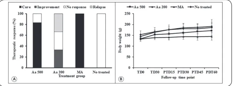
Therapeutic response and toxicity of Artemisia annua L. leaf powder in hamsters with experimental cutaneous leishmaniasis
The effectiveness of A. annua L. leaf powder was determined in hamsters based on the evolution of the lesions after treatment, in comparison with treatment with MA or no treatment, wherein
cure was deined as the resolution of the infection with complete
skin re-epithelialization (Figure 1B). The treatment of hamsters with CL caused by L. (V) panamensis with A. annua L. leaf powder at 500 mg/kg/day for 30 days resulted in cure in 5 up 6 hamsters (3 males and 2 females; 83.3%) at the end of the study period (PTD60; Figure 2A). Notably, cure was observed in some hamsters early after treatment, and the number of those cured increased over time. Thus, at the end of the treatment period, 2 up 6 hamsters were cured, whereas at PT30, 4 up 6 hamsters (66.6%) were cured. The parasitic load in the hamster that was not cured was 188,47 parasites per mg of tissue. In contrast, treatment with A. annua L. leaf powder, at a dose of 200 mg/ kg/day for 30 days, led to cure in 2 up 6 hamsters (1 male and 1 female) at the end of the treatment period; however, at PTD30, both these animals experienced a relapse. At the end of the study (PTD60), improvement in lesion size, treatment failure, and relapse were each observed in 33.3% cases (Figure 2A). The parasitic load in this group of hamsters at the end of the study was 272.15 ± 164.4 parasites/mg tissue. MA treatment
A B
FIGURE 2 - In vivo therapeutic response and effect on body weight in hamsters with cutaneous leishmaniasis treated with Artemisia annua L. leaf powder. The treatment outcome was evaluated at the end of treatment period (PTD60) in hamsters treated with Artemisia annua L. leaf powder at dose of 500 or 200mg/kg/day (oral) versus meglumine antimoniate (intramuscular injection) versus no treatment. In (A), the bars represent the percentage of animals showing
cure, improvement, failure, or relapse indings after 60 days of treatment. The differences between the groups were not signiicant (p = 0.3679). In (B), the lines represent the evolution of body weight in grams in hamsters orally treated with 500 or 200mg/kg/day of Artemisia annua L. leaf powder, in those treated
intramuscularly with meglumine antimoniate (MA) at 120mg/kg/day, and in those without any treatment. The differences between the groups were not signiicant
(p > 0.05). Aa 500: A. annua 500mg/kg/d; Aa 200: A. annua 200mg/kg/d; MA: meglumine antimoniate; TD0: treatment day 0; TD30: treatment day 30; PTD15:
post-treatment day 15; PTD30: post-treatment day 30; PTD45: post-treatment day 45; PTD60: post-treatment day 60.
was found to have cured all the animals of this group at the end of the study, and no live parasites were detected in any of these animals. In contrast, none of the hamsters in the non-treated group were cured during the study period (Figure 2A). Three hamsters
showed a reduction in their lesion size during the irst month after
infection, although they experienced a relapse subsequently; thus, all the hamsters had larger lesions at the end of the study period as compared to those at baseline. The parasitic load in this group was 541.456.8 ± 490.728.0 parasites/mg of tissue. In the present study, no differences in the response to treatment (either A. annua
or MA) and the sex of the animal were observed.
At the end of the study, the effectiveness of oral treatment with A. annua L. leaf powder, administered at 500mg/kg/day for 30 days, was similar to that observed with the intramuscular injection of MA, in terms of the clinical outcome and parasite load, at the end of the study (p > 0.05). However, the effectiveness of the treatment was different when A. annua L. leaf powder was administered at a dose of 200mg/kg (p < 0.05). None of the animals in the treatment groups experienced weight loss (Figure 2B). In contrast, weight gain was observed in all the hamsters during the study, which suggests that no toxic effect was produced in hamsters due to treatment with A. annua L. leaf powder.
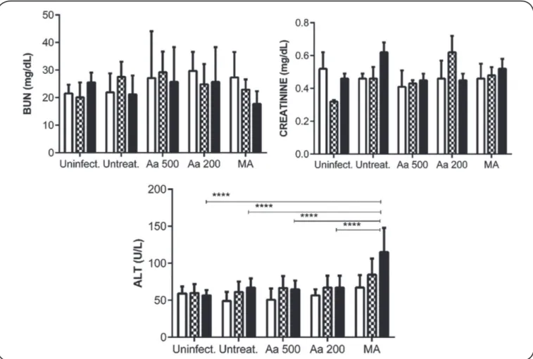
signiicant differences (p = 0.9988; Figure 3). In contrast, the treatment with MA increased the ALT levels at TD8 and PTD45, in comparison with those observed in the other treatment groups;
this difference was signiicant (p < 0.0001).
Histopathological analyses of the kidney indicated mild vacuolar degeneration, atrophy, hyperplasia, protein cylinders, and congestion in 1–2 animals in each treatment group, including the non-treated controls. Similarly, cloudy and vacuolar degeneration, karyomegaly, binucleation, and congestion were observed in the liver of few animals in each treatment group. As the histological changes were also observed in the non-treated animals, these alterations can be considered to be associated with the parasitic infection rather than the treatment.
Response to Artemisia annua L. leaf powder in two patients with uncomplicated cutaneous leishmaniasis treated with
Patients treated with a total of 30g of A. annua L. leaf powder for 20 days responded very well. At the end of treatment (TD21), the ulcers decreased in size by approximately 20-35%,
FIGURE 3 -Serum levels of BUN, creatinine, and ALT in hamsters before and after treatment with Artemisia annua L. leaf powder. Golden hamsters (n = 6 per group) were orally treated for 30 days with A. annua L. leaf powder at 500mg/kg/day or 200mg/kg/day. A third group of hamsters was intramuscularly treated for 10 days with meglumine antimoniate at 120mg/kg/day. Two additional groups of hamsters were included as controls: uninfected hamsters and hamsters that were infected but not treated. The bars represent the mean value of the corresponding metabolite detected before (white bar), on treatment day 8 (black and
white squares bar), and on day 45 after treatment (black bar). The differences in the serum level of ALT of hamsters treated with MA were signiicant between the
groups (p < 0.0001). Data represent the median values ± SD of 6 animals per treatment group. Aa 500: A. annua 500mg/kg/d; Aa 200: A. annua 200mg/kg/d;
MA: meglumine antimoniate; BUN: blood urea nitrogen;ALT: alanine amino transferase; SD: standard deviation. **** p<0.0001
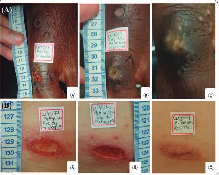
as compared to that before treatment. In both patients, complete cure (100% closure of the ulcer and presence of scar in the lesion site) was observed on day 45 after the end of treatment (PTD45); however, no good resolution images that indicated the cure could be obtained. The patients remained disease-free after 2 years of follow-up. Figure 4A shows the appearance of 2 lesions in patient P01 at TD0 (a), TD20 (b), and 12 months after the end of treatment (c). Similarly, Figure 4B shows the appearance of the ulcer in patient P02 at TD0 (a), TD10 (b), and 12 months after the end of treatment. Both patients remained disease-free at 26 and 24 months after treatment completion. Moreover, both patients did not develop any side-effects or
adverse reactions. In the irst patient, only the amylase level
was mildly elevated before, during, and after treatment (133, 156, and 144U/L on day 0 and 10 of treatment and day 45 post-treatment, respectively). The drug showed no impact on body function. The increased amylase levels prior to treatment did not affect patient management, but appears to be an incidental
FIGURE 4. Lesion evolution in patients after treatment with Artemisia annua L. leaf powder. The Figure shows the evolution of lesions in patient P01 (panel A) and P02 (Panel B at TD0 (before treatment) (a), TD10 (b), and 12 months after treatment completion (c). This patient had 4 ulcers. All the ulcers were cured at day 45 after treatment with a total of 30g of Artemisia annua L. leaf powder; patient P01 and P02 remained disease-free over 26 and 24 months of follow-up, respectively. No relapses were observed during this time. P01: patient 01; P02: patient 02; TD0: treatment day 0; TD10: treatment day 10.
A B C
A B C
DISCUSSION
CL is an infectious disease that can lead to serious psychological and social stigma, particularly in cases where the face and other visible areas are affected. Thousands of plants, have been used as medicines for several human diseases. Over the last decade, the development of drugs from medicinal herbs and phytochemicals from plants has been receiving increased attention through standardized and systematic research. Hence, currently, medicinal herbs and their derivative phytocompounds are being increasingly used as primary or complementary treatments for several diseases, including cancer. In particular, A. annua L. has been used for more than 2000 years in Chinese traditional herbal medicine for the treatment of fever, without any apparent emergence of resistance to artemisinin.
In the present study, the effectiveness and safety proile of A. annua L. leaf powder was found to be highly satisfactory.
A. annua L. leaf powder showed in vitro antileishmanial activity with no evidence of cytotoxicity or genotoxicity. As genotoxicity analyses based on only chromosomal aberration studies and
with low compound concentrations are not suficiently sensitive
to detect genotoxicity and mutagenicity, others tests such as Comet, Ames, and Vitotox tests, which would be more sensitive,
should be performed to identify signiicant dose-dependent
genotoxicity and mutagenicity of A. annua L. Although the IS of amphotericin B was higher than that of A. annua L. leaves powder, a value of 8 is considered important to identify a compound as promising to continue the evaluation phases in drug discovery and development.
levels were not only slightly increased in hamsters treated with
A. annua L. leaf powder and MA, but also in infected and non-treated hamsters. These results suggest that such an increase may probably be associated with the infection process of L. (V) panamensis. In addition, the ability of A. annua L. leaf powder to kill the intracellular amastigotes of L. (V) panamensis present in the ulcer, and cure cutaneous leishmaniasis was demonstrated
in hamsters; nevertheless, the eficacy may be affected by the
dose, since the same product administered at a dose of 200mg/ kg body weight/day for 30 days produced a cure, but at a very low rate and with relapse during the follow-up period.
Various products derived from A. annua L. have been reported to have activity against Leishmania species. For example, the n-hexane extracts of A. annua L. leaves and seeds induce apoptosis in intracellular amastigotes of L. (L) donovani without any cytotoxic effects on mammalian macrophages and the treatment of infected mice with these
n-hexane extracts signiicantly decreased the hepatic and splenic
parasitic load, and reduced spleen weight15. In another study,
the essential oils of A. annua L. leaves exhibited signiicant in
vitro leishmanicidal activity against intracellular amastigotes of
L. (L) donovani, without any cytotoxicity on murine macrophages, and the intra-peritoneal administration (200mg/ kg body weight) to infected BALB/c mice reduced the parasitic
burden by almost 90% in the liver and spleen and signiicantly
reduced the weight of these organs as well. No hepato- or nephrotoxicity was observed in these cases, as evidenced by the normal levels of the serum enzymes16. However, complete
cure of VL with A. annua n-hexane extracts nor essential oils has not been observed in these in vivo studies.
Finally, we assessed the potential usefulness of A. annua L. leaf powder to treat CL with a short evolution history, at a low dose. Rapid evolution of the lesion with complete healing was observed 1.5 months after treatment with A. annua L. leaf powder. Although the spontaneous healing of lesions caused by L. (V) panamensis has been reported in CL, this phenomenon usually occurs after several months of evolution and not during the initial stages.
These results are consistent with previous studies, wherein
the eficacy of the whole plant of A. annua L. was demonstrated. The use of the whole plant was found to be more effective in the treatment of a rodent malaria model, as compared to the use of a comparable dose of pure artemisinin26. Moreover,
the consumption of A. annua L. tea once a week over long periods (2.6 ± 1.4 years) by farm workers in Africa was found
to signiicantly reduce the episodes of malaria, in comparison
with workers who did not drink the tea; no adverse effects of such tea consumption on the health of workers was observed2728.
The content of artemisinin in the capsules containing A. annua L. leaf powder is approximately 0.1%; hence, it can be estimated that 1-3mg of artemisinin was administered daily. As the capsules only contain leaves, the possibility of the sub-therapeutic doses to generate resistance in malaria is low29. The advantage of A. annua L. powder, in comparison
to the tea preparation, is that the powder may supply all the molecules present in the plant, particularly the polysaccharides, coumarins, saponins, phytosterols, essential oils, polyphenols,
and lavonoids30. These molecules were reported to enhance
the action of artemisinin31 and may also have their own speciic
antimalarial action32.
The consumption of tea infusion and dried leaves of the
plant may have prophylactic and therapeutic eficacy, due
to the presence of a complex matrix of chemicals within the leaves, which appears to enhance both the bioavailability
and eficacy of artemisinin. Rodent studiesindicated longer
pharmacokinetic parameters in Plasmodium chabaudi-infected mice treated with A. annua dried leaves as compared to those in healthy mice. In healthy mice, the serum levels of artemisinin were >40-fold greater among those fed with dried leaves as compared to those fed with pure artemisinin. Human trial data also showed that when artemisinin was delivered in the form of dried leaves, 40-fold less artemisinin was required to obtain a therapeutic response, as compared to when pure artemisinin was administered33. However, future studies need to assess whether
an increase in the dose will enhance the eficacy.
In conclusion, to our knowledge, this is the irst study to
describe the therapeutic response of CL patients to A. annua
L. leaf powder, and to report the in vitro and in vivo activity of this compound against L. (V) panamensis. Overall, the bioassay testing results showed that the administration of A. annua L. leaf powder is easy and apparently safer and more effective than consumption as tea or pure drug infusion, and hence, it may serve as an adequate option for the treatment of non-complicated
CL. Although these indings need to be conirmed, we observed
that the therapeutic response to this herbal product might be enhanced by increasing the dose or frequency of administration. The potential effectiveness and safety of A. annua L. leaf powder observed in the present study could serve as fundamental evidence for considering this herb product as an alternative for CL treatment, and presents opportunities for future explorations of herb formulations in the treatment of CL.Moreover, further investigations of the utility of A. annua L. totum in the treatment of
CL is consistent with the strategy deined by the WHO to use and
apply traditional medicine and its products over the next decade.
Conlict of interest
The authors declare no conlict of interest.
Acknowledgements
The authors thank the patients for agreeing to participate in the study.
Financial support
Departamento Administrativo de Ciencia, Tecnología e Innovación-COLCIENCIAS (project number CT-695-2014) and University of Antioquia (Centros de Excelencia).
REFERENCES
2. Alvar J, Velez ID, Bern C, Herrero M, Desjeux P, Cano J. Leishmaniasis Worldwide and Global Estimates of Its Incidence. PloS One. 2012;7(5):e35671.
3. den Boer M, Argaw D, Jannin J, Alvar J. Leishmaniasis impact and treatment access. Clin Microbiol Infect. 2011; 17(10):1471-7.
4. Mishra BB, Tiwari VK. Natural products: an evolving role in future drug discovery. Eur J Med Chem. 2011;46(10):4769-807.
5. World Health Organization WHO traditional medicine strategy: 2014-2023. Geneva, 2014. http://www.who.int/medicines/ publications/traditional/trm_strategy14_23
6. Klayman DL. Qinghaosu (artemisinin): an antimalarial drug from China. Science. 1985;228(4703):1049-55.
7. Hsu E. The history of qing hao in the Chinese materia medica. Trans R Soc Trop Med Hyg. 2006;100(6):505-8.
8. Efferth T, Herrmann F, Tahrani A, Wink M. Cytotoxic activity of secondary metabolites derived from Artemisia annua L. towards cancer cells in comparison to its designed active constituent artemisinin. Phytomed. 2011;18(11):959-69.
9. Helal EGE, Abou-Aouf N, Khattab AM, Zoair MA. Anti-diabetic effect of Artemisia annua (kaysom) in alloxan-induced diabetic rats. Egyp J Hosp Med. 2014;57:422-30. doi:10.12816/0008476.
10. Efferth T, Romero M, Wolf D, Stamminger T, Marin J, Marschall M. The antiviral activities of artemisinin and artesunate. Clin Infect Dis. 2008;47(6):804-11.
11. Lubbe A, Seibert I, Klimkait T, van der Kooy F. Ethnopharmacology in overdrive: The remarkable anti-HIV activity of Artemisia annua. J Ethnopharm. 2012;141(3):854-59.
12. Mishina YV, Krishna S, Haynes RK, Meade JC. Artemisinins inhibit Trypanosoma cruzi and Trypanosoma brucei rhodesiense in vitro growth. Antimicrob Agents Chemother. 2007;51(5):1852-4.
13. Sen R, Bandyopadhyay S, Dutta A, Mandal G, Ganguly S, Saha P, et al. Artemisinin triggers induction of cell-cycle arrest and apoptosis in Leishmania donovani promastigotes. J Med Microbiol. 2007;56(9):1213-8.
14. Yang D, Liew F. Effects of qinghaosu (artemisinin) and its derivatives on experimental cutaneous leishmaniasis. Parasitol. 1993;106(1):7-11
15. Islamuddin M, Farooque A, Dwarakanath BS, Sahal D, Afrin F. Extracts of Artemisia annua leaves and seeds mediate programmed cell death in Leishmania donovani. J Med Microbiol. 2012;61(12):1709-18.
16. Islamuddin M, Chouhan G, Tyagi M, Abdin MZ, Sahal D, Afrin F. Leishmanicidal activities of Artemisia annua leaf essential oil against visceral leishmaniasis. Front Microbiol. 2014;5:626. doi: 10.3389/fmicb.2014.00626.
17. Onimus M, Carteron S, Lutgen P. The Surprising Eficiency of Artemisia annua powder capsules. Med Arom Plants. 2013;2:125. doi:10.4172/2167-0412.1000125.
18. Weathers PJ, Arsenault PR, Covello P, McMickle A, Reed D, Teoh KH. Artemisinin production in Artemisia annua – studies in plant and a novel delivery method for treating malaria and other neglected diseases. Phytochem Rev 2011;10(2):173-83.
19. Mesa CV, Blandón GA, Muñoz DL, Muskus CE, Flórez AF, Ochoa R, et al. In silico screening of potential drug with antileishmanial
activity and validation of their activity by in vitro and in vivo studies. J Chem Chem Eng. 2015;9:375-402. doi:10.17265/1934-7375/2015.06.002.
20. Organisation for Economic Co-operation and Development (OECD). Test No. 473: In Vitro Mammalian Chromosomal Aberration Test, OECD Guidelines for the Testing of Chemicals, Section 4, OECD Publishing, Paris. 2014.
21. Robledo SM, Carrillo LM, Daza A, Restrepo AM, Muñoz DL, Tobón J, et al. Cutaneous leishmaniasis in the dorsal skin of hamsters: a useful model for the screening of anti-leishmanial drugs. J Vis Exp. 2012;62:3533. doi: 10.3791/3533.
22. Carrillo LM, Montoya EA, Arbeláez N, Cadena H, Ramírez J, Robledo SM. Migration of Leishmania (Viannia) panamensis and its persistence in healthy skin of hamster. Rev UDCA Act Div Cient. 2014;17(2):341-50
23. Reynolds KM. The Kodak Ektachem dry layer technology for clinical chemistry. Upsala J Med Sci. 1986;91(2):143-6.
24. Montalvo AM, Fraga J, Rodríguez O, Blanco O, Llanos-Cuentas A, García AL, et al. Detection of Leishmania spp. based on the gene encoding HSP20. Rev Per Med Exp Salud Publica. 2014;31(4):635-43.
25. Finney JD. Statistical Method in Biological Assay.3rd ed. London: Charles Grifin & Co. Ltd, 1978; p. 508.
26. Elfawal MA, Towler MJ, Reich NG, Golenbock D, Weathers PJ, Rich SM. Dried whole plant Artemisia annua as an antimalarial therapy. PLoS One. 2012;7(12):e52746.
27. Ogwang PE, Ogwal JO, Kasasa S, Olila D, Ejobi F, Kabasa D, et al. Use of Artemisia annua L. infusion for malaria prevention: mode
of action and beneits in a Ugandan community. Br J Pharm Res.
2011;1(4):124-32.
28. Ogwang PE, Ogwal JO, Kasasa S, Olila D, Ejobi F, Kabasa D, et al. Artemisia annua L. infusion consumed once a week reduces risk of multiple episodes of malaria: a randomised trial in a Ugandan community. Trop J Pharm Res. 2012;11(3):445-53.
29. Elfawal MA, Towler MJ, Reich NG, Weathers PJ, Rich SM. Dried whole-plant Artemisia annua slows evolution of malaria drug resistance and overcomes resistance to artemisinin. Proc Nat Acad Sci. 2015;112(3):821-6.
30. Bilia AR, Santomauro F, Sacco C, Bergonzi MC, Donato R. Essential Oil of Artemisia annua L.: an extraordinary component with numerous antimicrobial properties. Evid Based Complement Alternat Med 2014; 2014(1):159819. doi: 10.1155/2014/159819.
31. Ferreira JF, Luthria DL, Sasaki T, Heyerick A. Flavonoids from Artemisia annua L. as antioxidants and their potential synergism with artemisinin against malaria and cancer. Molecules. 2010;15(3):3135-70.
32. Goulart HR, Kimura EA, Peres VJ, Couto AS, Aquino-Duarte FA, Katzin AM. Terpenes Arrest Parasite Development and Inhibit Biosynthesis of Isoprenoids in Plasmodium falciparum. Antimicrob Agents Chemother. 2004;48(7):2502-9.