Original Article
Rev. Latino-Am. Enfermagem2011 Jan-Feb;19(1):3-10
www.eerp.usp.br/rlae
Corresponding Author: Emilia Campos de Carvalho
Universidade de São Paulo. Escola de Enfermagem de Ribeirão Preto. Departamento de Enfermagem Geral e Especializada
Av. dos Bandeirantes, 3900 Campus Universitário
CEP: 14040-902 Ribeirão Preto, SP, Brasil E-mail: [email protected]
Clinical Application of Chamomilla Recutita in Phlebitis:
Dose Response Curve Study
1Paula Elaine Diniz dos Reis
2Emília Campos de Carvalho
3Paula Carolina Pires Bueno
4Jairo Kenupp Bastos
5This experimental and dose-response curve study aimed to carry out the quality control of
the Chamomilla recutita sample, as well as to estimate the ideal dose, for anti-inflammatory
effect, of the extract of its capitula, in patients with phlebitis due to peripheral intravenous
infusion of antineoplastic chemotherapy and to evaluate the toxicity of this extract in human
beings. The therapeutic efficacy, concerning the anti-inflammatory potential, of different
doses of Chamomilla recutita extract were analyzed and compared in 25 patients. The time
of regression of phlebitis was shorter for groups with 2.5% concentration (mean=29.2h,
standard deviation = 8.98) and 5% concentration (mean = 38.8h, standard deviation =
17.47). Local toxicity was almost not observed. This research contributes to the innovation
of the nursing clinical practice, since it suggests an alternative for the treatment of phlebitis
through the clinical use of phytotherapeutic drugs. (ClinicalTrials.gov Identifier: NCT
00989599).
Descriptors: Matricaria; Phlebitis; Dose-Response Relationship, Drug.
1 Paper extracted from Doctoral Dissertation “Uso tópico terapêutico da Chamomilla recutita em flebites decorrentes de terapia
intravenosa periférica”, presented to Programa de Pós-graduação em Enfermagem Fundamental, Escola de Enfermagem de Ribeirão Preto, Universidade de São Paulo, WHO Collaborating Centre for Nursing Research Development, SP, Brazil.
2 RN, Ph.D. in Nursing, Adjunct Professor, Universidade de Brasília, DF, Brazil. E-mail: [email protected].
3 RN, Ph.D. in Nursing, Full Professor, Escola de Enfermagem de Ribeirão Preto, Universidade de São Paulo, WHO Collaborating Centre
for Nursing Research Development, SP, Brazil. E-mail: [email protected].
4 Pharmacy-Biochemistry, M.Sc. in Pharmaceutical Sciences E-mail: [email protected].
5 Pharmacist, Ph.D. in Organic Chemistry of Natural Products, Full Professor, Faculdade de Ciências Farmacêuticas de Ribeirão Preto,
Aplicação clínica da Chamomilla recutita em lebites: estudo de curva dose-resposta
Neste estudo, buscou-se realizar o controle de qualidade da amostra de Chamomilla recutita, bem como estimar a dose ideal, para efeito anti-inlamatório, do infuso dos seus capítulos lorais, em pacientes com lebite, decorrente de infusão intravenosa periférica de quimioterapia antineoplásica, e avaliar a toxicidade desse infuso em seres humanos.
Trata-se de estudo experimental, do tipo curva dose-resposta, no qual foi analisada e comparada a eicácia terapêutica, quanto ao potencial anti-inlamatório, de diferentes doses do infuso da Chamomilla recutita, em 25 pacientes. O tempo de regressão da lebite foi menor para os grupos com concentração 2,5% (média=29,2h, desvio padrão=8,98)
e 5% (média=38,8h, desvio padrão=17,47) e praticamente não se observou toxicidade
local. Esta pesquisa contribui para a inovação da prática clínica em enfermagem, uma vez que sugere alternativa para o tratamento de lebites, por meio da utilização clínica de itoterápicos (ClinicalTrials.gov Identiier: NCT 00989599).
Descritores: Matricaria; Flebite; Relação Dose-Resposta a Droga.
Aplicación clínica de la Chamomilla recutita en lebitis: estudio de la curva dosis-respuesta
En este estudio, se buscó realizar el control de calidad de la muestra de Chamomilla recutita, así como estimar la dosis ideal, para efecto antiinlamatorio, de la infusión de sus inlorescencias, en pacientes con lebitis proveniente de introducción intravenosa periférica de quimioterapia antineoplásica y evaluar la toxicidad de esta infusión en los
seres humanos. Se trata de estudio experimental, del tipo curva dosis-respuesta, en el cual fue analizada y comparada la eicacia terapéutica, en cuanto al potencial antiinlamatorio, de diferentes dosis de la infusión de la Chamomilla recutita en 25 pacientes. El tiempo de regresión de la lebitis fue menor para los grupos con concentración 2,5% (promedio = 29,2h, desviación estándar = 8,98) y 5% (promedio = 38,8h, desviación estándar =
17,47) y prácticamente no se observó toxicidad local. Esta investigación contribuye para
la innovación de la práctica clínica en enfermería, una vez que sugiere una alternativa para el tratamiento de la lebitis por medio de la utilización clínica de itoterapéuticos. (ClinicalTrials.gov Identiier: NCT 00989599)
Descriptores: Matricaria; Flebitis; Relación Dosis-Respuesta A Droga.
Introduction
Phlebitis can be considered a temporary or
permanent limiting factor for treatment continuity in
cancer patients, as, after its occurrence, the peripheral
venous catheter should be immediately removed(1). Depending on the extent of the inlammatory process established at the venipuncture site, the vascular
endothelium suffers irreversible injuries, such as
phlebosclerosis, so that the venous segment cannot be
used for new intravenous infusion punctures, nor even
for simple blood collection(2). Thus, phlebitis prevention,
control of its evolution after its occurrence and reversal of its characteristic inlammatory signs are needed, mainly in patients whose venous network is already very
sensitive as a result of antineoplastic chemotherapy.
Conventionally, as a nursing intervention for
Reis PED, Carvalho EC, Bueno PCP, Bastos JK.
compresses alone is not enough to improve the inlammatory process, leading to the need to for medical prescription of systemic anti-inlammatory agents, which constitutes an additional factor for the immune system commitment of cancer patients. Therefore, it is
fundamental for Brazilian nursing to start to research, within its professional competency area, on alternative and more effective phlebitis treatment forms, using phytotherapy for example. The Federal Nursing Council
establishes and recognizes this practice, through COFEN Resolution 197/1997(3), as a specialty and/or qualiication for nursing professionals.
According to the “Guia para a realização de estudos de toxicidade pré-clínica de itoterápicos”(4) [Guide for preclinical phytotherapeutic drug toxicity studies], any and all phytotherapeutic or vegetal drugs under analysis should obligatorily be submitted to toxicological tests.
Toxicity studies need to be conducted with standardized samples of the phytotherapeutic or vegetal drug based on which it is produced. According to the same standard, toxicity should be assessed after the user’s exposure to a
single or fractionated dose, which should be administered to the patient within 24 hours.
The Lista de Registro Simpliicado [Simpliied
Registry] by the Brazilian Health Surveillance Agency(5)
already contains Chamomilla recutita (L.) Rauschert (Asteraceae), so that proving eficacy and safety is not needed. Infusion is not considered a pharmaceutical form yet in Brazil, however – as opposed to what happens in
other countries like Germany, which considers C. recutita
infusion a pharmaceutical form. This research aimed to estimate the ideal dose, for anti-inlammatory purposes, of C. Recutita loral capitula infusion, in patients with
phlebitis due to peripheral intravenous infusion of antineoplastic chemotherapy, as well as to assess the toxicity of this infusion in human beings. Besides, the vegetal species used was especially grown, standardized and characterized with a view to certiication for medicinal use and later validation of its therapeutic eficacy and safety, as a secondary study goal.
Method
Cultivation, standardization and characterization of
C. recutita
“Cultivar Mandirituba” C. recutita seeds were used
for planting, donated by Empresa de Assistência Técnica
e Extensão Rural do Paraná (EMATER-PR) [Paraná State
Rural Technical Assistance and Service Company].
Organic planting was followed, in an aviary bed, located
in a nursery in the Medicinal Plant Garden of the Federal
University of Mato Grosso do Sul (HPM-UFMS). Sowing
occurred in May 2005. During the cultivation cycle, weed
control, performed with the help of a hoe, and irrigations
through sprinklers were done whenever necessary. No
pesticides were used directly on the plants to control
plagues or diseases. Manual harvesting occurred in
September 2005 by technicians from HPM-UFMS. The drying of the collected loral capitula was done in a forced-air circulation glasshouse with temperature
ranging between 36º ± 2ºC, to constant mass, resulting
in 56.57 million ha-1, equivalent to 8kg of loral capitula of
C. recutita. After drying, the loral capitula of C. recutita
were stored in lidded glass pots and kept refrigerated.
The physical-chemical evaluation of C. recutita
inlorescences was done in accordance with the methods described in Farmacopéia Brasileira(6) and USP 28(7). A
sample of about 500.0 g was grinded and submitted
to physical-chemical characterization, including identiication, purity and integrity tests and marker dosage. This dosage consisted in the quantiication of total lavonoids in the loral capitula and α-bisabolol in essential oil.
After being submitted to the describe cultivation, harvesting, drying and storage processes, the dry loral capitula of C. recutita were destined for laboratory
analysis, when the excellent quality of the raw material was veriied, as evidenced in the physical-chemical characterization of the sample (Table 1).
Table 1 – Physical-chemical characterization of C. Recutita inlorescences
Test Result References
Aspect Dehydrated, slightly erased inflorescences. Presence of little powder. USP 28(7)
Organoleptic characteristics Aromatic, pleasant, sweet scent. Bitter taste. USP 28(7)
Humidity content 13.0% m/m Farm. Bras.(6)
Total Ashes 7.4% m/m Farm. Bras.(6)
Ashes insoluble in HCl 1.1% m/m Farm. Bras.(6)
Rev. Latino-Am. Enfermagem 2011 Jan-Feb;19(1):3-10.
Test Result References
Strange material 4.8% m/m Farm. Bras.(6)
Total flavonoid content 2.5% m/m Dowd(8); Jay(9)
Essential oil content 0.4% v/m, bluish-colored oil Farm. Bras.(6)
70% ethanol-extractable material 16.9% m/m Farm. Bras.(6)
Table 1 – (continuation)
Total lavonoid content in the loral capitula of chamomile was determined by 425 nm
spectrophotometry, according to an adapted method
described in literature(8-9). Total lavonoid concentration
(expressed in quercetin) was calculated per 100.0
g of the sample. The same procedure was applied to determine total lavonoid contents in the four infusion dosages (Table 1), with a view to better characterization
and comparison with future study data.
α-bisabolol content was determined by gas chromatography with lame ionization detector (GC-FID). Therefore, the essential C. recutita oil was previously
extracted, using hydrodistillation with a Clevenger
apparatus. Triple analysis was performed, using an
analytic α-bisabolol curve ranging from 200.0 to 1000.0
µg/mL, using piperonal as internal standard.
Finally, the main constituent elements present in the essential oil were identiied, using gas chromatography with mass detector (GC-MD).
α-bisabolol content in the essential oil could be quantiied through the equation of the straight line obtained from the analytic curve with internal
standardization (R = 0.9993), corresponding to 10.9%
m/m. According to GC-MD analysis of the essential oil, ive major peaks of the plant’s main active principles could be clearly identiied, which should be highlighted: α-bisabolol oxide B (19.6%); α-bisabolone oxide (5.2%);
α-bisabolol (9.0%); camazulene (1,3%); α-bisabolol oxide A (40.7%), besides spathulenol, β-elemene,
limonene oxide, β-farnesene and d-nerolidol.
Clinical Phase
A dose response curve trial was carried out, in which the therapeutic eficacy of different C. recutita infusion doses was analyzed and compared in cancer patients
with phlebitis deriving from peripheral intravenous
infusion of antineoplastic chemotherapy, looking at the anti-inlammatory potential. Dose response curve studies demonstrate the relation between the dose
(concentration) of an administered drug and the produced
tissue response or effect, permitting knowledge on the
adequate dose based on the wanted and unwanted
effects obtained during the clinical application(10).
The research was carried out at a public hospital in
the Federal District, which is a tertiary referral institution
for cancer care and offers 18 hematology and clinical
oncology beds. The sample comprised 25 patients who
formally agreed to participate by signing the Informed
Consent Term (ICT). All patients had been diagnosed with
degree-2 phlebitis according to the staging proposed by
the Infusion Nursing Society(11) and were between 20
and 30 years old. Thirteen participants were women and
12 men, whose white blood cell count showed adequate
normality levels for neutrophil (2000 – 7500/µl) and
monocyte (100 - 800/µl)(12) counts, medically diagnosed
with Acute Myeloid Leukemia (AML), submitted to the irst, second or third cycle of the chemotherapy protocol IDA + ARA-C (idarubicin and cytarabine), through
peripheral intravenous infusion.
The exclusion criteria were the patients’ afirmative response when asked about previous adverse reactions to
chamomile or any plant in the Asteraceae or Compositae
family, medical prescription of systemic or topical anti-inlammatory drug on the phlebitis site and refusal to continue participating in the study.
The primary researcher established the criterion
to allocate the subjects to the groups as follows: the irst selected patient was automatically allocated to trial group A, the second to group B, the third to group C, the fourth to group D and the ifth to the control group. This process was repeated until 25 patients had been
allocated.
Data were collected between September and
December 2005. Patients who complied with the selection criteria were allocated in ive groups, one of which was the control group. A 20 cm2 cotton compress
was used for the intervention, moistened with the C.
recutita infusion when applied in the trial groups (Table
1) or with lukewarm water in the control group. In all
Reis PED, Carvalho EC, Bueno PCP, Bastos JK.
The graphs showing the dose-response curve are
semi-logarithmic(10), with the dose axis showing the
drug concentration in exponential progress. Hence, in
this study, the researchers took care to establish the following dose concentrations, speciied in Table 1.
Table 2– Distribution of patient groups according to infusion dose
Group Portion (g) of dehydrated floral capitula of C. recutita in 400 mL of water Dose (%) Total flavonoid content (mg/mL)
A 5 1.25% 0.02
B 10 2.5% 0.04
C 20 5.0% 0.08
D 40 10.0% 0.19
After removing the peripheral venous device,
the moistened compress was applied on the phlebitis
evidenced in the subject’s upper limb, three times per
day (morning, afternoon and night), according to the
dose established in the group the patient had been
allocated to, i.e. trial group A, B, C, D or control. As
soon as the compress had been applied, the limb was wrapped in transparent PVC ilm to preserve local heat. Compress application time was set at 20 minutes for all
groups and compresses were changed every 5 minutes.
Only the main researcher applied the intervention in
all patient groups. After the phlebitis diagnosis, the application time of the irst compress was registered. The treatment site was assessed daily at three distinct
times: at 8, 13 and 19 hours, with a view to uniform
readings. The intervention was continued until the
complete disappearance of the erythema, considered
the main outcome.
The erythema was chosen as the parameter to assess the inlammatory regression as this is a classical sign of any inlammation and an objective data, present in degree-2 phlebitis (local pain, erythema
and/or edema), according to the staging proposed by
the Infusion Nursing Society(11). Thus, the erythema
regression time constituted a safe parameter to monitor the inlammation, offering the advantage that nursing professionals know and identify clinical phlebitis
staging criteria very well, entailing additional security
for assessment precision. To measure to erythema,
transparent paper ruled in one-centimeter squares was
used, based on which the erythema area was calculated
very precisely, so that any alteration in the dependent variable could be identiied.
Like the subject allocation procedure to the
respective groups, the same researcher performed the
intervention and evaluation.
Toxicity was investigated through visual assessment
of the application site, looking for any signal that would
indicate any reaction to the infusion, and also for
symptoms. For toxicological evaluation, a compress was
applied with the dose that presented the best
dose-response effect in four other subjects with the same
characteristics as the subjects but who did not present
phlebitis, in compliance with (ANVISA) [National
Health Surveillance Agency] Resolution RE Number 90,
issued on March 16th 2004 – about toxicity evaluation
of phytotherapeutic interventions – which suggests
additional toxicity evaluation in healthy individuals(3).
One-way variance analysis (ANOVA) was used for
statistical analysis of the results, followed by multiple
comparison test – Bonferroni test(13-14), using Graph
Pad Prism* software, demo version, 5.0 for Windows. Signiicance was set at 5% in all tests. Approval for this research project was obtained from the Institutional
Review Board of the Federal District Health Secretary,
opinion number 062/2004.
Results
Intergroup comparison showed a statistically signiicant difference in the groups that received 2.5% and 5% infusions (i.e. corresponding to total lavonoid contents of 0.04 and 0.08 mg/mL, respectively)
regarding the phlebitis regression time in comparison
with the other groups (concentrations of 1.25% and
10%) and the control group (Figure 1)
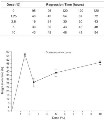
Erythema regression time in the study sample
ranged between 19 and 120 hours for different dose
concentrations used. The group in which the 2.5%
concentration compress was used showed the shortest
regression times: 19 and 24 hours (Table 3). The
dose-response curve (Figure 2) evidences that the average
phlebitis regression time was shorter for the 2.5%
concentration group (mean = 29.2h, standard deviation
Rev. Latino-Am. Enfermagem 2011 Jan-Feb;19(1):3-10.
= 8.98), followed by the 5% concentration group (mean
= 38.8h, standard deviation = 17.47), and longer for
the 1.25% (mean = 57.8h, standard deviation = 11.10)
and 10% concentration groups (mean = 49.4h, standard
deviation = 4.67). Mean regression time in the control
group was 110.4h and standard deviation 13.15.
Figure 1 – Effect of different C. recutita infusion
concentration on the reduction of phlebitis in patients
submitted to peripheral intravenous chemotherapy
R e g re ssi o n t ime (h ) 125 100 75 50 25 0
Control 1.25% 2.5% 5% 10% a
b
c
Dose (%)
Legend: a = p<0.001 related to control; p<0.01 related to 1.25%; p<0.05 related to 10%; p>0.05 related to 5%, b = p<0.001 related to control; p>0.05 related to 10%, c = p<0.001 related to control; p>0.05 related to 10%. Statistical analysis using ANOVA followed by Bonferroni multiple comparison test.
Table 3 – Regression time, in hours, of phlebitis according
to infusion dose group of C. recutita loral capitula
Dose (%) Regression Time (hours)
0 96 96 120 120 120
1.25 48 48 54 67 72
2.5 19 24 30 30 43
5 30 30 43 43 48
10 43 48 48 48 54
Figure 2 – Dose-response curve according to mean
regression times, in hours and infusion concentrations
of 1.25%, 2.5%, 5% and 10%, respectively, of C.
Recutita loral capitula, obtained through ANOVA with 60 56 52 48 44 40 36 32 28 24 20 16 12 8 4 0
1 2 3 4 5 6 7 8 9 10
Dose (%) R e g re ssi o n t ime (h ) Dose-response curve
As for toxicity, moderate to severe itching was
reported on the left forearm of one of the patients
allocated in trial group C, whose compress had been
applied on the anterior front forearm. As the itching expanded, the transparent PVC ilm used on the entire forearm could have caused this. The subject was
forwarded to the medical team, medicated with an
anti-histaminic drug and showed complete regression of the
itching within two hours. Local treatment was continued
with warm water compresses at 38° C until the complete
regression of the erythema.
With regard to toxicity evaluation in subjects without
phlebitis (n=4), no manifestations of hypersensitivity
reactions occurred, nor reports of burning, itching or any
other symptoms related with possible hypersensitivity to
the drug.
Discussion
It is fundamental to determine the total lavonoid contents when assessing a plant’s quality, especially in studies that use lavonoids for therapeutic purposes(15). Tests involving about 100 samples of 12 chamomile varieties cultivate in identical conditions showed lavonoid contents ranging between 1.0 and 2.6%, while twenty other samples of different origins showed total lavonoid content levels varying between 0.3 and 3.0%(15). The
sample used in this study showed 2.5% m/m of total lavonoids, in line with previously found results. As for α-bisabolol content in essential oil, the level identiied in the sample (10.9% m/m) surpassed literature
recommendations according to the used temperature,
which is 7%(15). Qualitative analysis of the plant’s active principles conirmed available literature data about the chemical composition of C. recutita, describing terpenes
(α-bisabolol, bisabolol oxide A and B, camazulene and sesquiterpenes), lavonoids (apigenin-7-glucoside, luteolin, quercetin), coumarins (umbelliferone) and
steroids(16). It should be reminded that these elements,
terpenes, steroids and sesquiterpenes exert anti-inlammatory effects, generally inhibiting the classical route of the complement system, interfering, in turn, in
arachidonic acid metabolism(17).
With regard to the clinical phase, research subjects
in all groups were comparable, as the main confounding
factors, including age, sex, white blood cell count and
baseline disease were similar. Among the 25 participants,
those in group A (dose 1.25%) showed the longest inlammation process regression time (range: 48 – 72h), while those in groups B and C showed the shortest
Reis PED, Carvalho EC, Bueno PCP, Bastos JK.
longer regression time than groups B and C because, despite a higher dose – 40 g of loral capitula of C. recutita – the compress could not be totally moistened
with the quantity of solvent used. This quantity did not
need to be adjusted though, as lower doses had already
demonstrated an excellent effect in terms of regression time of the inlammatory process.
Other clinical trials have conirmed the anti-inlammatory effect of C. recutita in radiation dermatitis, through the use of ointments with chamomile extract
(Kamilosan®)(18), in mucositis, through oral solution
(Kamilosan® Solução Oral)(19), in contact dermatitis
and eczema, through ethanolic extract(20-21), showing even superior eficacy results when compared with steroidal and non-steroidal anti-inlammatory drugs(21). Researchers(22) have assessed the eficacy of medical
plant infusions (chamomile, salvia and calendula)
for topical application in the treatment of Hand-Foot
Syndrome resulting from intravenous capecitabine
infusion, an antineoplastic chemotherapy drug used
in breast cancer patients. The sample comprised 11
patients who immersed their hands and feet into the infusion daily. Signiicant regression of the inlammatory process was found in all cases.
As for the dose, literature recommends
concentrations between 3 and 10% for external use
in compresses(23-24). It was observed, however, that
with 2.5% concentrations, results were as satisfactory
as with 5% concentrations, to the extent that the shorted regression time of the inlammation process was obtained in two patients, i.e. 19 and 24 hours. Both concentrations showed statistically signiicant results when compared mutually (p<0.05) and with
the control group (p<0.001). When compared with the
other concentrations (1.25% and 10%), the 2.5% dose showed a statistically signiicant difference (p<0.05), while the 5% dose showed no statistically signiicant difference with the other doses.
Therefore, the researchers decided to choose the 2.5% concentration (10 g/400 mL) for loral capitula infusions of as the standard dose for this study, although
literature recommends using concentrations between 3
and 10% for compresses. This small difference of -0.5%
between the result obtained in the dose-response curve
and the concentration recommended in literature (at
least 3%) can be attributed to the excellent quality of
the test sample, of Brazilian origins. Besides, it should be
highlighted that literature itself shows different variations in terms of quantity deinitions and even measurement units. The US Pharmacopeia, for example, indicates the
use of two dessert spoons for external use, equivalent to approximately 6g of dry loral capitula of C. recutita
in 250 mL of water(25), i.e. 2.4%, which is basically the
same standard dose found in this research phase. The
same source highlights that concentrations between 3
and 10% are indicated for ointments and gels.
It is also important to highlight that using less grams of loral capitula with better results in terms of time for phlebitis regression generates a better cost-beneit relation and, hence, greater advantage for consumers,
due to the obvious economy of the main resource.
Itching was reported in one of the patients allocated
in trial group C, with a 5% infusion concentration. The
reported hypersensitivity reaction – itching – occurred across the subject’s left forearm, who classiied it as moderate to intense when the researcher asked about the
intensity, although the compress had only been applied
on the anterior front middle third of the left forearm.
With regard to this episode, although allergic reactions
to C. recutita are quite rare, these can happen, to the
extent that one of the exclusion criteria was exactly the patient’s afirmative response as to any adverse reaction to chamomile or any plant from the Asteraceae
or Compositae family.
It should be clariied that the sites where the compresses were applied, in the trial as well as in the
toxicity control group, were assessed for an additional
two days after the application, with a view to investigating late signs and symptoms of toxicity. This was not veriied in any of the other subjects who accepted to participate
in the research. As for the itching in one of the sample
subjects, the symptom regressed within 72 hours,
without the need for any medication intervention.
Conclusion
The standardization of vegetal raw material, ranging
from the selection of the species, seeding, cultivation,
harvesting, drying, storage and quality assessment
is fundamental, mainly when used for therapeutic
purposes, like in the case of this research.
Based on the results, it can be inferred that, at
α = 5%, the C. recutita infusion presents minimal or almost zero toxicity for topical application. This study also demonstrated that the 2.5% concentration for loral capitula infusion of C. recutita, when applied for anti-inlammatory purposes in case of phlebitis deriving from peripheral intravenous chemotherapy infusion, is as
effective as the concentration suggested by literature,
Rev. Latino-Am. Enfermagem 2011 Jan-Feb;19(1):3-10.
This research contributes to the innovation of clinical
nursing practice, as it suggests a treatment alternative
for phlebitis deriving from peripheral intravenous
infusion during antineoplastic chemotherapy. Moreover,
theoretical support is provided regarding the methods to
adopt for the clinical use of phytotherapeutic drugs.
References
1. Machado AF, Pedreira MLG, Chaud MN. Adverse events
related to the use of peripheral intravenous catheters
in children according to dressing regimens. Rev.
Latino-Am. Enfermagem. Maio-junho 2008; 16(3):362-7.
2. Reis PED, Capucho CR, Vasques CI, Carvalho EC.
Efeitos adversos identificados em local de infusão
intravenosa periférica por drogas quimioterápicas. Cienc
Enferm. 2008; 14(2):55-64.
3. Conselho Federal de Enfermagem. Resolução COFEN
197/1997 [acesso em: 23 setembro 2009]. Disponível
em: http://www.portalcofen.gov.br/2007/materias.
asp?ArticleID=7041
4. Ministério da Saúde (BR). Agência Nacional de
Vigilância Sanitária. Resolução RE n. 90, de 18 de março
de 2004. Determina publicação do “Guia para a realização
de estudos de toxicidade pré-clínica de fitoterápicos”.
Diário Oficial da República Federativa do Brasil, Brasília
(DF): Imprensa Oficial; março 2004. Seção 1.
5. Ministério da Saúde (BR). Agência Nacional de
Vigilância Sanitária. Resolução de Diretoria Colegiada
(RDC) n. 89, de 16 de março de 2004. Determina
a publicação da Lista de Registro Simplificado de
Fitoterápicos junto ao Sistema de Vigilância Sanitária.
Diário Oficial da República Federativa do Brasil. Brasília
(DF): Imprensa Oficial; março 2004. Seção 1.
6. Farmacopéia Brasileira. 4a. ed. São Paulo: Atheneu;
1996. Parte 2, Fascículo 1.
7. . United States Pharmacopeia (USP) 28. 28ª. ed.
Rockville:United State Convention; 2005.
8. Dowd LE. Spectrophotometric Determination of
Quercetin. Anal Chem 1959; 31(7):1184-7.
9. Jay M, Gonnet J, Wollenweber E, Voirin B. Sur
L’analyse. Qualitative des Aglycones Flavoniques dans
Une Optique Chimiotaxinomique. Phytochemistry 1975;
14:1605-12.
10. Page C, Curtis M, Sutter M, Walker M, Hoffman B.
Farmacodinâmica e a quantificação da droga. In: Page C,
Curtis M, Sutter M, Walker M, Hoffman B. Farmacologia
integrada. 2ª ed. Barueri (SP): Manole; 2004. p. 57-68.
11. Infusion Nursing Society. Infusion Nursing Standards
of Practice. J Infusion Nurs. 2006; 29(1):S58-60.
12. Coutinho V, Coutinho MA. Análise do exame
hematológico. In: Zago MA, Falcão RP, Pasquini P.
Hematologia: fundamentos e prática. São Paulo (SP):
Atheneu; 2001. p. 1081.
13. Armitage P, Berry G. Statistical methods in medical
research. 3ª ed. Boston: Blackwell Scientific Publications;
1994.
14. Hamilton LC. Statistics with STATA. Califórnia:
Thomson – Brooks/Cole; 2004.
15. Franke R, Schilcher H. Chamomile: industrial profiles.
New York (NY): Taylor and Francis Group; 2005.
16. Szelenyi IO, Thiemer K. Pharmacological
experiments with compounds of chamomile:
Experimental studies of the ulcerprotective effect of
chamomile. Planta Med. 1979, 35: 218-27.
17. Schulz V, Hänsel R, Tyler VE. Fitoterapia racional: um
guia para as ciências da saúde. 4ª ed. São Paulo(SP):
Manole; 2002.
18. Maiche AG, Grohn P, Maki-Hokkonen H. Effect
of chamomile cream and almond ointment on acute
radiation skin reaction. Acta Oncol. 1991;30(3):395-6.
19. Carl W, Emrich LS. Management of oral mucositis
during local radiation and systemic chemotherapy: a study
of 98 patients. J Prosthet Dent. 1991;66(3):361-9.
20. Santoro M. Efeito terapêutico do extrato etanólico
das flores de camomila em base cremosa no tratamento
da dermatite das fraldas – estudo multicêntrico. Rev
Paul Pediatr. 1998;16(2):69-76.
21. Aertgeerts P, Albring M, Klaschka F, Nasemann T,
Patzelt-Wenczler R, Rauhut K, et al. Vergleichende
prüfung von Kamillosan® Creme gegenüber steroidalen
(0,25% Hydrocortison, 0,75% Fluocortinbutylester)
und nichtsteroidalen (5% Bufexamac) externa in der
erhaltungstherapie von ekzemerkrankungen. Z Hautkr
1985;60(3):270-7.
22. Kern E, Schmidinger M, Locker GJ, Kopp B.
Management of capecitabine-induced hand-foot
syndrome by local phytotherapy. Wien Med Wochenschr.
2007;157(13-14):337-42.
23. Blumenthal, M. The complete german commission
E monographs – therapeutic guide to herbal medicines.
Boston: American Botanical Council; 1998.
24. WHO. WHO monographs on selected medicinal
plants. Genebra: World Health Organization; 1999.
25. PDR for herbal medicines. 2ª ed. Montvale: Medical
Economics Company; 2000.
Received: Oct. 7th 2009