Caso Clínico / Radiological Case Report
Correspondência
Alexandre Gomes Martins Batista Rua dos almocreves, nº16, Aldeia Nova da Azoia 2970-085 Sesimbra
e-mail: [email protected]
1 Hospital José Joaquim Fernandes, Beja 2 -Instituto Português de Oncologia de Lisboa Francisco Gentil, Lisboa
Serviço de Radiologia do Instituto Português de Oncologia de Lisboa Francisco Gentil Diretor: Dr. José Venâncio
Recebido a 16/07/2014 Aceite a 13/09/2014
POST-MENOPAUSAL METRORRHAGIA – AN OVARIAN
THECOMA PRESENTATION
METRORRAGIA PÓS-MENOPAUSA – UMA APRESENTAÇÃO DE
TECOMA OVÁRICO
Alexandre Batista1, Teresa Margarida Cunha2
Resumo
Os tecomas são tumores raros do ovário, do grupo dos tumores dos cordões sexuais, de natureza sólida e frequentemente unilaterais. Têm maior incidência no período pós-menopausa e normalmente são silenciosos. Quando sintomáticos traduzem-se por dor pélvica e metrorragia (condicionada pela habitual natureza produtora de estrogénios do tumor). Podem ser concomitantes a síndrome de Meigs e/ou de Golin-Goltz e associarem-se a transformação benigna ou maligna do endométrio. Embora a ecografia possa ser inespecífica neste contexto, uma avaliação multiparamétrica abrangente em ressonância magnética, incluindo por estudo dinâmico e com ponderação em difusão, permite frequentemente orientar de modo favorável a marcha diagnóstica.
Apresentamos um caso raro de tecoma do ovário, com espessamento associado do endométrio, avaliado por ecografia ginecológica por vias supra-púbica e transvaginal bem como tomografia computorizada e ressonância magnética, confirmado cirurgicamente. Tratou-se de uma examinada caucasiana de 61 anos de idade, apresentando-se com metrorragia pós-menopáusica, sem outros sintomas nem contexto familiar relevante. Procedeu-se, a este propósito, a uma revisão da literatura focada no diagnóstico multimodal diferencial, apresentação clínica, tratamento e prognóstico destes tumores.
Palavras-chave
Tecoma; Ovário; Ecografia; Tomografia Computadorizada; Ressonância Magnética.
Abstract
Thecomas represent rare, solid sex-cord stromal ovarian tumors, often unilateral, asymptomatic and occurring in postmenopausal patients. When symptomatic, they most commonly present with pelvic pain and metrorrhagia (due to their frequent estrogenic releasing nature). Thecomas can occur concomitantly with Meigs and/or Golin-Goltz syndrome and may also be associated with benign or malign endometrial transformation. Although gynecologic transabdominal and transvaginal ultrasound can be quite unspecific in this particular solid ovarian tumor presentation, magnetic resonance imaging including diffusion and dynamic data can frequently suggest the diagnosis and significantly facilitate the diagnostic work-up.
We report a rare case of ovarian thecoma, with concomitant endometrial thickening, demonstrated by gynecologic transvaginal ultrasonography, computed tomography and magnetic resonance contrasted imaging, surgically confirmed. The patient was a 61 years old caucasian female presenting with postmenopausal metrorrhagia, without other associated symptoms nor family medical context. On this regard, we performed a review of the literature, focused on multimodal differential diagnosis imaging, clinical presentation, treatment and prognostic of this pathological finding.
Key-words
Thecoma; Ovary; Ultrasound; Computed Tomography; Magnetic Resonance.
ACTA RADIOLÓGICA PORTUGUESA
Setembro-Dezembro 2014 nº 103 Volume XXVI 61-65
Clinical history
61 years old caucasian female patient presenting with postmenopausal metrorrhagia for a period of four months, without other associated symptoms and with no relevant family medical context. The patient was not on hormone replacement therapy. Physical examination was unremarkable, with no evident abdominal or pelvic masses, nor localized pain. Laboratory findings included an elevated CA 125 value (86,7 U/ml), normal
CEA (0,9 ng/ml) and CA 19.9 (11,8 U/ml), and normal hemoglobin (13,2 g/dL) and hematocrit (37,8%).
Image findings
The patient was then referred to our Department and evaluated through a dedicated pelvic magnetic resonance (MR) study, revealing an exofitic solid growth originating in the posterior wall of the right ovary, measuring 110 x 100 x 80 mm (craneo-caudal x latero-lateral x antero-posterior maximum diameters). The tumor had regular and well delimited contours, demonstrated isosignal with adjacent muscle on T1 weighted images and discrete hypersignal on T2 weighting. There were intratumoral regions of high cellularity with diffusion restriction, as observed on the b = 1000 s/mm2 images and apparent diffusion coefficient (ADC) map. The dynamic tumoral evaluation showed gradual gadolinium uptake, including in the interstitial late phase (180 seconds post injection), in a type 1 time-signal intensity curve [1], with evident hypovascularity comparing with the more avid arterial enhancement of adjacent myometrium. Additionally, a probable endometrial polyp with 76 x 43 x 17 mm (craneo-caudal x latero-lateral x antero-posterior maximum diameters) was observed. There was also a small amount of free intraperitoneal fluid within the recto-uterine cul de sac (Figs. 3-10).
These findings, in agreement with the presenting clinical history and laboratory values, suggested primarily a thecoma. Hysterectomy with bilateral salpingo-oophorectomy was performed. On macroscopy, the tumor was solid, with uniform an endometrial thickening of 12,5 mm was noted (Fig. 1-B).
There was a discrete volume of free fluid in the right ovarian fossa. There were no enlarged pelvic lymph nodes.
Subsequently, a computed tomography (CT) scan with i.v. administration of iodinated contrast media was performed, confirming the previously identified right adnexal mass (with mild vascular enhancement on venous phase) and endometrial thickening (Fig.2). No pelvic or para-aortic lymph node enlargement was identified.
A
B
Fig. 1 - Transvaginal ultrasonography. Right adnexal mass, heterogeneous, mostly hypoechoic, measuring 108 x 71 mm (yellow arrow). Concomitant endometrial thickening of 12,5 mm (white arrow).
Fig. 2 - Computed tomography scan with i.v. administration of iodinated contrast media (venous phase). Well delimitated right adnexal solid mass with mild vascular enhancement. Endometrial thickening can also be detected (white arrow).
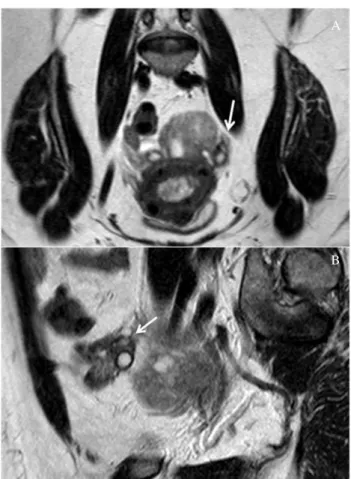
Fig. 3 - Axial T2-weighted magnetic resonance. Right adnexal tumor with well defined contours and discrete hypersignal on T2-weighted sequence. Note the endometrial thickening and a small amount of free fluid in the recto-uterine recess (white arrow).
texture (excluding a few cystic spaces), well-circumscribed, and measured 120 x 110 x 70 mm. Sectional analysis of the tumor revealed a yellow and fascicular mass (Fig.11). The tumor had more than 80% of its cellular content positive for alpha-inhibin and a low proliferation index (<10%). There were two endometrial polyps (40 and 30 mm of maximum diameter). The pathological findings were consistent with thecoma.
Discussion
Thecomas represent rare, solid sex-cord stromal ovarian tumors, accounting for approximately 0.5 – 1% of all primary ovarian
A
B
Fig. 5 - Coronal (A) and sagital (B) T2-weighted magnetic resonance depicting normal left ovary appearance (white arrow).
Fig. 6 - Axial T1-weighted magnetic resonance. The mass demonstrates isosignal with adjacent muscle on T1-weighted sequence.
Fig. 7 - Fat-suppressed axial T1-weighted MR image after gadolinium administration (arterial phase). There is little mass enhancement comparing to myometrium.
Fig. 8 - Fat-suppressed axial T1-weighted MR image after gadolinium administration (intersticial phase). Gradual gadolinium uptake with discrete enhancement in the interstitial late phase (180 seconds post injection), in a type 1 time-signal intensity curve, revealing the lesion´s fibrin rich territory.
ovarian medulla and should be considered apart form fibromas, which originate from the cortex [4]. Although being uncommon, they represent the most frequent solid primary ovarian tumor [5].
Thecomas of the ovary can present with pleural effusion or ascites (Meigs syndrome) and may also be associated with basal cell nevus Golin-Goltz syndrome (large bilateral fibrotic ovarian tumors, basal cell carcinomas of the skin, odontogenic keratocysts and other abnormalities) [6], though they usually are asymptomatic, and when symptomatic, they are most commonly manifested by pelvic pain and metromenorrhagia (due to their frequent estrogen releasing nature) [7,8]. Estrogen stimulation by a pre-existing thecoma may also induce the development of endometrial hyperplasia and endometrial polyps and presumably induce mesenchymal and mixed epithelial / mesenchymal uterine tumors, namely adenosarcoma [9, 10].
They are often unilateral and occur in postmenopausal patients but can develop in younger patients (mean ages in the fifth and sixth decades) [11, 12]. As most adnexal tumors do, the laboratory workup on a thecoma presentation can reflect elevation of particular tumor markers, such as CA-125 [13].
Histologically thecomas are similar to theca interna cells of the ovary and have a mixed lipid and colagenic composition, the later derived from spindle, oval or round cells [6].
The management approach to thecomas is surgical in larger lesions, with excellent prognosis as they are mainly benign tumors [14].
TVUS is commonly the first choice imaging modality for suspected pelvic tumor, due to its easy access and safety profile, nonetheless being often non specific. Thecomas usually present as a solid mass or, if larger, predominantly solid with few cystic areas, being the solid component iso or hypoecogenic in relation with adjacent stroma, frequently with significant posterior attenuation, without identifiable tumoral calcification. On color Doppler evaluation thecomas usually have neglectable vascularity [9, 15]. On CT evaluation tumor attenuation is often similar to adjacent myometrium, with evident hypovascularity on arterial phase enhancement study, sometimes being evident progressive delayed contrast uptake due to fibrin rich tumoral areas [11]. This same fibrous tumoral component is responsible for the low T1-weighted and very low T2-weighted MR signal (excluding scattered areas of high signal intensity corresponding to cystic or edematous change) [5, 11]. If fatty elements are present, they can be identified as hyperintense on T1-weighted and translate in a decreased signal intensity on selective fat-saturation or out-of-phase gradient echo sequences [9]. On diffusion-weighted imaging (DWI), most thecomas display intermediate signal similar to that of myometrium, attributable to their cellular content of fibroblasts and thecal cells, but lower than the usually observed in malignant, high cellular ovarian tumors. The ADC of thecomas and other adnexal masses has no described relevant difference, although the ADC of thecomas has been reported to be significantly lower than that of leyomiomas [16].
The differential diagnosis of ovarian thecoma primarily encompasses masses with a fibrous component, as uterine leyomioma, Brenner tumor and mature cystic teratoma [17]. When considering uterine leyomioma, the most common pitfall lies in broad ligament leyomioma and pedunculated leyomioma. However, their origin can usually be traced, namely due to the
Fig. 11 - Tumor macroscopic surface section. Two sections of the right oophorectomy specimen revealed a yellowish tone and fascicular solid mass.
neoplasms [2]. Ovarian sex cord tumors are those who arise from granulosa, theca, Leydig, Sertoli or stromal fibroblast cells [3]. Notwithstanding sometimes having an interspersed fibrous component, thecomas are presumably originated from the
A
B
Fig. 10 - Diffusion-weighted MR image. Intratumoral areas of high cellularity with diffusion restriction, as observed in the
b = 1000 s/mm2 images (A) and apparent diffusion coefficient (ADC)
bridging vascular sign [18], that clarifies their feeding by uterine arteries, opposed to thecomas, supplied by ovarian arteries or ovarian branches of uterine arteries [17].
Brenner tumours are usually very small (<2 cm) and benign epithelial ovarian lesions, mostly composed of fibrous dense tissue and urothelium-like transitional cells. They most commonly present as a multilocular cystic tumor or a smaller predominantly solid mass. Their dense fibrous stroma has low signal on T2-weighted MR imaging, and an expected gradual gadolinium uptake. Usually they comprise extensive amorphous calcifications [19]. Their small size compared to the median thecoma size of approximately 13 cm, more prominent calcifications and extensive cystic component usually allows the differential diagnosis [9].
Mature teratomas most frequently occur in pre-menopausal women, and present as a unilocular cystic tumour (in 88% of the cases), filled with sebaceous content and lined with epithelium. Due to their origin in two or more germinal layers, they can contain hair and teeth, that when present are commonly encompassed by a wall protuberance (the Rokitansky nodule or dermoid plug) [20]. The most common imaging US features for mature teratoma are the presence of a cystic lesion with a densely echogenic shadowing mural nodule (dermoid plug); an
ecogenic (sebaceous) mass with very strong posterior attenuation (“tip of the iceberg” sign) and a dermoid mesh, with multiple thin echogenic lines (hairs) passing within the cyst. On CT and MRI imaging, macroscopic fat within a cyst, with or without mural calcification, is diagnostic. Less common monodermal dermoids include struma ovarii (with a predominance of thyroidal tissue) and carcinoid [21].
Our patient presented with a predominantly solid right adnexal mass, hypoechoic on ultrasound, with regular and defined contours and no evident vascularization on color Doppler. On MRI interrogation the tumor showed to be originated from the posterior right ovarian wall, had gradual intersticial enhancement, revealing its fibrous component, and small pockets of cellularity in DWI. There was no accompanying significant ascites, peritoneal lesions or pelvic enlarged lymph nodes. There was also an endometrial polyp. These findings, in a postmenopausal metrorrhagia context, and in conjunction with the described clinical and laboratory data suggested primarily a thecoma, with mixed theca cells and fibrous composition and expected estrogenic endometrial effect.
The lesion was surgically confirmed as thecoma.
References
1 - Thomassin-Naggara, I.; Daraï, E.; Cuenod, C. A.; Rouzier, R.; Callard, P.; Bazot, M. - Dynamic contrast-enhanced magnetic resonance imaging: a useful tool for characterizing ovarian epithelial tumors. J Magn Reson Imaging, 2008, 28(1):111-20.
2 - Chen, V. W.; Ruiz, B.; Killeen, J. L.; Coté, T. R.; Wu, X. C.; Correa, C. N. - Pathology and classification of ovarian tumors. Cancer, 2003, 97:2631-42. 3 - Wilkinson, N.; Osborn, S.; Young, R. H. - Sex cord stromal tumours of the ovary: A review highlighting recent advances. Diagn Histopathol, 2008, 14(8):388-400.
4 - Nocito, A. L.; Sarancone, S.; Bacchi, C.; Tellez, T. - Ovarian thecoma: Clinicopathological analysis of 50 cases. Ann Diagn Pathol, 2008, 12:12-6. 5 - Troiano, R. N.; Lazzarini, K. M.; Scoutt, L. M.; Lange, R. C.; Flynn, S. D.; McCarthy, S. - Fibroma and fibrothecoma of the ovary: MR imaging findings. Radiology, 1997, 204:795-8.
6 - Scully, R. E.; Young, R. H.; Clement, P. B. - Atlas of Tumor Pathology,
Tumors of the Ovary, Maldeveloped Gonads, Fallopian Tube, and Broad Ligament.
Third series, Fascicle 23. Washington, DC: Armed Forces Institute of Pathology, 1998.
7 - Sivanesaratnam, V.; Dutta, R.; Jayalakshmi, P. - Ovarian fibroma: clinical and histopathological characteristics. Int J Gynaecol Obstet, 1990, 33:243-7. 8 - Leung, S. W.; Yuen, P. M. - Ovarian fibroma: a review on the clinical characteristics, diagnostic difficulties, and management options of 23 cases. Gynecol Obstet Invest, 2006, 62:1-6.
9 - Hricak, H. - Diagnostic imaging: gynecology, 1st ed. Salt Lake City, UT, Amirsys/Elsevier, 2007, 7:28-31.
10 - Nomura, K.; Aizawa, S.; Ushigome, S. - Adenosarcoma of the uterine corpus associated with ovarian thecoma. Pathol Int, 2001 Sep, 51(9):735-8. 11 - Bazot, M.; Ghossain, M. A.; Buy, J. N. et al. - Fibrothecomas of the ovary: CT and US findings. J Comput Assist Tomogr, 1993, 17:754-9.
12 - Chechia, A.; Attia, L.; Temime, R. B.; Maklouf, T.; Koubaa, A. - Incidence, clinical analysis and management of ovarian fibromas and fibrothecomas. Am J Obstet Gynecol., 2008, 199(5):473. e1-4.
13 - Takemori, M.; Nishimura, R.; Hasegawa, K. - Ovarian thecoma with ascites and high serum levels of CA125. Arch Gynecol Obstet, 2000 Jul, 264(1):42-4.
14 - Jung, S. E.; Rha, S. E.; Lee, J. M. et al. - CT and MRI findings of sex cord-stromal tumor of the ovary. AJR Am J Roentgenol, 2005, 185(1):207-15. 15 - Atri, M.; Nazarnia, S.; Bret, P. M.; Aldis, A. E.; Kintzen, G.; Reinhold, C. - Endovaginal sonographic appearance of benign ovarian masses. Radiographics, 1994, 14(4):747-60, discussion 761-2.
16 - Zhang, H.; Zhang, G. F.; Wang, T. P. - Value of 3.0 T diffusion-weighted imaging in discriminating thecoma and fibrothecoma from other adnexal solid masses. Journal of ovarian research, 2013, 6:58.
17 - Seung, E.; Lee, J. M.; Rha, S. E.; Byun, J. Y.; Jung, J. I.; Hahn, S. T. - CT and MR Imaging of Ovarian Tumors with Emphasis on Differential Diagnosis. RadioGraphics, 2002, 22:1305-25.
18 - Kim, J. C.; Kim, S. S.; Park, J. Y. - Bridging vascular sign” in the MR diagnosis of exophytic uterine leiomyoma. J Comput Assist Tomogr, 2000 Jan-Feb, 24(1):57-60.
19 - Moon, W. J.; Koh, B. H.; Kim, S. K. et al. - Brenner tumor of the ovary: CT and MR findings. J Comput Assist Tomogr, 2000, 24:72-6.
20 Comerci, J. T. Jr; Licciardi, F.; Bergh, P. A.; Gregori, C.; Breen, J. L.
-Mature cystic teratoma: a clinicopathologic evaluation of 517 cases and review of the literature. Obstet Gynecol, 1994, 84:22-8.