18e
Rev Bras Med Esporte _ Vol. 13, Nº 1 – Jan/Fev, 2007 * Pontifícia Universidade Católica de São Paulo-Sorocaba. Centro deCiên-cias Médicas e Biológicas (CCMB). Sorocaba, SP – Brasil. 1. Aluno de graduação do curso de Medicina.
2. Professor Doutor do Departamento de Morfologia e Patologia. 3. Professor Doutor do Departamento de Ciências Fisiológicas. Approved in 25/7/06.
Correspondence to: Beatriz Lavras Costallat, Rua Ezequiel Magalhães, 26 – J. Paineiras – 13092-522 – Campinas, SP. E-mail: [email protected]
Insulin resistance with creatine supplementation
in laboratory animals
*
Beatriz L. Costallat1, Lísia Miglioli1, Phelipe A.C. Silva1, Neil F. Novo2 and João L.G. Duarte3
O
RIGINALA
RTICLEKeywords: Glucose; Dietetic supplements; Sports. ENGLISH VERSION
ABSTRACT
Introduction and objective: Creatine supplementation has been
used in order to improve muscular performance. This substance affects glucose metabolism and stimulates the in vitro as well as the in vivo insulin secretion. Nevertheless, long-term insulin hyper-secretion may also induce insulin resistance. The present work analyzed the effects of creatine oral supplementation in order to evaluate the possibility of occurrence of resistance to in vivo insu-lin. Methods: Forty-eight Wistar rats (24 female / 24 male) were divided in two groups of 24 (control and study) and subdivided in six groups of eight. They were fed with standard food during four weeks, having water ad libitum. Moreover, the study group received dietetic supplement of creatine (0.4 g creatine for 30 ml of water per rat / day). In the 7th, 14th, 21st and 28th day of the experiment,
12 rats were anesthetized (sodium thiopental 0.15 mL / 100 g) af-ter six hour-fasting, being submitted to intravenous insulin toler-ance test (0.5 mL of 30% regular human insulin and 70% saline solution). The blood samples were collected from the tail veins of the rats, in the basal, three, six, nine, 12 and 15 minutes after insulin administration times. The glucose measurement was per-formed through the glucose oxidase method. The study was previ-ously approved by the Research Ethics Committee of CCMB-
PUC-SP. Results: The mean of the glucose decrease constant (KITT) was
calculated through the formula 0.693 / T1 / 2. The study group, when compared with the control group, presented insulin resistance at day 21 (p < 0.0004) and day 28 (p < 0.0001). Conclusion: This study shows that extended creatine supplementation may lead to insulin resistance. Besides that, it should be carefully used in indi-viduals with glucose metabolism disturbances.
INTRODUCTION
Oral creatine supplementation is generalized in professional as well as amateur athletes in several age groups and it is recom-mended by the American College of Sports Medicine for perfor-mance improvement in short-term exercises and maximal power (1-2). Creatine is a natural nutrient of animal origin, which is found in
meat and fish. It is endogenously synthesized in the liver, pancre-as and kidneys, from some amino acids (glycine, arginine, methion-ine). It is an important energy reservoir for muscular contraction, since around 95% of the body creatine is stored in the skeletal muscle under free or phosphorylated forms (as creatine phosphate – PCr). When the energy demands increase, the creatine phos-phate supplies the phosphos-phate for the adenosine diphosphos-phate (ADP) with the aim to synthesize adenosine triphosphate (ATP). This kind of reaction rapidly occurs and results in energy for high-intensity
and short-duration physical activities. Creatine supplementation has the objective to increase the content of muscular phosphocreat-ine; although the results concerning its efficiency are controversial yet(3). Besides increasing muscular creatine storage, the creatine
dietetic supplementation may increase creatine phosphate resyn-thesis(4), although it has not always been observed(5).
Despite being considered licit and safe by the International Olym-pic Committee, there are certainly some risks with this supple-mentation(6). There are papers which show unwanted effects of
creatine chronic supplementation(7) leading to hepatic and renal
overload. However, it has not been found in another study(8) renal
overload in healthy individuals, even if it shows that those with renal dysfunction should not use the supplementation.
Creatine also affects the metabolism of carbohydrates. When intraperitoneally injected, it leads to hypoglycemia(9) and its
sup-plementation in the diet has shown improvement when there is alteration in the glucose tolerance(10). Since the 70’s, studies with
animal models as well as ‘in vitro’ have demonstrated that insulin increases the blood creatine transportation to the skeletal muscle of rats(11-12). It has also been demonstrated that creatine increases
the muscular storages of glycogen(13). Glycogen resynthesis(14) and
increase of the glucose-carrier protein(15) have been observed in
the creatine supplementation, altering the regulation of the glu-cose metabolism(16-17).
Possibly, the mechanism through which creatine affects glucose metabolism is the stimulation of insulin pancreatic secretion, since, although glucose is the greatest stimulator of insulin secretion, it can also be induced by proteins and amino acids. The role of creat-ine as stimulator of insulin secretion has been demonstrated in in vitro(18-19) as well as in vivo studies, confirming the very same
alter-ations(16-17).
Nevertheless, this long-term insulin hypersecretion may induce also to insulin resistance(20), which has probably not been
previous-ly studied in creatine supplementation. The aim of the present work was to analyze the effects of oral creatine supplementation on the metabolism of carbohydrates, especially to evaluate the possibility of occurrence of in vivo insulin-resistance in animal models (rats) while receiving creatine supplementation for four weeks.
METHODS
The tests were conducted in the Physiology Laboratory of the Center of Medical and Biological Sciences (CCMB), PUC of São Paulo, Sorocaba Campus. Forty-eight albino Wistar rats were se-lected, 24 males and 24 females, from the CCMB bio cemetery. These animals were divided in eight cages of polyvinyl chloride (PVC) filled with sterilized white pine tree sawdust (approximately 65 cm2 of area for each animal), with six animals of the same sex
50-Rev Bras Med Esporte _ Vol. 13, Nº 1 – Jan/Fev, 2007
19e
55% was kept as ideal for the animals. All animals were fed withthe same food (Labina, by Purina) in pellets, 30 g/day per animal) as well as with water ad libitum for the Control group and water with solved creatine for the Study group. The Study group received creatine supplementation in the proportion of 0.4 g of creatine for 30 mL of water per rat/day for four weeks. The experiments oc-curred on the 7th; 14th; 21st and 28th days of the research. On each day of the experiment, six rats from the Control group and six from the Study group were chosen from the cages (three males and three females each). They were all put on six-hour fasting, beginning at seven o’clock in the morning of the experiment day. For the Study group water with creatine was also removed and later replaced by plain water.
Estimation of in vivo insulin action using insulin-tolerance
test of 15 minutes (ITT): At 1 p.m. of the experiment day the
insulin-tolerance test began, being the rats intraperitoneally anes-thetized (Thiopental Sodium 0.15 mL/100 g), using disposable sy-ringe of 1 mL and 27.5 G ½ needle. Whenever necessary, anesthe-sia was completed with ethyl ether. A small section in the distal extremity of the rats’ tail was performed for blood samples collec-tion. The first collection was conducted prior to the insulin admin-istration (basal). The drops were pipetted with 20 µl pipettes and disposable pipette tips. The sample was placed in ependorff tubes
ment, the animals were sacrificed by the section of the diaphragm muscle.
Blood glucose concentration: The enzymatic method was used
for measurement of the blood glucose concentration, using a glu-cose oxidase kit (Laborlab brandname).
Bioethics: The work was approved by the Ethics in Research
Committee of the CMBB-PUCSP according to the specific resolu-tions for experiments with animals on September 29, 2003.
Statistical analysis: Variance Analysis for repeated values was
used(21), with the aim to separately compare for each group the
values observed in the basal; three; six; nine; 12 and 15 minutes’ times in each of the experiments. T-Student test was used for the comparison of the Study and Control groups concerning the ob-served values in each of the times mentioned above. One-way Variance Analysis was applied with the purpose to compare the four days for each group in each of the considered times(21).
The ratio of the decrease constant (angular coefficient) of glu-cose (KITT) was calculated through the formula 0.693/(T1/2), being T1/2 the time necessary to reduce the basal glycemia in half. The T1/
2 of the plasma glucose was calculated from the inclination of the
decrease curve during its linear phase(22).
The p value was established at 0.05 or 5% the significance lev-el.
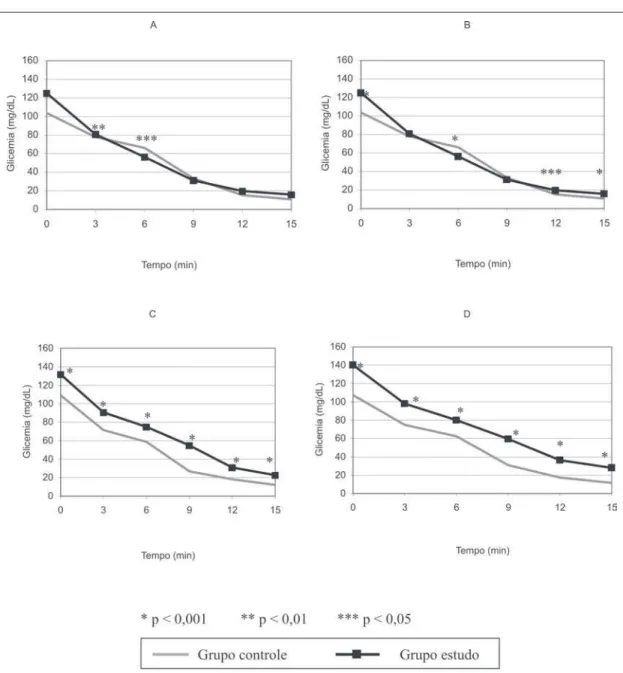
Figura 1 – Curvas glicêmicas médias nos dias de experimento 7 (A), 14 (B), 21 (C) e 28 (D) nos tempos estudados nos grupos controle e estudo
with 100 µl of 5% acid trichloroacetic acid solu-tion (TCA) slightly shaken and stored in a container with ice. Such procedure was identically performed for all groups. Later, 0.5 mL of a 30% regular insu-lin (Biohuinsu-lin U-100) and 70% saline solution was administered to both groups in a 1 mL (100 U) sterile and disposable sy-ringe, BD Plastipak brand-name, after animal’s lap-arotomy, with the aim to administer the solution in the inferior cava vein. This solution was always kept cold. The blood samples were collected from both groups in the basal (0 minute); three; six; nine; 12 and 15 minute’s times, after insulin administra-tion.
All the samples ob-tained were centrifuged in a centrifuge refrigerated at –4oC, at 3000 rpm for 10
minutes in the Medical Bi-ology Sector; Microbacte-ria Section, Regional Soro-caba Laboratory of the Institute Adolfo Lutz. The supernatant was pipetted and the glucose measure-ments were later per-formed. The remaining of the supernatant was fro-zen in a conventional freezer at –20oC. At the
experi-20e
Rev Bras Med Esporte _ Vol. 13, Nº 1 – Jan/Fev, 2007Figura 2 – Evolução da curva glicêmica média nos grupos controle e estu-do
RESULTS
The variance analysis showed a significant decrease of the gly-cemia mean values during the time for both groups (p < 0.001) at days seven; 14; 21 and 28. For day 7, the t-Student test showed significant difference between the glycemia means in the three (p < 0.01) and 6 (p < 0.05) times, with higher values for the Control group. For day 14, the t-Student test showed significant difference between the glycemia means in the basal (p < 0.001); six; (p < 0.001); 12 (p < 0.05) and 15 (p < 0.001) times; with higher value for the Control group in time six, and higher values for the Study group in the basal, 12 and 15 times. For day 21, the t-Student test showed significant difference between the glycemia means in all times (p < 0.001); with higher values for the Study group in all times. The same situation occurred on day 28: significant difference was shown between the glycemia means (p < 0.001), with higher val-ues for the Study group in all times. These data can be seen in figure 1.
The variance analysis for each of the times and days of the ex-periment showed that in the Control group significant difference was not observed between the mean values of glycemia in each time on the different days. For the Study group, the analysis re-vealed that in all times the mean values after 28 days were signif-icantly higher than the mean values after days seven and fourteen, and equivalent to the mean values after 21 days (p < 0.001). Like-wise, the mean values after 21 days were significantly higher than the mean values after seven and 14 days (p < 0.001). The values after 14 days were equivalent to the ones after seven days. These data can be seen in figure 2.
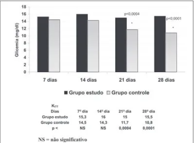
The KITT according to what was mentioned by Bonora et al.(22)
was used for the estimation of the glucose decrease constant in both groups in the 28 days of experiment. Figure 3 shows that
there was significant decrease of the glucose concentration in the Control group when compared with the Study group at day 21 (p < 0.0004) as well as at day 28 (p < 0.0001), showing that insulin-resistance occurred in the two last weeks in the group which re-ceived creatine supplementation when compared with the Control group.
Figura 3 – Decaimento da glicose em ambos os grupos nos dias de expe-rimento (KITT)
DISCUSSION
The creatine administration affects the glucose homeostasis as well as the insulin levels. It has been demonstrated in vitro(18-19)
that creatine affects the metabolism of the carbohydrates when directly stimulates the insulin secretion of isolated pancreatic is-lets. Such fact was confirmed in vivo(16) in a work studying the
creatine supplementation in rats in order to observe the long-term effects in the glucose transportation and storage in the skeletal muscle. Moreover, it showed high insulin secretion and alteration in the glucose homeostasis after eight weeks of supplementation when compared with the controls. Thus, it has been demonstrat-ed that there is a relation between the effects of the extenddemonstrat-ed creatine supplementation and its action in the glucose metabo-lism, with increase of the insulin pancreatic secretion concomitant with a hyperglycemia state. Insulin hypersecretion was not ob-served with the use of 5 g of creatine(23) or after three days of
creatine supplementation in humans(24); however, these studies do
not answer the question of the chronic use of creatine, since the supplementation for a period longer than three days is usual.
Although it has been demonstrated in vivo(16) that chronic
sup-plementation of creatine leads to hypersecretion of insulin, the fact is that the long-term hypersecretion of insulin may also induce in-sulin-resistance(20), which is the topic of the present study. Having
this hypothesis as starting point, we investigated the effects of this hypersecretion of insulin facing chronic supplementation of creatine, searching for evidences of insulin-resistance in rats weekly followed during seven, 14, 21 and 28 days.
Rev Bras Med Esporte _ Vol. 13, Nº 1 – Jan/Fev, 2007
21e
As previously described, the KITT was used in order to confirmthe glucose decrease in the 28 days of experiment in both groups(22).
Moreover,it was observed that in the Study group the glucose decrease does not occur as in the Control group, showing that insulin-resistance occurred in animals which received creatine from the last two weeks on.
In a diabetes experimental model, it has been observed that cre-atine supplementation can improve the sensibility to insulin in ex-trapancreatic sites(25). Other works which study the relationship
between creatine use and glucose metabolism show that such supplementation, besides the expected benefits in the application in sports medicine, could have other applications. Long-term cre-atine supplementation improves insulin pancreatic secretion as well as glucose tolerance, it could therefore be applied in patients with diabetes type 2(16). Although there is no clear definition of the
cre-atine role in the treatment of this disease, two studies comparing the creatine use with medication traditionally used in diabetes – sulphonylurea and metformin – observed that the use of creatine has an effect similar to sulphonylurea in the glycemic control of patients with diabetes type 2(26); moreover, the use of creatine may
be similar to the treatment with metformin in these patients(27).
Conversely, this present study showed that the oral creatine supplementation in rats led to the development of insulin-resis-tance derived from chronic use of creatine, which causes insulin hypersecretion which would be responsible for the insulin-resis-tance itself(20). Although further experimental studies should be
developed in order to clarify whether the found metabolic alter-ations are permanent, as well as studies in humans should be de-veloped for the confirmation of these observations, the hypothe-sis that creatine supplementation could be benefic for the prevention or treatment of diabetes type 2 becomes incorrect if development of insulin-resistance occurs.
Based on these experimental findings, one may suppose that there may be the development of insulin-resistance in humans in response to its hypersecretion, stimulated by extended creatine supplementation. Professional athletes frequently make use of oral creatine supplementation and, besides suitable physical fitness; they are under medical supervision and monitoring. Nonetheless, it is important to highlight that amateur athletes also use creatine for the improvement of their muscular performance, perhaps with no reliable information sources(28) neither the required medical
fol-low-up, though. Although no important alterations in the metabo-lism of carbohydrates with short-term use of creatine occurs(29),
the information that chronic use of this protein may lead to insulin-resistance should guide evaluation medical protocols of sports ac-tivities practitioners, either amateur and/or professional. Such pro-cedure would better control and suitably indicate supplementation, especially of those with previous alterations of glucose metabo-lism, which would make the use of creatine a considerable risk.
ACKNOWLEDGMENTS
Scientific initiation scholarship to the students from PIBIC-CEPE-PUC-SP.
All the authors declared there is not any potential conflict of inter-ests regarding this article.
REFERENCES
1. Terjung RL, Clarkson ER, Eichner P, Greenhaff PJ, Hespel RG, Israel WL, et al. The physiological and health effects of oral creatine supplementation. Med Sci Sports Exerc. 2000;32:706-17.
2. Volek JS, Rawson ES. Scientific basis and practical aspects of creatine supple-mentation for athletes. Nutrition. 2004;20:609-14.
3. Mendes RR, Tirapegui J. Considerações sobre o exercício físico, creatina e nu-trição. Revista Brasileira de Ciências Farmacêuticas. 1999;35(2):196-209. 4. Greenhaff PL, Bodin K, Söderlund K, Hultman E. Effect of oral creatine
supple-mentation on skeletal muscle phosphocreatine resynthesis. Am J Physiol. 1994; 266:E725-30.
5. Vanderberghe K, Van Hecke P, Van Leemputte M, Vanstapel F, Hespel P. Phos-phocreatine resynthesis is not affected by creatine loading. Med Sci Sports Ex-erc. 1999;31:236-42.
6. Brudnak MA. Creatine: are the benefits worth the risk? Toxicol Lett. 2004;150(1): 123-30.
7. Edmunds JW, Jayapalan S, De Marco NM, Saboorian MH, Aukema HM. Creat-ine supplementation increases renal disease progression in Han:SPRD-cy rats. Am J Kidney Dis. 2001;37(1):73-8.
8. Poortmans JR, Francaux M. Long-term oral creatine supplements does not im-pair renal function in healthy athletes. Med Sci Sports Exerc. 2000;31(8):379-85. 9. Hill R. The effect of administration of creatine on the blood sugar. J Biol Chem.
1928;78:iv[abstr].
10. Ferrante RJ, Andreassen OA, Jenkins BG, Dedeoglu A, Kuemmerle S, Kubilius JK, et al. Neuroprotective effects of creatine in a transgenic mouse model of Huntington’s disease. J Neurosci. 2000;20:4389-97.
11. Koszalka TR, Andrew CL, Brent RL. Effect of insulin on the uptake of creatine-1-14 C by skeletal muscle in normal and X-irradiated rats. Proc Soc Exp Biol Med. 1972;139(4):1265-71.
12. Haugland RB, Chang DT. Insulin effect on creatine transport in skeletal muscle. Proc Soc Exp Biol Med. 1975;148(1):1-4.
13. Zehnder M, Rico-Sanz G, Kuhne G, et al. Muscle phosphocreatine and glycogen concentrations in humans after creatine and glucose polymer supplementation measured non-invasively by P and C-MRS. Med Sci Sports Exerc. 1998;30:S264. 14. Robinson TM, Sewell DA, Hultman E, Greenhaff PL. Role of submaximal exer-cise in promoting creatine and glycogen accumulation in human skeletal mus-cle. J Appl Physiol. 1999;87:598-604.
15. Op’t Eijnde B, Urso B, Richter EA, Greenhaff PL, Hespel P. Effect of oral creatine supplementation on human muscle GLUT4 protein content after immobilization. Diabetes. 2001;50(1):18-23.
16. Rooney K, Bryson J, Phuyal J, Denyer G, Caterson I, Thompson C, et al. Creat-ine supplementation alters insulin secretion and glucose homeostasis in vivo. Metabolism. 2002;51:518-22.
17. Young JC, Young RE. The effect of creatine supplementation on glucose uptake in rat skeletal muscle. Life Sci. 2002;71:1731-7.
18. Alsever RN, Georg RH, Sussman KE. Stimulation of insulin secretion by guanid-inoacetic acid and other guanidine derivatives. Endocrinology. 1970;86:332-6. 19. Marco J, Calle C, Hedo JA, Villanueva ML. Glucagon-releasing activity of
guani-dine compounds in mouse pancreatic islets. FEBS Lett. 1976;64:52-4. 20. Ferranini E, Natali A, Bell P, Cavallo-Perin P, Lalic N, Mingrone G. Insulin
resis-tance and hypersecretion in obesity. European Group for Study of Insulin Resis-tance (EGIR). J Clin Invest. 1997;100:1166-73.
21. Sokal RR, Rohlf FJ. Biometry: the principles and practice of statistics in biologi-cal research. New York: WH Freeman and Co., 1981;776-8.
22. Bonora E, Moghetti P, Zancanaro C, Cigolini M, Querena M, Cacciatori V, et al. Estimates of in vivo insulin action in man: comparison of insulin tolerance tests with euglycemic and hyperglycemic glucose clamp studies. J Clin Endocrinol Metab. 1989;68:374-8.
23. Green AL, Hultman E, Macdonald IA, Sewell DA, Greenhaff PL. Carbohydrate ingestion augments skeletal muscle creatine accumulation during creatine sup-plementation in humans. Am J Physiol. 1996;271;E821-6.
24. Green AL, Simpson EJ, Littlewood JJ, Macdonald IA, Greenhaff PL. Carbohy-drate ingestion augments creatine retention during creatine feeding humans. Acta Physiol Scand. 1996;158:195-202.
25. Eijnde BO, Jijakli H, Hespel P, Malaisse WJ. Creatine supplementation increas-es soleus muscle creatine content and lowers the insulinogenic index in an ani-mal model of inherited type 2 diabetes. Int J Mol Med. 2006;17(6):1077-84. 26. Rocic B, Znaor A, Vucic M., Profozic V, Rocic P, Aschroft SJH, et al. The effect of
creatine on glycemic control in NIDDM patients on sulfonylurea therapy. Diabe-tes. 1999;48:358.
27. Bajuk NB. Therapeutic comparison of metform and creatine in the glycemic con-trol of patients with type 2 diabetes mellitus. Diabetes. 2001;50(1):430. 28. Froiland K, Koszewski W, Hingst J, Kopecky L. Nutritional supplement use among
college athletes and their sources of information. Int J Sport Nutr Exerc Metab. 2004;14(1):104-20.