Efects of the supplementation with alpha-lipoic acid on
muscular antioxidant biomarkers of trained mice
Guilherme Vannucchi PortariI,II, Ruan Carlos Macêdo de MoraesIII, Rafael DeminiceIV, Fábio Lera OrsattiI, Susana
MerinoV
DOI: 10.5935/MedicalExpress.2017.01.05
I Universidade Federal do Triângulo Mineiro, Instituto de Ciências da Saúde, Exercise Biology Research Group (BioEx), Uberaba, MG, Brasil.
II Universidade Federal do Triângulo Mineiro, Instituto de Ciências da Saúde, Departamento de Nutrição, Laboratório de Nutrição Experimental, Uberaba, MG, Brasil. III Universidade de São Paulo, Instituto de Ciências Biomédicas, Departamento de Fisiologia e Biofísica, São Paulo, SP, Brasil.
IV Universidade Estadual de Londrina, Departamento de Educação Física, Londrina, PR, Brasil.
V Universidade de São Paulo, Faculdade de Medicina de Ribeirão Preto, Departamento de Clínica Médica, Laboratory of Molecular Studies in Nutrition and
Me-tabolism, Ribeirão Preto, SP, Brasil.
BACKGROUND: Performing high intensity or exhaustive exercise can lead to muscle damage such as injuries, chronic fatigue and overtraining, partly due to the high synthesis of reactive oxygen species. The α-lipoic acid (ALA) and its reduced form, dihydrolipoic acid, act as potent antioxidant and eliminate free radicals. Although this response depends on the type of exercise and supplementation, animal and human studies have shown the beneits of antioxidant supplementation on the recovery of damages caused by exhaustive exercise, either by restoring antioxidant levels or by decreasing the damage.
OBJECTIVE: We evaluated the efect of ALA supplementation on muscular biomarkers of oxidative stress following exhaustive exercise of trained mice.
METHODS: Sixty mice were trained to swim for 6 weeks. On the last week, half of the animals were supplemented daily with 100 mg/kg of oral gavage of ALA in soy oil as a vehicle. The other half received just the vehicle. On the last day 20 animals from each group were submitted to an exhaustion protocol with 10% overweight attached to tail. Animals were euthanized on 3 moments: basal, just after the exhaustive protocol (0 h) and, 4 h after the exhaustive protocol. The gastrocnemius muscle was promptly excised and homogenized. The homogenates were used to estimate oxidative stress biomarkers.
RESULTS: There was a simultaneous decrease of non-protein thiols and vitamin E after 4 h of exhaustive exercise in the ALA group (p<0.05) compared to the control group, suggesting the consumption of these compounds in the process of lipid peroxidation. Interestingly, there was an increase of nitrate and nitrite in ALA group (p<0.05) and a decrease in the control (p<0.05) compared to basal moment, possibly by activation of endothelial nitric oxide synthase. The total antioxidant capacity remained unchanged in the ALA group.
CONCLUSION: The supplementation with ALA resulted in a protection against oxidative stress caused by exhaustive exercise.
KEYWORDS: Alpha-Lipoic acid; Antioxidants; Oxidative stress; Exercise.
Merino S, Moraes RCM, Deminice R, Orsatti FL, Portari GV. Efects of the supplementation with alpha-lipoic acid on muscular antioxidant biomarkers of trained mice. MedicalExpress (São Paulo, online). 2017 Feb;4(1):M170105.
Received for Publication on September 29, 2016; First review on November 4, 2016; Accepted for publication on February 4, 2017; Online on February 17, 2017
E-mail: [email protected]
■
INTRODUCTIONPhysical exercise, in its different variations of intensity or type, increases the production of free radicals in the body1 which can play an important role in muscle
fatigue or damage.2 Strenuous exercise can increase, by
(Hopar SA-333, ZhongShan, China). This device allowed to place 6 groups of animals to swim simultaneously, while keeping the water at the desired temperature for longer. Mice were carefully towel-dried after each training session. The animals swam in groups of 5 for more vigorous exercise promotion when compared with individual swimming.12
During the last week of the training protocol (8th week), half of the animals received a daily load of 100 mg/kg body weight (by gavage, always after the training sessions), of
α-lipoic acid (Zhejiang Chemicals, Hangzhou, Zhejiang,
China) diluted in soybean oil. The other half of the animals received the same protocol but only with soybean oil (Control group).
On the last day of the protocol, the animals were submitted to an exhaustion protocol consisting of the addition of 10% of body weight attached to the animal’s tail. The exhaustion was determined by the inability of the animal to remain on the surface of the water for about 8 to 10 seconds.12 The exercise time was monitored and the
exhaustion was confirmed by blood lactate levels.
Animals were sacrificed by decapitation forming
3 subgroups: subgroup 1) Baseline: 10 controls, 10 tests,
euthanatized but not subject to the exhaustion protocol; subgroup 2) zero hours post-exhaustion: 10 controls, 10 tests, euthanized immediately after they arrived at exhaustion; and sub group 3) four hours post-depletion; 10 controls, 10 tests, euthanized 4 h after exhaustion protocol.
The gastrocnemius muscle was promptly excised and
homogenized in Potter-Elvehjem tissue grinder, centrifuged at 3000 rpm for 15 min to remove tissue debris; the
supernatant was stored at -20 ° C for later analysis.
Analysis of biomarkers of oxidative stress
The oxidative stress biomarkers were analyzed in
homogenates by methods already used in our laboratory. All the reagents used were of analytical grade or better. The dosage of protein carbonyls was performed by the method proposed by Odetti et al. (1996).13 Lipid peroxidation was
measured by thiobarbituric acid (TBARS) according to the methodology proposed by Buege and Aust (1978).14 The
quantification of nitrate and nitrite was obtained following
the method described by Miranda et al. (2001).15
Non-protein thiols, whose main representative in biological systems is the reduced glutathione (GSH), was assayed by the method of Sedlak and Lindsay (1968).16 Vitamin E analysis
was performed on HPLC after the extraction recommended by Arnaud et al. (1991).17 The total antioxidant capacity
was determined in muscle homogenates by commercial kit from Sigma Co. (St. Louis, MO, USA) following the
manufacturer instructions: briefly, the antioxidant assay
is based on the formation of a ferryl myoglobin radical
from myoglobin and hydrogen peroxide, which oxidizes the ABTS (2,2′-azino-bis(3-ethylbenzthiazoline-6-sulfonic
acid)) to produce a radical cation ABTS+, a soluble green
related with exercise is nitric oxide (NO). NO is a free radical generated mainly by nitric oxide synthase (NOS) from L-arginine, while a secondary but important source in tissues is its bound form, stored as S-nitrosothiols. Basically,
3 isoforms of NOS have been indentified, i.e., an endothelial
(eNOS), a neuronal (nNOS) and, an inducible (iNOS) related
with inflammatory processes.4
More than three decades of studies on the production
of free radicals by aerobic exercise have yielded is insufficient
evidence on the role of antioxidant supplementation to prevent or restore damage after an exhaustive exercise session.5 Although this depends on the type of exercise and
supplementation, animal and human studies have shown
benefits of antioxidant supplementation on the recovery of
damages caused by exhaustive exercise, either by restoring antioxidant levels or by decreasing the damage.6-8
The α-lipoic acid (ALA) is a disulfide compound with eight carbon atoms which acts as the natural cofactor in the pyruvate dehydrogenase and α-ketoglutarate complexes.9
The exogenous α-lipoic acid is absorbed and taken to the cells where it is reduced to dihydrolipoic acid (DHLA) which has been shown by presenting potent antioxidant activity.10
DHLA is known as a strong reducing agent that regenerates the main physiological antioxidants of lipid and aqueous
phases, such as vitamin E, ascorbic acid, and glutathione (GSH), which are oxidized by ROS.10,11
Because DHLA has a potent reducing power and works synergistically with other antioxidant systems because of its very low toxicity, ALA supplementation
has been the object of research regarding its therapeutic
potential. Therefore, in this study, we decided to evaluate
the effect of supplementation of α-lipoic acid on oxidative
stress biomarkers in exhaustive post-exercise period of trained mice.
■
MATERIALS AND METHODSThe experimental design was approved by the Ethics
Committee on Animal Use of Federal University of Triangulo Mineiro case # 219/12. Male swiss mice (n = 60), weighing 18.9 to 28.7 g were acclimated to the environment for 1 week (ambient temperature of 22-230 C and photoperiod of 12/12 hours of light/darkness). All animals had free access to water and food (Nuvilab CR1, Nuvital Nutrients
Ltda, Curitiba, PR, Brazil). Before the training protocol began the animals were adapted for five consecutive days (day 1: 5 min; day 2: 15 minutes; day 3: 30 min; day 4: 45 min; day 5: 60 min) to the aquatic environment to minimize
the stressor effects of the water. After a two-day break the animals were submitted to 6 weeks of a swimming protocol, 60 min/dail, 5 days/week (monday to friday). The training
was performed in 40 cm high, 20 cm diameter drilled PVC cylinders immersed in a polystyrene box filled with 30 cm
color chromogen that can be determined at 405 nm. In the
presence of antioxidants, the radical cation is suppressed to an extent dependent on the activity of the antioxidant and the color intensity is decreased proportionally. Trolox,
a water-soluble vitamin E analogue, serves as a standard
or a control antioxidant.
Statistical treatment
The normality of the data was verified by the
Kolmogorov-Smirnov test. Because all data had normal distribution they are presented as mean±standard deviation. ANOVA was used for detecting differences in results in the same group at different times, while differences between two groups at the same time were determined using Student’s t test. A p value of less than 0.05
was set as the level of significance. Statistical analyses were carried out using GraphPad Instat (GraphPad Software, Inc.,
San Diego, California USA).
■
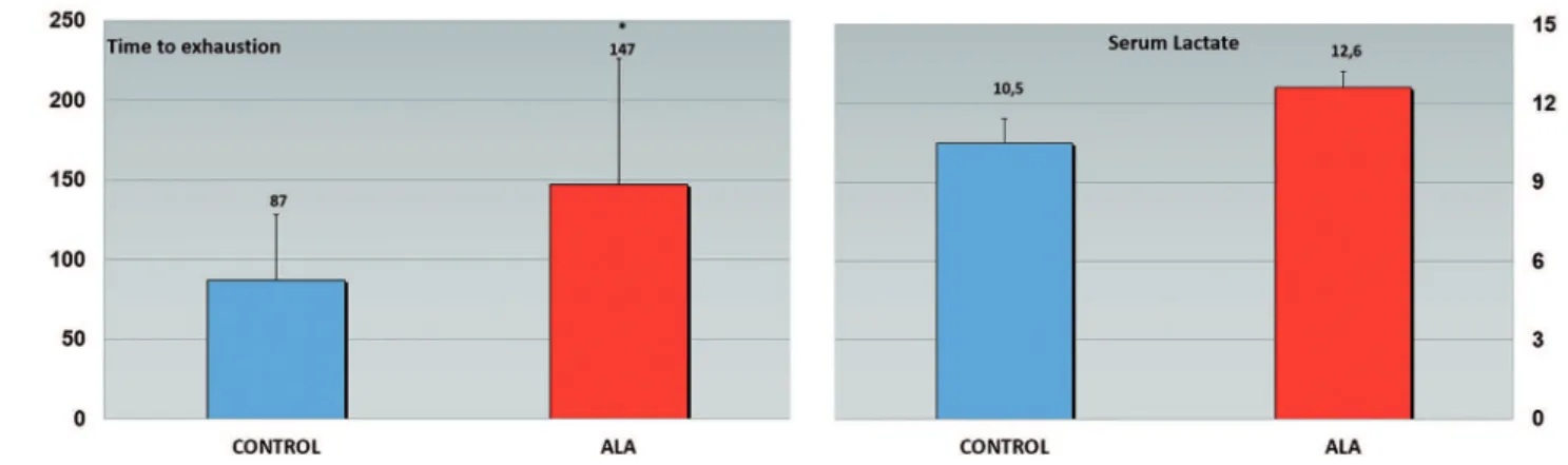
RESULTSThe exhaustive exercise session was effective, as evidenced by similar lactecemia values found in group 0 h post-exhaustion of both the Control group (10.5 ± 0.9 mmol / L) as the ALA group (12.6 ± 0.5 mmol / L) with no difference between them (p> 0.05). However, the ALA group
remained longer in exercise compared to control, 147.0 ± 79.0 vs. 87.0 ± 41.2 s (p <0.05), respectively, showing greater
exhaustive capacity (Figure 1).
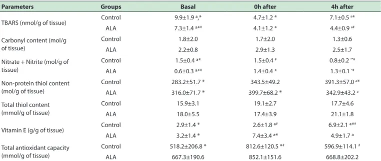
Table 1 shows the values of oxidative damage biomarkers and antioxidants. Note that in the ALA group lipid, peroxidation estimated by TBARS decreased soon after the exhaustion protocol and remained low within
4 hours, while in the control group a decrease at 0h was followed by an increase in TBARS after 4 hours of
exhaustion. No differences were found between the protein carbonyl values for the groups or tested times, although the
Figure 1 - Time to exhaustion (left) and lactate levels (right) in treated and control mice. Exhaustion was signiicantly delayed in treated mice, whereas lactate levels were equivalent.
values were higher for the supplemented group (almost 60% higher in 0h after exhaustive test). Regarding nitric oxide, tested by the sum of nitrate and nitrite, there was a
decrease from baseline in the period of exhaustion after 4
hours in the control group while the reverse was observed for the supplemented group. However, at baseline the
control group was significant higher compared with ALA
group. For non-protein thiols, there was an increase after the exhaustion for the supplemented group while this increased in the control group was observed only in the
period 4 hours after exhaustion. There were no changes
in relation to the total thiols over time or between groups.
Similarly to non-protein thiols, vitamin E increased in the
ALA group soon after exhaustion while in the control group
this increase was only seen 4 hours after exhaustion. The
only variation in the total antioxidant capacity was observed in control group that increased was observed immediately after exhaustion.
■
DISCUSSIONThe objective of this study was to evaluate the effect
of supplementation with ALA on oxidative stress in trained
mice muscle just after the exhaustion test and 4 hours after
this same test.
In global terms, we observed a benefit of
supplementation with ALA in terms of oxidative stress on the longer time to reach exhaustion. Although certainly several other adaptations promoted by the training may be involved such as enhanced cardiac output and VO2, capillary density, mitochondrial biogenesis, our experimental
design was proposed to equalize these effects by training
all groups with the same intensity and therefore the only different variable was the supplementation with a supposed antioxidant substance.
The parameters of oxidative injury analyzed have
Table 1 – Muscle biomarkers of oxidative stress in the diferent times relative to exhaustive exercise
Parameters Groups Basal 0h after 4h after
TBARS (nmol/g of tissue) Control 9.9±1.9 ª,* 4.7±1.2 * 7.1±0.5
a*
ALA 7.3±1.4 ª*# 4.1±1.2 * 4.4±0.9 a#
Carbonyl content (mol/g of tissue)
Control 1.8±2.0 1.7±2.0 1.3±0.6
ALA 2.2±0.8 2.9±1.3 2.5±1.7
Nitrate + Nitrite (mol/g of tissue)
Control 1.5±0.4 ª* 1.5±0.4 # 0.8±0.2 ª*#
ALA 0.6±0.3 ª*# 1.4±0.4 * 1.3±0.1 ª#
Non-protein thiol content (mol/g of tissue)
Control 283.2±51.7 * 343.5±49.2 391.3±57.0 a*
ALA 316.0±71.7 * 399.7±68.2 * 342.9±43.2 a
Total thiol content (mmol/g of tissue)
Control 15.9±3.1 19.1±2.7 17.7±4.6
ALA 18.0±5.5 17.4±3.9 21.1±1.8
Vitamin E (g/g of tissue) Control 2.9±1.4 * 2.6±1.8 ª
# 6.9±2.1 ª*#
ALA 3.2±1.4 * 7.4±3.4 ª* 4.9±1.7 ª
Total antioxidant capacity (mmol/g of tissue)
Control 518.2±206.8 * 812.6±120.5 *# 596.9±114.1 #
ALA 667.3±190.6 852.1±151.6 668.8±202.2
lipid peroxidation. However, although without statistical differences, the values of carbonylated protein were higher for the group supplemented with ALA. This unexpected
finding may be a metal-catalyzed prooxidant effect of ALA
supplementation. Actukeren et al (2010)18 demonstrated in
vitro the increase of carbonylated protein in the presence
of high concentrations of ALA (100µM). The vitamin E, a
potent antioxidant located on lipophilic compartments19,
was increased in the ALA group after the exhaustion while in the control group the increase was only observed after
4 hours in the same test. These results seem an effect of recruit circulating vitamin E in order to protect membranes from oxidative damage. In addition, it is possible that there was an ALA interaction in restoration of vitamin E.
ALA can also recycle the GSH or even spare it.10
An increase was observed in the concentration of
non-protein thiols 4h post-exhaustion in the control group
was observed compared to animals that did not undergo
exhaustive exercise. It was also observed and an increase
after exhaustion at the ALA group than those who did not undergo exhaustive exercise indicating that the recovery of cellular homeostasis remains several hours after exercise. N O p o s s e s s e s a n i m p o r t a n t ro l e i n l o c a l hemodynamics, and it’s physiologically caused by activation of eNOS4, which we observed in the control group at baseline while the supplemented group was possibly suppressed at the same time. However, NO reacts with superoxide anion producing a more reactive species, peroxynitrite.20
Soon after exercise nitric oxide levels increased to the supplemented group and decreased in control indicating a possible protective role in order to restore muscle
hemodynamics in the first case and possibly the reaction
of nitric oxide with superoxide anion, in the latter.
The total antioxidant capacity indicates the cumulative effect of all the antioxidants and is used to evaluate a number of physiological conditions in humans and animals.21 In our study, no differences were found
between the control group and ALA. However, in the control group noticed an increase in the concentration of
total antioxidant capacity in animals that were euthanized
soon after exhaustion than those who did this exercise
and then a decrease in the animals that were euthanized 4h after exhaustion in relation to that were euthanized 0h post-exhaustion. This finding agrees with a study by
Michailidis et al. (2007)22, that although measured in the
serum of untrained humans, found an immediate increase in the total antioxidant capacity after exhaustive treadmill running and a decline after 3 hours of exercise. This increase in total antioxidant capacity after exercise suggests that the acute exercise actives the body’s antioxidant defenses.
Our study has some limitations which have to be pointed out. First, there was no sedentary group as a control to test the effect of exercise per se as an antioxidant. However, this untrained group could bring false positive results due to inability to perform the exhaustion test. Additionally, another limitation is the fact that we used the whole gastrocnemius muscle without separating its red and white portions due to the minute amount of sample present in mice. Certainly this may be relevant for terms of comparison with other studies, such as, glutathione values.. We had no other methodology than TBARS to access lipid
peroxidation, although we know of the lack of specificity
of this methodology.
In conclusion, we have demonstrated that
supplementation with ALA induced a protective effect on oxidative stress in the post-exercise exhaustive of trained
suplementação com antioxidante na recuperação de danos
causados pelo exercício exaustivo, seja restaurando os
níveis de antioxidantes ou diminuindo os danos.
OBJETIVO: Avaliar o efeito da suplementação com ácido
α-lipóico sobre biomarcadores musculares de estresse oxidativo após o exercício exaustivo de camundongos treinados.
METODOLOGIA: Os camundongos (n = 60) foram treinados em natação por 6 semanas. Na última semana, metade dos animais foram suplementados diariamente com gavagem oral de 100 mg / kg de ácido α-lipóico
em óleo de soja como veículo. A outra metade recebeu
apenas o veículo. No último dia 20 animais de cada grupo foram submetidos ao protocolo de exaustão com 10% de sobrepeso atado à cauda. Os animais foram
eutanasiados em 3 momentos: basal, logo após o protocolo
de exaustão (0 h) e 4 h após o protocolo de exaustão. O músculo gastrocnêmio foi imediatamente coletado e
homogeneizado. Os homogeneizados foram usados para
acessar os biomarcadores de estresse oxidativo.
RESULTADOS: Houve diminuição simultânea de
tióis não protéicos e vitamina E após 4 h de exercício
exaustivo no grupo ácido α-lipóico (p <0,05) em relação ao grupo controle, sugerindo o consumo destes compostos
no processo de peroxidação lipídica. Interessantemente,
houve aumento de nitrato e nitrito no grupo ácido α-lipóico
(p <0,05) e diminuição no controle (p <0,05) em relação ao momento basal, possivelmente pela ativação da óxido
nítrico sintase endotelial. A capacidade antioxidante total permaneceu inalterada no grupo ácido α-lipóico.
CONCLUSÃO: A suplementação com ácido α-lipóico resultou em proteção contra o estresse oxidativo causado pelo exercício exaustivo.
PALAVRAS-CHAVE: Ácido alfa-lipóico; Antioxidantes;
Estresse oxidativo; Exercício.
■
REFERENCES1. Powers SK, Nelson WB, Hudson MB. Exercise-induced oxidative stress
in humans: cause and consequences. Free Radic Biol Med. 2011 Sep
1;51(5):942-50. DOI: 10.1016/j.freeradbiomed.2010.12.009. 2. Andrade FH, Reid MB, Allen DG, Westerblad H. Effect of hydrogen
peroxide and dithiothreitol on contractile function of single skeletal
muscle fibres from the mouse. J Physiol. 1998;509 (Pt 2):565-75. 3. Oztasan N, Taysi S, Gumustekin K, Altinkaynak K, Aktas O, Timur H, et
al. Endurance training attenuates exercise-induced oxidative stress in erythrocytes in rat. Eur J Appl Physiol. 2004;91(5-6):622-7. DOI: 10.1007/s00421-003-1029-6
4. Dyakova EY, Kapilevich LV, Shylko VG, Popov SV, Anfinogenova Y.
Physical exercise associated with NO production: signaling pathways
and significance in health and disease. Front Cell Dev Biol. 2015;3:19. DOI: 10.3389/fcell.2015.00019.
5. Powers SK, DeRuisseau KC, Quindry J, Hamilton KL. Dietary
antioxidants and exercise. J Sports Sci. 2004;22(1):81-94. DOI:
10.1080/0264041031000140563
6. Polotow TG, Vardaris CV, Mihaliuc AR, Goncalves MS, Pereira B, Ganini D, et al. Astaxanthin supplementation delays physical exhaustion and prevents redox imbalances in plasma and soleus muscles of Wistar
rats. Nutrients. 2014;6(12):5819-38. DOI:10.3390/nu6125819
mice, perceived through the simultaneous increase of
non-protein thiols and vitamin E in the process of lipid
peroxidation. The recovery of cellular homeostasis continues for several hours after exercise, as perceived
by the increase in both vitamin E and non-protein thiols 4h post-exhaustion in the control group, although the role of the enzymatic antioxidant system (eg, superoxide
dismutase, catalase, glutathione peroxidase, among others) can not be ruled out. Only the total antioxidant capacity remained unchanged in the supplemented group, and nitrate and nitrite increased with supplementation.
■
ACKNOWLEDGEMENTSThe authors would like to thank the Coordenação
de Aperfeiçoamento de Pessoal de Nível Superior (CAPES),
by granting of the scholarship to SM. This study was partially supported by a grant (CDS - APQ-01606-10) of
the Fundação de Amparo à Pesquisa do Estado de Minas Gerais (FAPEMIG).
■
AUTHOR CONTRIBUTIONSMerino S: conception, design, intellectual and
scientific content of the study, technical procedures, acquisition of data, critical revision, manuscript writing; Moraes RCM: critical revision, manuscript writing; Deminice R: intellectual and scientific content of the study, critical revision, manuscript writing; Orsatti FL: intellectual and scientific content of the study, critical revision, manuscript writing; Portari GV: conception, design, intellectual and scientific content of the study, technical procedures, critical
revision, manuscript writing. Supervisor.
■
CONFLICT OF INTERESTThe authors declare that no conflict of interest with respect to tis project.
EFEITOS DA SUPLEMENTAÇÃO COM ÁCIDO ALFA-LIPÓICO SOBRE BIOMARCADORES ANTIOXIDANTES MUSCULARES DE CAMUNDONGOS TREINADOS
CONTEXTO: A realização de exercício de alta intensidade ou exaustivo pode levar a danos musculares, como lesões, fadiga crônica e overtraining, em parte devido
à alta síntese de espécies reativas de oxigênio. O ácido α-lipóico e sua forma reduzida, o ácido dihidrolipóico, atuam
como potentes antioxidantes e eliminam os radicais livres. Apesar de depender do tipo de exercício e suplementação,
7. Slattery KM, Dascombe B, Wallace LK, Bentley DJ, Coutts AJ. Effect of N-acetylcysteine on cycling performance after intensified training. Med sci in sports and exerc. 2014;46(6):1114-23. DOI: 10.1249/
MSS.0000000000000222
8. Zembron-Lacny A, Slowinska-Lisowska M, Szygula Z, Witkowski K, Stefaniak T, Dziubek W. Assessment of the antioxidant effectiveness of
alpha-lipoic acid in healthy men exposed to muscle-damaging exercise.
J Physiol Pharmacol . 2009 Jun;60(2):139-43.
9. Khanna S, Atalay M, Laaksonen DE, Gul M, Roy S, Sen CK. Alpha-lipoic
acid supplementation: tissue glutathione homeostasis at rest and after
exercise. J Appl Physiol. (1985). 1999:86(4):1191-6.
10. Chae CH, Shin CH, Kim HT. The combination of alpha-lipoic acid supplementation and aerobic exercise inhibits lipid peroxidation in
rat skeletal muscles. Nutr Res. 2008;28(6):399-405. DOI: 10.1016/j.
nutres.2008.02.010.
11. Coombes JS, Powers SK, Rowell B, Hamilton KL, Dodd SL, Shanely RA, et al. Effects of vitamin E and alpha-lipoic acid on skeletal muscle contractile properties. J Appl Physiol (1985). 2001;90(4):1424-30.
12. Donatto FF, Prestes J, Frollini AB, Palanch AC, Verlengia R, Cavaglieri CR. Effect of oat bran on time to exhaustion, glycogen content and serum cytokine profile following exhaustive exercise. J Int Soc Sports Nutr. 2010;7:32. DOI: 10.1186/1550-2783-7-32. 13. Odetti P, Garibaldi S, Gurreri G, Aragno I, Dapino D, Pronzato MA, et
al. Protein oxidation in hemodialysis and kidney transplantation.
Metabolism. 1996;45(11):1319-22. DOI: 10.1016/S0026-0495(96)90108-0
14. Buege JA, Aust SD. Microsomal lipid peroxidation. Methods in enzymology. 1978;52:302-10.
15. Miranda KM, Espey MG, Wink DA. A rapid, simple spectrophotometric
method for simultaneous detection of nitrate and nitrite. Nitric Oxide.
2001;5(1):62-71. DOI: 10.1006/niox.2000.0319
16. Sedlak J, Lindsay RH. Estimation of total, protein-bound, and nonprotein sulfhydryl groups in tissue with Ellman’s reagent. Anal Biochem. 1968;25(1):192-205.
17. Arnaud J, Fortis I, Blachier S, Kia D, Favier A. Simultaneous
determination of retinol, alpha-tocopherol and beta-carotene in serum
by isocratic high-performance liquid chromatography. J Chromatogr. 1991;572(1-2):103-16.
18. Atukeren P, Aydin S, Uslu E, Gumustas MK, Cakatay U. Redox
homeostasis of albumin in relation to alpha-lipoic acid and
dihydrolipoic acid. Oxid Med Cell Longev. 2010;3(3):206-13. DOI: 10.4161/oxim.3.3.11786.
19. Urso ML, Clarkson PM. Oxidative stress, exercise, and antioxidant
supplementation. Toxicology. 2003;189(1-2):41-54. DOI: 10.1016/ S0300-483X(03)00151-3
20. Powers SK, Jackson MJ. Exercise-induced oxidative stress: cellular
mechanisms and impact on muscle force production. Physiol Rev.
2008;88(4):1243-76. DOI: 10.1152/physrev.00031.2007
21. Ghiselli A, Serafini M, Natella F, Scaccini C. Total antioxidant capacity
as a tool to assess redox status: critical view and experimental data.
Free radical biology & medicine. 2000;29(11):1106-14. DOI: 10.1016/ S0891-5849(00)00394-4