ALVES LS ETAL.
500 REV ASSOC MED BRAS 2017; 63(6):500-503
ORIGINAL ARTICLE
Concomitant testicular infection by Zika virus and
Schistosoma
mansoni
in a Brazilian young boy
LEONARDO SOUZA ALVES1*, CESAR ESTANISLAU2, LUCIO BARRETO3, FRANCISCO BATISTA3, NIVALDO TOPPA4
1MD, Urologist. Sociedade Brasileira de Urologia. American Urologic Association. Procriar - Instituto de Urologia e Andrologia, Belo Horizonte, MG, Brazil 2Biologist. Departament of Biologic Sciences, Faculdade Newton de Paiva, Belo Horizonte, MG, Brazil
3MD, Faculdade Ciências Médicas de Minas Gerais (FCM-MG), Belo Horizonte, MG, Brazil 4Patologist. Ex-Professor of Pathology, UFMG, Belo Horizonte, MG, Brazil
S
UMMARYStudy conducted by Procriar – Instituto de Urologia e Andrologia, Belo Horizonte, MG, Brazil
Article received: 10/29//2016
Accepted for publication: 11/20/2016
*Correspondence:
Address: Rua da Bahia, 2.696, sala 1.303 Belo Horizonte, MG – Brazil Postal code: 30120-016 [email protected]
http://dx.doi.org/10.1590/1806-9282.63.06.500
The identification of a escrotal mass without pain or report of trauma should be investigated to rule out scrotal cancer. We report the case of a young Brazilian boy who underwent orchiectomy after magnetic resonance imaging (MRI) and duplex scan (DS) indicating a high possibility of cancer. Blood exams ruled out the possibility of cancer. Testicular biopsy was not indicated. After surgery the
diagnostic was extensive orchiepididymitis by Schistosoma. In endemic areas
orchiepididymis by Schistosoma should be investigate to avoid unnecessary surgeries. This patient was also infected with Zika virus.
Keywords: Zika virus, Schistosoma mansoni, testicle, orchiepididymitis.
C
ASEWe report the case of a young Brazilian boy, who was referred with inflammation of the right testicle and epididymis. The patient presented with volumetric expansion but no pain, without any apparent cause. Scrotal trauma or unpro-tected sexual activity were not reported. The preliminary scans, magnetic resonance imaging (MRI) and duplex scan determinated a possible testicular cancer. Analysis of sper-mogram showed the presence of immobile and dead sperm. The analysis of sperm culture showed no infection. Labora-tory exams included: Alpha-fetoprotein (AFP) blood test, lactate dehydrogenase (LDH) and beta-human chorionic gonadotropin (hCG), all normal. The patient was prescribed the use of antibiotic and anti-inflammatory medication for 10 days, but no reduction in testicular mass size was observed. After talking to the patient about the diagnostic difficulties, we opted for orchiectomy.
Preoperative tests were all normal. The surgery was uneventful, and the patient was discharged on the same day. The examination of the surgical piece demonstrated extensive right testicular and epididymal fibrosis. The microscopic examination of the testis ruled out the pos-sibility of cancer, but confirmed the diagnosis of extensive loss of testicular structure and schistosome egg-induced granulomas (Figure 1). After surgery, the patient’s relatives reported that he had had contact with stagnant water
approximately 60 days before the symptoms started. Ac-cording to the family, the boy was admitted to the hospi-tal with flu-like symptoms, low fever and rashes on chest and lower limbs. At the time of hospitalization, infection with Dengue virus was suspected. Symptomatic treatment was initiated with analgesics. The patient was hospitalized for 48 hours and then discharged. The blood test (RT-PCR) performed at the time of admission confirmed later that the etiology was a Zika virus infection. The patient was discharged with instructions for frequent hydration and no more. After surgery, the patient received oral treatment with oxamniquine to treat infection with Schistosoma, being instructed to only have sexual intercourse with the use of condoms for a period of 6 months.
Z
IKA VIRUSZika virus (ZIKV) is an RNA virus of the Flaviviridae fam-ily, related to the Dengue and Yellow fever viruses. It causes Zika fever, a disease with symptoms similar to Dengue fever and Chikungunya, but more lenient (Figure 2).
CONCOMITANTTESTICULARINFECTIONBY ZIKAVIRUSANDSCHISTOSOMAMANSONIINA BRAZILIANYOUNGBOY
REV ASSOC MED BRAS 2017; 63(6):500-503 501
2014 FIFA World Cup, also being carried by immigrants from Africa.2,3,8,11 Symptoms include: mild fever, pruritus
and cutaneous rash, fatigue, myalgia, dehydration, diar-rhea, photophobia, dizziness and loss of appetite. These symptoms may remain for 7 to 30 days. Unlike the Den-gue virus, the Zika does not usually cause blood dyscra-sia.5-8 ZIKV is transmitted through the bite of an infected Aedes aegypti mosquito, which is also a vector of Dengue virus and is endemic to the regions affected.2-8,19
Transmis-sion through saliva and/or semen is still controversial, although the presence of live ZIKV has been detected in
these secretions.6-8 The virus has also been detected in
urine. The diagnosis of the virus is usually done clini-cally. Blood tests are not always available, are costly and take 2 to 7 days to confirm the diagnosis. Enzyme-linked immunosorbent assay (ELISA)-based serological tests can be used for the diagnosis of ZIKV. The test detects the presence of antibodies against ZIKV and, therefore, at the beginning of the symptoms, the test is negative. It should be requested five days after the first signs appear. Real time polymerase chain reaction (RT-PCR) is a method of DNA and RNA amplification that does not use living
organisms and can detect ZIKV as symptoms occur.6,9-11
ZIKV can be detected in samples of semen or saliva using PCR, but these virus identification kits are not widely available in all countries. The clinical picture triggered by ZIKV resembles a flu-like condition, but chronic com-plications are more important. Guillain-Barré syndrome (GBS) is a neurological complication that can be fatal, with progressive paralysis of the muscles of the body. In microcephaly, which is brain malformation due to
infec-tion with the virus, the cephalic perimeter is smaller than usual, leading to permanent neurologic loss. It occurs in
fetuses of mothers who were infected with ZIKV.2
Treat-ment is usually supportive and depends on the severity of symptoms. The patient usually remains in the hospital for 24-48 hours, under observation with analgesic and antipyretic drugs only. Despite the production of antibod-ies against ZIKV and Dengue virus, it is not known wheth-er this is lasting immunity or not.9-11,19 In cases like GBS
or microcephaly, the patients will need hospitalization and continuous care.
S
CHISTOSOMASchistosomes are trematode parasites approximately 1 cm (male) to 1.5 cm (female) long. Schistosome infection is frequent in developing countries with low socioeco-nomic conditions. Schistosomes are found as three dif-ferent species, and are considered endemic in Central and
South America (S. mansoni), and in the Middle East and
Asia (S. haematobium and S. japonicum). In the Americas,
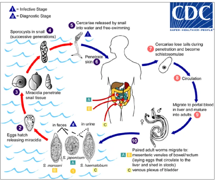
the disease is known as water belly or snail fever. The par-asite is found in B. glabrata snails, the intermediate host, in freshwater ecosystems.1,13-15,17
In the reproductive cycle, the parasite infects the snail, where self-reproduction takes place. The eggs are then discarded in standing water. At that point, the parasite is eliminated (cercaria stage) in water, where its definitive
host (human) is infected. Once in the human body, S.
mansoni has tropism for mesenteric plexus, veins in the pelvic region. In these areas, the parasite reproduces once again and the eggs reach the entire body through veins FIGURE 1 Granuloma in ibrosis phase with a viable S. mansoni egg
in the middle. (Photo by Nivaldo H. Toppa).
ALVES LS ETAL.
502 REV ASSOC MED BRAS 2017; 63(6):500-503
and lymphatic vessels, especially the liver and the spleen (Figure 3). There are reports of eggs found in the kidneys, bladder, prostate, epididymis and testes, which are usu-ally discovered post mortem.15 The infectious process that
follows is due to the intense immune reaction against the eggs of Schistosoma. The typical lesion is that of granulomas with spiculated egg in a central position (Figure 1).
The granuloma involves the Schistosoma with mast cells, macrophages, multi-nucleated giant cells (Langhans’ cells), eosinophils, lymphocytes and fibroblasts. In the process, there is extensive inflammation and fibrosis, egg deposi-tion, and destruction of organ architecture.17,18
D
ISCUSSIONThis is the case of a Brazilian boy, living in an urban area, which starts with a history of testicular increase, with no
apparent cause. The initial clinical report, laboratory tests and imaging methods led to a diagnosis of malignant tes-ticular injury. In cases of testes-ticular lesions with suspicion of testicular cancer, testicular biopsy should not be done, as it can promote the spread of malignant cells and loss of surgical healing potential. In this context, the difficulties related to diagnosis and treatment were properly explained to the patient. After the postoperative diagnosis of schis-tosomal orchitis, and in the absence of malignant tumor of the testicle, it became clear that the collection of clinical data was incomplete. Contact with still waters and wheth-er it was infested with the intwheth-ermediate host S. mansoni was not investigated. Contact with stagnant water 60 days before the onset of symptoms was later confirmed by the patient. We did not inquire about hospitalization for treatment of ZIKV infection, despite the existence of a positive RT-PCR.
CONCOMITANTTESTICULARINFECTIONBY ZIKAVIRUSANDSCHISTOSOMAMANSONIINA BRAZILIANYOUNGBOY
REV ASSOC MED BRAS 2017; 63(6):500-503 503
We cannot say if the first infection was that of Schis-tosoma or Zika. However, literature shows that ZIKV can remain viable in the testicle due to the natural barrier that prevents the formation of testicular antibodies for
up to 6 months.14-19 Schistosome can remain alive in a
definitive host for up to 20 years.18 However, with Schis-tosoma infection and the intense inflammation with granuloma formed around the eggs, this protective bar-rier to ZIKV was lost, which leads us to believe that the virus was “attacked” by the host’s natural defense cells. Unfortunately, we were not able to identify the presence of the virus in the surgical piece due to lack of resources at that time. The literature reports the identification of ZIKV in secretions such as saliva, semen and urine.3-5,7,8,10,19
Test kits for these diagnoses are not always available, but lack of identification does not mean necessarily that the microorganism is not present. Examinations can yield false positives due to a window period.10-12,19 The patient
was told to use condoms for a period of 6 months, in ac-cordance with the guidelines of health agencies such as the CDC, WHO and Anvisa.3,6,8,10,11,19
C
ONCLUSIONConcurrent infection with the two agents may or may not have been a coincidence. We emphasize that all inhabitants of risky areas, as well as people traveling to these places, can be infected with ZIKV and Schistosoma. As the risky areas are
endemic for the vectors Aedes aegypt (ZIKV) and
Biomphal-aria glabrata (Schistosoma), the risk of contracting the two diseases concomitantly is reasonable. The potential com-plications of two concomitant parasitic infections should determine the efforts to be made by all in order to decrease the prevalence of these diseases. Ultimately, it all comes down to education, basic sanitation and personal hygiene.
R
ESUMOInfecção testicular associada com Zika vírus e Schistosoma
mansoni em jovem brasileiro
A identificação de massa escrotal sem relato de dor ou trauma deve ser investigada a fim de afastar a possibili-dade de câncer escrotal. O artigo reporta o caso de um jovem brasileiro que apresentou massa escrotal com essas características. Ressonância nuclear magnética (RNM) e ultrassonografia (US) indicaram grande possibilidade de câncer. Os marcadores tumorais sanguíneos estavam normais, e a biópsia não poderia ser realizada. O resulta-do anatomopatológico diagnosticou extensa fibrose es-quistossomótica, associada a quadro clínico e sorológico
de Zika vírus concomitantemente. Em regiões endêmicas, pacientes com alterações escrotais devem ser pesquisados a fim de evitar cirurgias desnecessárias.
Palavras-chave: vírus Zika, Schistosoma mansoni, testícu-lo, orquiepididimitis.
A
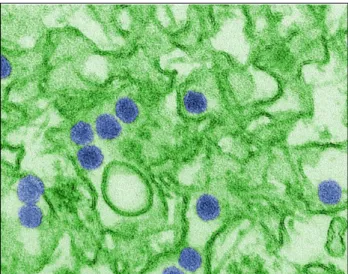
CKNOWLEDGMENTSWe thank Cynthia Goldsmith, CDC/USA, for the ZIKV microphotography. Also, the US CDC for allowing the use of ZIKV and Schistosome lifecycle figures, and F.G., the patient, who allowed us to write this manuscript.
R
EFERENCES1. Nussenzweig RS. Parasitic disease as a cause of immunosuppression. New Engl J Med. 1982; 306(7):423-4.
2. Oehler E, Watrin L, Larre P, Leparc-Goffart I, Lastere S, Valour F, et al. Zika virus infection complicated by Guillain-Barré syndrome--case report, French Polynesia, December 2013. Euro Surveill. 2014; 19(9).pii:20720. 3. Musso D, Roche C, Robin E, Nhan T, Teissier A, Cao-Lormeau VM. Potential
sexual transmission of Zika virus. Emerg Infect Dis. 2015; 21(2):359-61. 4. Gourinat AC, O’Connor O, Calvez E, Goarant C, Dupont-Rouzeyrol M.
Detection of Zika virus in urine. Emerg Infect Dis. 2015; 21(1):84-6. 5. Oliveira Melo AS, Malinger G, Ximenes R, Szejnfeld PO, Alves Sampaio S,
Bispo de Filippis AM. Zika virus intrauterine infection causes fetal brain abnormality and microcephaly: tip of the iceberg? Ultrasound Obstet Gynecol. 2016; 47(1):6-7.
6. Oster AM, Brooks JT, Stryker JE, Kachur RE, Mead P, Pesik NT, et al. Interim guidelines for prevention of sexual transmission of Zika virus - United States, 2016. MMWR Morb Mortal Wkly Rep. 2016; 65(5):120-1.
7. Musso D, Roche C, Nhan TX, Robin E, Teissier A, Cao-Lormeau VM. Detection of Zika virus in saliva. J Clin Virol. 2015; 68:53-5.
8. Atkinson B, Hearn P, Afrough B, Lumley S, Carter D, Aarons EJ, et al. Detection of Zika virus in semen. Emerg Infect Dis. 2016; 22(5):940. 9. Hamel R, Dejarnac O, Wichit S, Ekchariyawat P, Neyret A, Luplertlop N, et
al. Biology of Zika virus infection in human skin cells. J Virol. 2015; 89(17):8880-96.
10. Mansuy JM, Dutertre M, Mengelle C, Fourcade C, Marchou B, Delobel P, et al. Zika virus: high infectious viral load in semen, a new sexually transmitted pathogen? Lancet Infect Dis. 2016; 16(4):405.
11. Hills SL, Russell K, Hennessey M, Williams C, Oster AM, Fischer M, et al. Transmission of Zika virus through sexual contact with travelers to areas of ongoing transmission—continental United States, 2016. MMWR Morb Mortal Wkly Rep. 2016; 65(8):215-6.
12. Oster AM, Russell K, Stryker JE, Friedman A, Kachur RE, Petersen EE, et al. Update: Interim guidelines for prevention of sexual transmission of Zika virus-United States, 2016. MMWR Morb Mortal Wkly Rep. 2016; 65(12):323-5. 13. Katz N, Almeida, K. Esquistossomose, xistosa, barriga d’água. Ciênc Cultura.
2006; 55(1):38-43.
14. De Silva S, Walsh J, Rown M. Symptomatic Schistosoma mansoni infection as an immune restoration phenomenon in a patient receiving antiretroviral therapy. Clin Infect Dis. 2006; 42(2):303-4.
15. Arban V. Lesions caused by Schistosoma mansoni in the genitourinary tract of men. Am J Clin Pathol. 1956; 26(9):1010-21.
16. Rambau PF, Chandika A, Chalya PL Jackson K. Scrotal swelling and testicular atrophy due to schistosomiasis in a 9-year-old boy: a case report. Case Rep Infect Dis. 2011; 2011:787961.
17. Francolugo-Vélez VA, Zarzosa-Alguiar J. Infección del tracto urinario por Schistosoma haematobioum. Un caso en Cuernavaca, Morelos. México. Rev Mex Urol. 2010; 70(3):187-92.
18. Iglesias JD. Aspectos médicos das parasitoses humanas. São Paulo: Guanabara Koogan; 1997. p. 186-210.