Mueller-Hillis maneuver and angle of progression: Are they correlated?
SOFIA MENDES1*, RITA SILVA1, INÊS MARTINS1, SUSANA SANTO1, NUNO CLODE11MD, Department of Obstetrics, Gynecology and Reproductive Medicine, Hospital de Santa Maria, Lisboa, Portugal
S
UMMARYStudy conducted at the Departamento de Obstetrícia, Ginecologia e Medicina da Reprodução, Hospital de Santa Maria, Lisboa, Portugal
Article received: 11/24/2016
Accepted for publication: 12/4/2016
*Correspondence:
Departamento de Obstetrícia, Ginecologia e Medicina da Reprodução, Hospital de Santa Maria Address: Av. Prof. Egas Moniz Lisboa – Portugal Postal code: 1649-035 [email protected]
http://dx.doi.org/10.1590/1806-9282.63.06.527
Objective: Mueller-Hillis maneuver (MHM) and angle of progression (AOP) measured by transperineal ultrasound have been used to assess fetal head descent during the second stage of labor. We aimed to assess whether AOP correlates with MHM in the second stage of labor.
Method: A prospective observational study including women with singleton pregnancy in the second stage of labor was performed. The AOP was measured immediately after the Mueller-Hillis maneuver. A receiver-operating characteristics (ROC) curve analysis was performed to determine the best discriminatory AOP cut-off for the identification of a positive MHM. A p-value less than 0.05 was considered statistically significant.
Results: One hundred and sixty-six (166) women were enrolled in the study and 81.3% (n=135) had a positive MHM. The median AOP was 143° (106° to 210°). The area under the curve for the prediction of a positive maneuver was 0.619 (p=0.040). Derived from the ROC curve, an AOP of 138.5° had the best diagnostic performance for the identification of a positive MHM (specificity of 65% and a sensitivity of 67%).
Conclusion: An AOP of 138° seems to be associated with a positive MHM in the second stage of labor.
Keywords: second stage of labor, angle of progression, Mueller-Hillis maneuver, intrapartum, ultrasonography.
I
NTRODUCTIONSpontaneous vaginal delivery is the desirable mode of de-livery for most pregnancies. However, some women fail to progress into second stage of labor and require obstetric intervention. Progression of labor is traditionally assessed by digital examination. In 1885, Mueller1 described a
ma-neuver in which an assistant applies fundal pressure and a second examiner determines descent of the fetal present-ing part. The technique was modified by Hillis2 in 1930 to
allow its execution by a single person, and later became known as Mueller-Hillis maneuver (MHM). According to March et al., a positive Mueller-Hillis maneuver should reassure the clinician of an exceptional likelihood of achiev-ing a vaginal delivery.3 Thorp et al.,4 however, failed to prove
its utility to predict dystocia. Moreover, for the determina-tion of fetal head stadetermina-tion, several other studies showed that digital vaginal examination5-7 is subjective with high
inter--observer variability. Intrapartum transperineal ultrasonog-raphy (ITU) has been suggested as a much more reliable method for assessing fetal head descent, as it provides
objective and reproducible results.8 It is neither
time-con-suming nor causes discomfort of the patient.9 However, it
may not be available everywhere. The angle of progression measured by transperineal ultrasound in the second stage of labor has been shown to be useful in predicting spon-taneous vaginal delivery.10,11
The aim of our study was to assess whether the angle of progression correlates with the Mueller-Hillis maneu-ver in the second stage of labor.
M
ETHODFrom November 2014 to September 2015, we conducted a prospective observational study at our unit. Pregnant women with a single fetus in cephalic presentation in the early second stage of labor were included.
op-posite hand on the uterine fundus. The descent of the head with reference to the interspinous line was evaluated. The examination was performed between contractions. A positive Mueller-Hillis maneuver was defined as descent of the fetal head of at least one centimeter. Any lesser degree of descent was defined as a negative result. After clinical evaluation, transperineal ultrasound was performed. The ultrasound transducer was placed on the perineum in a mid-sagittal position between the labia, below the pubic symphysis. A sagittal view with clear visualization of the pubic symphy-sis and of the fetal skull was obtained. Two lines were drawn: a line parallel to the long axis of the symphysis and a line tangential to the fetal head. The so-called angle of progres-sion between the constructed lines was then measured directly on the screen and registered on each patient file.10
The measurement was performed between contractions. Both examinations were performed by eight different clinicians with at least two years of experience. All of them received training on how to do transperineal ultrasound. MHM and AOP were measured by two different clinicians that were blinded to the findings of each other in order to avoid bias.
For ultrasound evaluation, we used a portable machine (ALOKA® IP-1233).
Baseline sociodemographic and clinical characteristics were summarized using descriptive statistics. The primary endpoint was to assess the correlation between AOP and a positive Mueller-Hillis maneuver, which was evaluated using a receiver-operating characteristic (ROC) curve. Ad-ditionally, an analysis to assess if AOP or MHM influenced the type of delivery (normal vs. instrumental) was performed. Chi-square test was used to evaluate nominal variables and ANOVA, t-test and Kruskall-Wallis were used for quantitative variables. Results were considered statisti-cally significant when p-value was < 0.05. The statistical analysis was performed using SPSS software version 20.0.
R
ESULTSOne hundred and sixty-six (166) women were enrolled during the study. The mean maternal age was 30.1 (±5.3) years. The mean gestational age was 39.1 (±1.4) weeks. One hundred and one (101/60.8%) women were nuliparous and 65 (39.2%) were multiparous. Oxytocin was admin-istered to 161 (97%) women and regional anesthesia was given to 164 (98.7%) women. Seventy-five (75) women had normal vaginal delivery, while 91 had instrumental vagi-nal delivery and, of these, two had a failed trial of instru-mental delivery and one had cesarean section. The mean birth weight of newborns was 3,305 (±392) grams. The patient characteristics are shown in Table 1.
TABLE 1 Description of study population – patient characteristics.
N (%) or median (range)
Age (years) 31 (15-43)
Race Caucasian African Other
141 (84.9) 22 (13.3) 3 (1.8) Parity
Nuliparous Multiparous
101 (60.8) 65 (39.2)
Gestational age (weeks) < 37
≥ 37
39 (32-41) 7 (4.2) 159 (95.8) Body mass index
< 30 ≥ 30
133 (80.1) 33 (19.9) Labor induction
Yes No
53 (31.9) 110 (66.3)
Labor augmentation with oxytocin Yes
No
16 (97) 5 (3)
Regional anesthesia 164 (98.7)
Delivery
Mode of delivery Normal
Instrumental vaginal delivery Prolonged 2nd stage of labor
Fetal distress
Shorten the 2nd stage of labor
Cesarean section
Failure to progress in 2nd stage of labor
75 (45.2) 89 (53.6) 70 (78.6) 15 (16.8)
4 (4.5) 2 (1.2)
Fetal birth weight (g) 3,282 (2,100-4,410)
Apgar score < 7 at the 5th minute 0
The median AOP was 143° (106° to 210°). One hundred and thirty-five (135/80.8%) women had a positive MHM. A ROC curve was plotted, evaluating the sensibility and
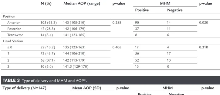
No differences were found in AOP considering fetal position, but the proportion of positive/negative MHM was significantly different between anterior and transverse positions. Regarding the AOP or MHM and fetal head stations no significant differences were noted (Table 2). Moreover, no differences between the type of delivery and AOP or MHM were found. However, when instrumental vaginal delivery for fetal distress or shortening of the 2nd
stage were excluded, the difference became statistically significant for both methods (Table 3).
D
ISCUSSIONThe main objective of the obstetrician is to promote the safe birth of a healthy baby. Most births occur vaginally and spontaneously; however, there are situations in which this is not possible. The success of an instrumental vagi-nal delivery depends on a proper obstetric assessment prior to the procedure. Patients who are inappropriately qualified for vaginal instrumental delivery (too high po-sition of fetal head) are at an increased risk of complica-tions after multiple vacuum traction or failed forceps procedure. The Mueller-Hillis maneuver constitutes one of the first attempts to predict dystocia. Thorp et al.4
performed MHM on 106 pregnant women after active labor was diagnosed. The authors couldn’t find any dif-ferences in the rate of abdominal delivery or operative
vaginal delivery whether the maneuver was positive or negative. On the other hand, March et al.3 concluded that
a positive maneuver in the second stage of labor is strong-ly associated with a vaginal delivery and that a negative one associates significantly with prolonged second stage, and higher cesarean section rate. Despite these conflicting results and lack of other studies, the Mueller-Hillis ma-neuver is still being used.
Intrapartum ultrasound has been suggested to over-come the subjectivity of clinical assessment. Various ultra-sound measurements have been proposed. The angle of progression was describe by Barbera et al.10 who showed
that an angle of at least 120° was always associated with subsequent spontaneous vaginal delivery with a good intra- and inter-observer variability. Torkildsen et al.11 and
Egg-ebo et al.12 reported that for primiparous women with
prolonged first stage of labor, the AOP could predict the probability of a vaginal delivery being the optimal cut-off 110°. Kalache et al.13 reported that for pregnant women in
a prolonged second stage of labor, there is a 90% chance of vaginal delivery with an AOP of 120°. However, the value of AOP associated with spontaneous delivery is not con-sensual, since studies pointed out higher values for AOP.14,15
Globally, in our study, AOP showed a poor correlation with MHM, indicated by a low AUC. Nevertheless, we have found that AOP over 138.5° had the best diagnostic per-formance for the identification of positive MHM in the early second stage of labor but with a low sensibility and specificity that preclude its use for clinical practice.
We included women with indication to expedite de-livery not only due to dystocia but also to fetal distress. None of the methods are expected to be used to predict situations that require prompt obstetric interventions. In fact, when excluding cases of fetal distress/shortened second stage of labor, both AOP and MHM seemed to be associated with type of delivery. The main purpose of our study, however, was to find a relation between the ma-neuver and AOP and not between both methods and the type of delivery. Nevertheless, a subanalysis was made excluding cases of fetal distress, and the results were similar (data not shown).
To our knowledge, ours is the first study to correlate MHM with AOP; however, there are important limitations. We made a single measurement of the AOP after MHM but, since labor is a dynamic process, repeating measure-ments of both angle and MHM every 30 minutes might be helpful. In fact, Ghi et al.14 reported that, in the first
40 minutes of the second stage of labor, AOP was useful for predicting mode of delivery but afterwards the differ-ence lost its statistical differdiffer-ence. Moreover, as
previ-FIGURE 1 ROC curve to determine the best discriminatory AOP
cut-off for the identiication of a positive MHM.
Sensitivity
Speciicity
Diagonal segments are produced by ties ROC curve
1.0
0.8
0.6
0.4
0.2
0.0
1.0 0.8
0.6 0.4
0.2 0.0
AUC (95CI) p-value
ously recorded,16 uterine contractions and active maternal
pushing seem to affect AOP in the second stage of labor. We made a single measurement of the AOP after the MHM
and not during the maneuver and therefore we do not know if this could change the results.
In our study, both examinations were performed im-mediately after full cervical dilation was determined, i.e., in the early second stage of labor. Consistent timing for the examination enables a more accurate correlation of both methods but it is difficult to exactly assess the mo-ment of full cervical dilation. Moreover, other variables might have affected the maneuver’s outcome, such as maternal habitus, position of the head, flexion/extension of the head, force applied or resistance of the maternal abdomen. Fetuses presenting as occiput posterior position are thought to follow different paths of descent16 and,
since we did not evaluate separately this subset of fetuses, which might have been a limitation of our study. Lastly, because ultrasound measurements and clinical assess-ments were performed by residents with different levels of experience, and this variable may influence the clinical evaluation,17 the inverse correlation between lower head
station and higher AOP was not seen in our population. If one method can predict spontaneous delivery with a high likelihood, it may prevent unnecessary obstetric
interventions. The ideal method for obstetric assessment is one that is available worldwide, easy to perform and reproducible. Regardless of the individual value of the MHM and AOP as previously reported to predict the type of delivery (which is debatable in both methods), our data failed to find a strong correlation between these methods.
C
ONFLICT OF INTERESTThe authors declare no conflict of interest.
R
ESUMOManobra de Mueller-Hillis e ângulo de progressão: eles estão correlacionados?
Objetivo: A manobra de Mueller-Hillis (MHM) e o ângu-lo de progressão da apresentação (AOP) medido através de ecografia transperineal têm sido utilizados para avaliar a descida do polo cefálico durante o segundo estágio do trabalho de parto. O objetivo do nosso trabalho foi avaliar se o AOP se correlaciona com a MHM no segundo estágio do trabalho de parto.
Método: Conduzimos um estudo observacional e pros-pectivo. Incluímos mulheres com gravidez unifetal com feto em apresentação cefálica, no segundo estágio do
TABLE 2 Position and head station – only assessed by clinical examination.
N (%) Median AOP (range) p-value MHM p-value
Positive Negative
Position
Anterior 105 (63.3) 143 (108-210) 0.288 90 14 0.020
Posterior 47 (28.3) 142 (106-179) 37 11
Transverse 14 (8.4) 141 (123-165) 8 6
Head Station
≤ 0 22 (13.2) 135 (123-163) 0.406 17 4 0.310
1 73 (43.7) 144 (106-210) 56 17
2 62 (37.1) 142 (113-179) 52 10
3 10 (6.0) 141.5 (129-170) 10 0
TABLE 3 Type of delivery and MHM and AOP*.
Type of delivery (N=147) Mean AOP (SD) p-value MHM p-value Positive Negative
Normal 145.33 (±13.7) 0.034++ 67 8 0.005**
Instrumental vaginal delivery 140.52 (±14.7) 67 22
Cesarean section 129.5 (±12.0) 1 1
*Excluding instrumental vaginal deliveries indications: “fetal distress” and “shorten the 2nd stage of labor”. ++ t-student, comparing normal vs. operative delivery (instrumental plus cesarean).
trabalho de parto. O AOP foi medido imediatamente após a manobra de Mueller-Hillis. Foi construída uma curva ROC (receiver-operating characteristics) para determinar o melhor AOP para a identificação de uma manobra posi-tiva. Um valor p inferior a 0,05 foi considerado estatisti-camente significativo.
Resultados: Cento e sessenta e seis mulheres (166) foram incluídas no estudo, e em 81,3% (n=135) a MHM foi po-sitiva. A mediana do AOP foi de 143° (106° a 210°). A área abaixo da curva para a previsão de uma manobra positiva foi 0,619 (p=0,040). Derivado da curva ROC, um AOP de 138,5° teve o melhor desempenho diagnóstico para a identificação de uma MHM positiva (especificida-de (especificida-de 65% e sensibilida(especificida-de (especificida-de 67%).
Conclusão: Um AOP de 138° parece estar associado com uma MHM positiva no segundo estágio de traba-lho de parto.
Palavras-chave: segundo estágio do trabalho de parto, ângulo de progressão, manobra de Mueller-Hillis, intra-parto, ecografia.
R
EFERENCES1. Mueller P. About the prognosis for delivery with a narrow pelvis. Arch Gynaekol. 1885; 27:311-2.
2. Hillis DS. Diagnosis of contracted pelvis by impression method. Surg Gynecol Obstet. 1930; 51:852-4.
3. March MR, Adair CD, Veille JC, Burrus DR. The modified Mueller-Hillis maneuver in predicting abnormalities in second stage labor. Int Journal of Gynecology and Obstetrics. 1996; 55(2):105-9.
4. Thorp JM, Pahel-Short L, Bowes WA Jr. The Mueller-Hillis maneuver: can it be used to predict dystocia? Obstet Gynecol. 1993; 82(4 Pt 1):519-22.
5. Dupuis O, Silveira R, Zentner A, Dittmar A, Gaucherand P, Cucherat M, et al. Birth simulator: reliability of transvaginal assessment of fetal head station as defined by the American College of Obstetricians and Gynecologists classification. Am J Obstet Gynecol. 2005; 192(3):868-74.
6. Buchmann E, Libhaber E. Interobserver agreement in intrapartum estimation of fetal head station. Int J Gynecol Obstet. 2008; 101(3):285-9.
7. Tutschek B, Torkildsen EA, Eggebo TM. Comparison between ultrasound parameters and clinical examination to assess fetal head station in labor. Ultrasound Obstet Gynecol. 2013, 41(4):425-9.
8. Chou MR, Kreiser D, Taslimi MM, Druzin ML, El-Sayed YY. Vaginal versus ultrasound examination of fetal occiput position during the second stage of labor. Am J Obstet Gynecol. 2004; 191(2):521-4.
9. Rozenberg P, Porcher R, Salomon LJ, Boirot F, Morin C, Ville Y. Comparison of the learning curves of digital examination and transabdominal sonography for the determination of fetal head position during labor. Ultrasound Obstet Gynecol. 2008; 31(3):332-7.
10. Barbera AF, Pombar X, Perugino G, Lezotte DC, Hobbins JC. A new method to assess fetal head descent in labor with transperineal ultrasound. Ultrasound Obstet Gynecol. 2009; 33(3):313-9.
11. Torkildsen EA, Salvesen KÅ, Eggebø TM. Prediction of delivery mode with transperineal ultrasound in women with prolonged first stage of labor. Ultrasound Obstet Gynecol. 2011; 37(6):702-8.
12. Eggebø TM, Hassan WA, Salvesen KÅ, Lindtjørn E, Lees CC. Sonographic prediction of vaginal delivery in prolonged labor: a two-center study. Ultrasound Obstet Gynecol. 2014; 43(2):195-201.
13. Kalache KD, Dückelmann AM, Michaelis SA, Lange J, Cichon G, Dudenhausen JW. Transperineal ultrasound imaging in prolonged second stage of labor with occipitoanterior presenting fetuses: how well does the ‘‘angle of progression’’ predict the mode of delivery? Ultrasound Obstet
Gynecol. 2009; 33(3):326-30.
14. Ghi T, Youssef A, Maroni E, Arcangeli T, De Musso F, Bellussi F, et al. Intrapartum transperineal ultrasound assessment of fetal head progression in active second stage of labor and mode of delivery. Ultrasound Obstet Gynecol. 2013; 41(4):430-5.
15. Kameyama S, Sato A, Miura H, Kumagai J, Sato N, Shimizu D, et al. Prediction of spontaneous vaginal delivery by transperineal ultrasound performed just after full cervical dilation is determined. J Med Ultrasonics. 2016; 43(2):243-8. 16. Tutschek B, Braun T, Chantraine F, Henrich W. A study of progress of labour using intrapartum translabial ultrasound, assessing head station, direction, and angle of descent. BJOG. 2011; 118(1):62-9.