SOCIEDADE BRASILEIRA DE ORTOPEDIA E TRAUMATOLOGIA
w w w . r b o . o r g . b r
Original
Article
Treatment
of
congenital
clubfoot
using
Ponseti
method
夽
Alceu
José
Fornari
Gomes
Chueire
∗,
Guaracy
Carvalho
Filho,
Otto
Yosuke
Kobayashi,
Leonardo
Carrenho
DepartmentofOrthopedicsandTraumatology,FaculdadedeMedicinadeSãoJosédoRioPreto(Famerp),SãoJosédoRioPreto,SP,Brazil
a
r
t
i
c
l
e
i
n
f
o
Articlehistory:
Received30May2015 Accepted18June2015 Availableonline4May2016
Keywords:
Clubfoot
Lowerextremitydeformities Congenital
Manipulation Orthopedic Treatmentoutcome
a
b
s
t
r
a
c
t
Objective:Toquantitativelyandqualitativelyanalyzetheresultsfromtreatmentof congen-italclubfootwithameanfollow-upof4.6years.
Methods:26patientswhounderwenttreatmentbymeansofthePonsetimethodwere ana-lyzed(totalof39feet).Themeanageatthestartofthetreatmentwas5.65months.The meanlengthofthefollow-upsubsequenttotenotomyoftheAchillestendonwas4.6years. Patientswithsecondaryclubfootwereexcluded.Epidemiologicaldata,radiographic mea-surementsontheKiteangleanddatafromasatisfactionquestionnaireandtheLaaveg questionnairewereanalyzed.
Results:Amongthe26patientstreated,onepresentedrecurrenceofthedeformityandhad toreturntothebeginningofthetreatment.Themeanscorefromthequestionnaireand physicalexaminationwas89.76points,andthisresultwasconsideredgood.99%ofthe patientsrespondedthattheirfeetneverhurtorhurtonlyupongreatactivity;88%saidthat theirfeetdidnotlimittheiractivities;and96%saidthattheywereverysatisfiedorsatisfied withtheresultsfromthetreatment.ThemeanKiteangleinanteroposteriorviewwas28.14◦ anditwas26.11◦inlateralview.
Conclusion: TreatmentforidiopathiccongenitalclubfootbymeansofthePonsetimethod bringsbetterresultstogetherwithlesssoft-tissueinjury,thusconfirmingtheeffectiveness andgoodreproducibilityofthismethod.
©2015SociedadeBrasileiradeOrtopediaeTraumatologia.PublishedbyElsevierEditora Ltda.ThisisanopenaccessarticleundertheCCBY-NC-NDlicense(http:// creativecommons.org/licenses/by-nc-nd/4.0/).
夽
StudyconductedattheDepartmentofOrthopedyandTraumatology,FaculdadedeMedicinadeSãoJosédoRioPreto(Famerp),São JosédoRioPreto,SP,Brazil.
∗ Correspondingauthor.
E-mail:[email protected](A.J.F.G.Chueire). http://dx.doi.org/10.1016/j.rboe.2015.06.020
Resultados: Dos 26 pacientes tratados, um apresentou recidiva da deformidade, foi necessárioretornaraoiníciodotratamento.Apontuac¸ãomédiadoquestionárioedoexame físicofoide89,76,resultadoconsideradobom;99%dospacientesresponderamqueospés nuncadoemoudoemsomenteaosgrandesesforc¸os;88%responderamqueopénãolimita asatividades;96%responderamqueestãomuitosatisfeitosousatisfeitoscomosresultados dotratamento.AmédiadoângulodeKitenaincidênciaanteroposteriorfoide28,14◦eno perfil26,11◦.
Conclusão:OtratamentoparapétortocongênitoidiopáticopelométodoPonsetiéoquetraz melhoresresultadosassociadoamenorlesãodepartesmoles,oqueconfirmaaeficáciae aboareprodutibilidadedométodo.
©2015SociedadeBrasileiradeOrtopediaeTraumatologia.PublicadoporElsevier EditoraLtda.Este ´eumartigoOpenAccesssobumalicenc¸aCCBY-NC-ND(http:// creativecommons.org/licenses/by-nc-nd/4.0/).
Introduction
Congenital clubfoot (CCF), also known as congenital tal-ipesequinovarus,isthemostcommonorthopedicdeformity thatrequiresintensivetreatment1andaffectsapproximately 1:1000livebirths.2
Itisacongenitaldysplasiaofallmusculoskeletalstructures (muscles,tendons,ligaments,osteoarticularand neurovascu-larstructures)distaltotheknee.1Thefootpresentsequinus, cavus,varusandadductedpositions,andissupinated.
CCF etiology may be associated with myelodysplasia, arthrogryposis,ormultiplecongenitalabnormalities,butthe mostcommonpresentationistheisolateddeformity,which isconsideredtobeidiopathic.Manytheorieshavebeen pro-posed to explain the etiology of idiopathic CCF. They are relatedwithvascularimpairment,externalfactors (intrauter-ine positioning), abnormal muscle insertions, and genetic factors.3 In normal fetal development of the lower limbs, betweenthe 6thand 8th weekofintrauterinelife,feet are similar to clubfeet (equinus, cavus, varus, adducted, and supinated),butbythe12thweekthe feetmovetothe nor-malposition.Thismeansthattheconditionmay bedueto thepermanenceofthefootpositionatthebeginningof devel-opment.ItissafetostatethattheCCFetiologyismultifactorial andmodulatedbychangesinembryonicdevelopment.1
CCF treatment has been a challenge to orthopedic sur-geons.Thefirsttreatmentreportscomefromthe19thcentury, withtheuseofdevicesforforcedmanipulations.Inthe1980s and1990s,soft-tissueposteromedial releasesurgeries were
performed.Thisproceduresyieldedpooroutcomes,with stiff-ness,pain,andfunctionalimpairmentofthefoot.4
ThePonsetimethodiswidespreadworldwide.Itconsists ofaseriesofmanipulationsandimmobilizations,aswellas AchillestenotomytocorrectCCFdeformities.Aftertenotomy, anorthosisisusedtomaintainthecorrectionobtainedand preventrecurrence.3,5–7
Thisstudyaimedtoquantitativelyandqualitatively ana-lyze the results of treatment for CCF performed by the PediatricOrthopedicsteamofourservice.Dataanalysisrefers topatientsonwithmeanfollow-upof4.6years.Throughthe dataobtained,thedegreeofefficiencyandsatisfactionwith thetreatmentinourservicewereassessed.
Methods
TheresearchprojectwasapprovedbytheMedicalEthics Com-mitteeoftheinstitution.
Thestudyretrospectivelyevaluated26patientsundergoing CCFtreatmentwiththetechniquedescribedbyPonseti,5from August2003toMay2012,comprisingatotalof39feet.The meanageoftreatmentonsetwas5.65months(1monthto3 yearsand10months).Themeanfollow-uptimeafterAchilles tenotomywas4.6years(3monthsto8.58years).
Fig.1–Kiteangleinanteroposteriorview.
numberofplastercastexchanges, ageat(anddateof) the Achillestenotomy,andtheneedforotherprocedures.
Afterreviewingthemedicalrecords,patientswerereferred foroutpatientfollow-up,whengaitandrangeofmotionwere assessed,andansweredtheevaluationquestionnaire devel-opedbyLaavegand Ponseti8 (Appendix1).Anteroposterior andlateralradiographicimagingofthetreatedfeetweremade tomeasuretheKiteangle(talocalcanealangle;Figs.1and2). Beforeansweringthequestionnaireandundergoingphysical examination,patientswereinformedaboutthestudyandthe parent/guardiansignedtheinformedconsent.
Ponsetimethod
This technique, developed by Ignacio Ponseti, combines manipulation, serial plaster cast immobilizations, percuta-neousAchillestenotomy,andabductionorthosis.
Manipulations and immobilizations were conducted by residentsof the OrthopedicSurgeryteam inan outpatient
Fig.2–Kiteangleinlateralview.
Fig.3–Longlegplastercastcorrectingcavus,varus,and adduction.
clinic, under the supervision of an experienced pediatric orthopedicsurgeon.
Thecastchangesweremadeweeklyoreverytwoweeks, accordingtotheevolutionaspect,andalwaysstartedbycavus correction,followedbygradualcorrectionoftheadduction, supination,andvarus(Fig.3).
After correction of the cavus, varus, adduction and supination,thepercutaneousAchillestenotomyforequinus correctionwasscheduled.
Achillestenotomieswereperformedinthesurgeryward withpatientsundergeneralanesthesiabytrainingresidents, underthesupervisionofanexperiencedpediatricorthopedic surgeon.
Afterasepsisandantisepsisofthesurgicalsite, percuta-neousAchillestenotomywasperformedwithaNo.11scalpel blade.Aftersutureanddressing,alonglegplastercastwas placedtomaintainthecorrectionachievedbysurgery.
After three weeks of tenotomy, the plaster cast was removedandtheuseaDenis-Browneabductionorthosiswas initiated,with70◦ofexternalrotationforthepathologicalfoot
and40◦forthenormalfoot,used23h/dayinthefirstthree
months,andthenonlyatnight(12–14h/day)untilfouryears ofage.
Results
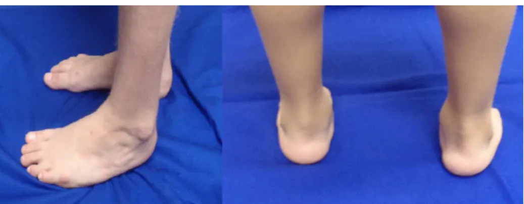
Fig.4–Seven-year-oldpatient,fiveyearsafterAchillestenotomy.
months).Themeannumberofplastercastusedwas8.3per patient,rangingfrom3to14.
Ofthe26patientstreated,onehaddeformityrecurrence. In this case, it was necessary to resume treatment from the beginning, with new serial casting and new Achilles tenotomy.
Themeanscoreofthequestionnaireandphysical exami-nationwas89.76,rangingfrom69to100(Appendix2).
Theresultswereclassifiedasexcellentwhenthescorewas between90and100points;good,80–89;fair,70–79;andpoor, lowerthan70.
Fiftysevenpercentofthepatientsansweredthattheirfeet neverached; 42%hadpainonexertion; 88%answeredthat thefootdidnotlimittheiractivities;7%reportedoccasional limitationsduringactivities;5%reportedfrequentlimitations duringactivities;73%saidtheywereverysatisfiedwiththe resultsoftreatment;23% were satisfied;4%reportedbeing neithersatisfiednordissatisfied(Fig.4).
Themeanpassivemotionscorewas7.61,rangingfrom2 to10.
ThemeananteroposteriorviewKiteangleofrightfeetwas 28.62◦andofleftfeet,27.68◦.MeanlateralviewKiteangleof
rightfeetwas27.34◦ andofleftfeet,24.93◦.Themean
over-allanteroposteriorviewanglewas28.14◦,andmeanoverall
lateralview26.11◦.
Discussion
ThePonsetitechniqueforthetreatmentofCCFhasbeen avail-ableandusedforover50years,butithasgainedpopularity onlyinrecentdecades.9
Boretal.9 conductedastudyinwhich74 patientswere treatedwiththePonsetimethodandwerefollowed-upfora meanof6.3years.TheyassessedfootmotionandappliedaDSI questionnaire,inwhichallpatientsshowedhighsatisfaction withtheendresultand89%presentedgoodfootmotion.In thepresentstudy,themeanpatientfollow-upaftertenotomy was4.6years,andthequestionnaireandphysical examina-tion developedby Laavegand Ponsetiwere assessed.8 The resultsofthe questionnaireandphysicalexaminationwas 89.76points,classifiedasagoodscore.
In a study that compared the results of surgical treat-mentbyopenCCFreleasewiththoseobtainedbyLaveegand Ponseti,8Dobbsetal.10observedameanscoreof65.3points
versus87.5points,andexcellentandgood resultsin33%of patientsversus74%respectively.Theyreportedthatpatients treatedwithopensurgerypresentedweaknessinthe tibio-tarsal andsubtalar joints,arthritis, lossofmuscle strength (especiallythesuraltriceps),painandresidualdeformity.They also reported need of furthersurgery in 47% of cases.11,12 Nonetheless,theauthorsindicatedthatthisistheonlyoption incaseoffailuretothePonsetimethod.
Ipolitoet al.13 comparedthe Ponsetimethodtoanother method(Marino-Zuco)anddemonstratedtheeffectivenessof theformerindeformitycorrectionusingonlyasimpleAchilles tenotomy,incontrastwiththelattermethod,whichrequired moreaggressivereleasesurgeriesthatinfluencedtheoutcome of thetwo groups (78%vs. 43% excellent and good results respectively).
Ponsetietal.6publishedastudywith322CCFcasestreated by their team, including not onlyidiopathic CCF, but also patients with neurological diseases and arthrogryposis, in which most had severe clubfeet. Due to the greater sever-ityofdeformities,56%presentedrecurrences;ofthese,18% hadasecondrecurrenceand10%hadathirdrelapse, need-ing notonlyAchillestenotomy, but alsomedialsofttissue release.6Inthepresentstudy,allpatientshadidiopathicCCF, whichisconsideredtobethemildestform,withthebest treat-mentoutcomes.Inourseries,onlyonepatienthaddeformity recurrence,requiringtorestartthetreatment,withnewserial castingandnewAchillestenotomy.
Conclusion
Conflicts
of
interest
Theauthorsdeclarenoconflictsofinterest.
Appendix
1.
CCF
Questionnaire
–
Ponseti
method
Patient: Chart:
Birth date:
Mother's Name:
Telephone:
Age at start of treatment:
Side : ___ Right ___ Left ___ Bilateral
Had been treated in another hospital before: ___ Yes ___ No
Comorbidities: ___ Yes ___ No
If yes, which?
Submitted to another surgical procedure:
Number of casts before tenotomy:
Date of tenotomy:
Age at tenotomy:
Required any other surgical procedures for correction of foot deformity:
___ No
___ Yes
Which? Date?
CCFfunctionalassessmentscale–Laaveg,Ponseti. Satisfaction(20points)
I’m
• Verysatisfiedwiththeendresult(20points)
• Satisfiedwiththeendresult(16points)
• Neither satisfied nor unsatisfiedwith the end result(12
points)
• Unsatisfiedwiththeendresult(8points)
• Veryunsatisfiedwiththeendresult(4points)
Function(20points) Inmydailylife,myclubfoot
• Doesnotlimitmyactivity(20points)
• Occasionallylimitsmystrenuousactivities(16points)
• Usuallylimitsmeinstrenuousactivities(12points)
• Limitsmeoccasionallyinroutineactivities(8points)
• Limitsmeinwalking(4points)
Pain(30) Myclubfoot
• Isneverpainful(30points)
• Occasionallycausesmildpainduringstrenuousactivities
(24points)
• Usuallyispainfulafterstrenuousactivitiesonly(18points)
• Isoccasionallypainfulduringroutineactivities(12points)
• Ispainfulduringwalking(6points)
Positionofheelwhenstanding(10points)
• Heelvarus0◦orsomevalgus(10points)
• Heelvarus0–5◦(5points)
• Heelvarus6–10◦(3points)
• Heelvarusgreaterthan10◦(0points)
Passivemotion(10points)
• Dorsiflexion–(1pointforevery5◦(maximum5points))
• Totalvarus–valgusmotionoftheheel(1pointforevery10◦
(maximum3points))
• Totalinversion–eversionoffoot(1pointper25◦(maximum
2points))
Gait
• Normal(6points)
• Cantoewalk(2points)
• Canheelwalk(2points)
• Limp(−2points)
• Noheelstrike(−2points)
• Abnormaltoe-off(−2points)
Physicalexam:
• Dorsiflexionankle
• Heelvarus–valgus
• Inversionandeversionoftheforefoot
• Heelorthostatismposition
• Metatarsaladductioninorthostaticposition
Radiologicalassessment(AP+lateral) Anteroposterior:
• Talocalcanealangle–assessesthevarus–valgusofthe
hind-foot
Lateral:
10
10 21/08/2003 1m bilatnão 9 18/01/2004 não 20 20 24 3+2+1=6 10 20 22 20 24 90 20
26 10
5+3+2=10
10
10
10
10
10
10
10
10
10
10 30 20 20 não
10/05/2005 7 não dir 20/12/2004 1m rho
11 100
12 11/07/2005 1m bilatnão 6 05/11/2005 não 20 16 24 5 3+3+2=8 10 21 20 22 24 83
13 13/03/2008 1m dir não 5 29/07/2008 não 20 20 30 5+3+2=10 10 32 43 100
14 11/05/2007 1m esq não 10 15/12/2007 não 20 20 24 5 3+2+1=6 10 22 25 85
15 28/06/2006 1m esq não 7 03/12/2006 não 20 20 30 5+3+2=10 10 32 29 100
16 10/08/2008 1m bilatnão 9 02/02/2009 não 20 20 30 5 5+2+2=9 10 28 30 38 42 94 17 13/01/2009 1m bilatnão 7 11/06/2009 não 20 20 30 5+3+2=10 10 22 28 25 30 100 18 11/05/2005 1a5m esq sim 6 15/07/2007 2a2m não 20 20 30 3+2+1=6 8 35,7 18,9 94 19 13/02/2009 2a3m bilatnão 14 23/04/2012 3a2m não 20 20 30 3+2+1=6 8 30 32 24 28 94 20 07/07/2003 1m bilatnão 10 07/01/2004 6m não 20 20 24 5+3+2=10 10 23 26 27 30 96 21wrgf 29/07/2007 2m bilatnão 14 04/05/2007 11m não 16 20 24 3 0+1+1=2 6 29,1 36,3 14,1 14,5 71 22 05/04/2004 2m esq não 10 09/12/2004 8m não 20 20 30 3+3+2=10 10 29,3 15,8 100 23 23/01/2007 1m bilatnão 7 24/05/2007 4m sim 12 16 30 5 3+3+1=7 6 32 17 28 26 76 24 13/09/2006 esq não 20 12/03/2007 6m não 16 12 24 5 2+3+2=7 10 32 15 74
25 13/04/2007 1m esq não 13 23/08/2007 4m não 16 20 30 0+3+2=5 6 27 15 87
26 26/06/2005 2a3m dir não 9 12/01/2008 2a7m não 20 20 24 2+3+2=7 8 30,3 20,4 89 2334 krm
lmb rmc
chbs
sff
jlpv
kns
acm gfs
dms rpcm
mpp hpb
2m
4m
4m
4m
4m
4m 5m 7m
6m
r
e
f
e
r
e
n
c
e
s
1. HerringJB.Congenitaltalipesequinovarus.In:TachdjianMO, editor.Tachdjian:pediatricorthopaedics.Philadelphia:W.B. SaundersCompany;2001.p.922–59.
2. Wynne-DaviesR.Familystudiesandaetiologyofclubfoot.J MedGenet.1965;2:227–32.
3. DobbsMB,GurnettCA.Updateonclubfoot:etiologyand treatment.ClinOrthopRelatRes.2009;467(5):1146–53. 4. AronsonJ,PuskarichCL.Deformityanddisabilityfrom
treatedclubfoot.JPediatrOrthop.1990;10(1):109–19.
5. PonsetiIV.Treatmentofcongenitalclubfoot.JBoneJointSurg Am.1992;74(3):448–54.
6. PonsetiIV.Congenitalclubfoot:fundamentalsoftreatment. Oxford:OxfordUniversityPress;1996.
7. PonsetiIV,SmoleyEN.Congenitalclubfoot:theresultsof treatment.JBoneJointSurgAm.1963;45(2):261–344.
8.LaavegSJ,PonsetiIV.Long-termresultsoftreatmentof congenitalclubfoot.JBoneJointSurgAm.1980;62(1):23–31. 9.BorN,CoplanJA,HerzenberbJE.Ponsetitreatmentof
idiopathicclubfoot:minimum5-yearfollowup.ClinOrthop RelatRes.2009;467(5):1263–70.
10.IppolitoE,FarsettiP,CateriniR,TudiscoC.Long-term comparativeresultsinpatientswithcongenitalclubfoot treatedwithtwodifferentprotocols.JBoneJointSurgAm. 2003;85(7):1286–94.
11.DobbsMB,NunleyR,SchoeneckerPL.Longtermfollow-upof patientswithclubfeettreatedwithextensivesoft-tissue release.JBoneJointSurgAm.2006;88(5):986–96. 12.CooperDM,DietzFR.Treatmentofidiopathicclubfoot.A
thirty-yearfollow-upnote.JBoneJointSurgAm. 1995;77(10):1477–89.