6 artigo 518
ORIGINAL ARTICLE
1 – Assistant Professor and Head of the Shoulder and Elbow Surgery Group, Department of Orthopedics and Traumatology, School of Medical Sciences, Santa Casa de São Paulo, São Paulo, SP, Brazil.
2 – Assistant Professor and Attending Physician in the Shoulder and Elbow Surgery Group, Department of Orthopedics and Traumatology, School of Medical Sciences, Santa Casa de São Paulo, São Paulo, SP, Brazil.
3 – Attending Physician in the Shoulder and Elbow Surgery Group, Department of Orthopedics and Traumatology, School of Medical Sciences, Santa Casa de São Paulo, São Paulo, SP, Brazil.
4 – Trainee in the Shoulder and Elbow Surgery Group, Department of Orthopedics and Traumatology, School of Medical Sciences, Santa Casa de São Paulo, São Paulo, SP, Brazil. 5 – Adjunct Professor, Academic Consultant and Member of the Shoulder and Elbow Surgery Group, Department of Orthopedics and Traumatology, School of Medical Sciences, Santa
Casa de São Paulo, São Paulo, SP, Brazil.
Work performed in the Department of Orthopedics and Traumatology, School of Medical Sciences, Santa Casa de São Paulo (DOT-FCMSCSP), Fernandinho Simonsen Wing, São Paulo, SP, Brazil. Director: Prof. Dr. Osmar Avanzi.
Correspondence: R. Dr. Cesário Mota Jr. 112, Vila Buarque, 01221-020 São Paulo, SP. E-mail: [email protected] / [email protected] Work received for publication: May 12, 2011; accepted for publication: July 27, 2011.
EVALUATION OF THE RESULTS FROM ARTHROSCOPIC SURGICAL
TREATMENT FOR TRAUMATIC ANTERIOR SHOULDER INSTABILITY
USING SUTURING OF THE LESION AT THE OPENED
MARGIN OF THE GLENOID CAVITY
Alberto Naoki Miyazaki1, Marcelo Fregoneze2, Pedro Doneux Santos3, Luciana Andrade da Silva3, Guilherme do Val Sella3, Clodoaldo Duarte4, Vinícius Botelho4, Sergio Luiz Checchia5
AbSTRACT
Objective: To evaluate the clinical results from patients with traumatic anterior shoulder instability that was treated surgi-cally through arthroscopic viewing, using bioabsorbable an-chors and a technique for remove the cartilage of the anterior glenoid rim for repairing a Bankart lesion. Method: Between March 2006 and October 2008, 27 shoulders in 27 patients with a diagnosis of traumatic anterior shoulder instability were operated. The patients’ mean age was 28 years and they had had between two and 25 previous episodes of disloca-tion. The patients were predominantly male (24; 89%). The minimum length of follow-up was 24 months and the mean was 36 months. None of the patients had previously under-gone surgery on the affected shoulder or had any significant bone lesion at the glenoid margin. The postoperative clinical assessment was done using the Rowe scale. To measure the
INTRODUCTION
Anterior post-traumatic shoulder instability is a disease related to injuries of the joint capsule, its li-gaments and the glenoid labrum. For its treatment to be successful, the surgical approach has to be su-fficiently flexible to deal with the variety of lesions encountered(1). Jakobsen et al(2) found via arthroscopy
that after the first episode of traumatic shoulder
dis-The authors declare that there was no conflict of interest in conducting this work
This article is available online in Portuguese and English at the websites: www.rbo.org.br and www.scielo.br/rbort
preoperative and postoperative joint range of motion, we used the method described by the American Academy of Ortho-paedic Surgeons (AAOS). Results: According to the Rowe criteria, 25 patients (93%) achieved excellent results and two (7%) had poor results. None of the patients presented good or fair results. Twenty-three patients were satisfied with the results obtained (85%), and returned to their activities without limitations, while four patients (15%) had some degree of limitation. There was recurrence of instability in two patients (7%). Conclusion: Treatment of traumatic anterior shoulder instability through arthroscopic viewing using a technique for remove the cartilage of the anterior glenoid rim for repairing a Bankart lesion provided excellent results for 93% of the patients operated.
Keywords – Shoulder Dislocation/therapy; Shoulder Dislo-cation/surgery; Arthroscopy
location, the capsule or glenoid labrum injury rate is
93.5%. From biomechanical analyses, Bigliani et al(3)
described stretching of the glenohumeral ligaments and joint capsule of the shoulder that occurs after re-peated dislocations. These structural abnormalities of the capsule and ligaments lead to a recurrent pattern of dislocations, and not just Bankart lesions(4).
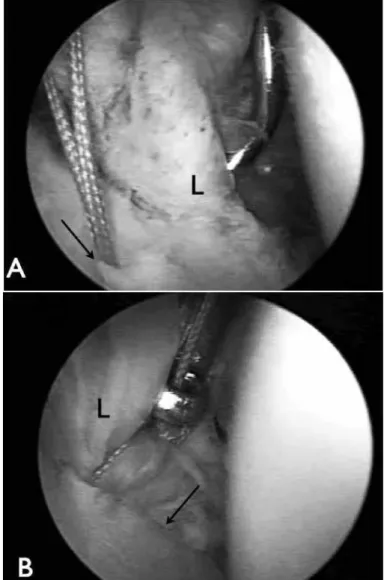
Figure 1 – View through the posterior portal of the right shoulder in deckchair position. L – Glenoid labrum A) anchor fixed in the carti-laginous rim of the glenoid cavity (arrow). B) Bankart lesion repaired over the glenoid cartilage (arrow).
these repairs are not free from complications, such as fractures of the anterior glenoid rim, violation of the tendon of the subscapularis muscle and prolonged duration of surgery(6).
The advent of arthroscopy gave rise to improved recognition of anatomopathological lesions, thus pro-viding better understanding of the etiology of ante-rior shoulder instability(7). Johnson(8) was the first to
propose a technique for arthroscopic viewing, for use in treating anterior shoulder instability. This techni-que used metal staples and the recurrence rate was
found to be 21%. In 1988, Morgan and Bodenstab(9)
introduced a transglenoid suturing technique with arthroscopic viewing, for repairing Bankart lesions.
In 1991, Wolf(10) introduced a repair technique using
anchors. Subsequent studies using this technique sho-wed encouraging results, with instability recurrence rates ranging from 8 to 12%(1,11-14 ).
In a randomized prospective study, Moore reported similar failure rates from treating anterior shoulder
instability using open and arthroscopic techniques(15).
In a recent meta-analysis, Hobby et al(16) found similar
results, with a mean failure rate of 8.9% and without any statistically significant difference in the failure rate in surgical techniques using anchors for lesion suturing, between open and arthroscopic routes.
In the traditional technique, suturing of the capsule and the glenoid labrum at the neck of the scapula is recommended. In recent studies, this has been des-cribed as a technique that provides worse results and
should be avoided(17). Alternatively, suturing on the
cartilage of the anterior glenoid rim has been
recom-mended(10) (Figure 1), which in our view adds
difficul-ty to healing, since there is no open bone surface for contact with the capsule-ligament structures. Burkhart
et al(18) described a repair technique for these lesions
in which 2 to 3 mm of the anterior glenoid rim was prepared using a curette or shaver blade to remove the cartilage, thus providing enough open surface for good healing (Figures 2 and 3).
The present study had the aim of evaluating the clinical results among patients with anterior shoulder instability that was treated surgically by means of ar-throscopic viewing, using bioabsorbable anchors, in accordance with the technique described by Burkhart for repairing Bankart lesions(18).
MATERIALS AND METHODS
Between March 2006 and October 2008, the Shoulder and Elbow Group of the Department of Or-thopedics and Traumatology of the School of Medical Sciences of Santa Casa de São Paulo performed ope-rations on 27 shoulders of 27 patients with a diagnosis of traumatic anterior shoulder instability.
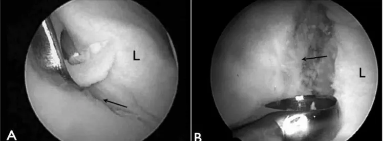
Figure 2 – Opening of the anterior joint margin of the glenoid cavity (glenoid rim) using a curette. L – Glenoid labrum. A) Opened joint margin viewed through the posterior portal (arrow). B) Opened joint margin viewed through the anterosuperior portal (arrow).
were regularly practicing some type of physical activity. We established the following as inclusion criteria: at least two episodes of traumatic anterior dislocation of the shoulder; at least 24 months of postoperative follow--up; and a surgical procedure performed under arthros-copic viewing, using the technique described previously. The exclusion criteria were: instability of non-traumatic etiology, uncontrolled epilepsy, follow-up for less than two years, previous surgery on the shoulder evaluated, bone lesion on the anterior glenoid rim greater than 20% and signs of capsule-ligament laxity.
All the patients presented signs of anterior apprehension with the shoulder at 90 degrees of abduction and external rotation in the preoperative clinical examination. Imaging examinations (radiographs, magnetic resonance or magnetic arthro-resonance) were performed to view the Bankart lesion and evaluate any significant bone deficiency at the anterior glenoid rim, which was not shown in any case.
The surgical procedure with arthroscopic viewing was performed by means of regional blockade and ge-neral anesthesia, with the patient in lateral decubitus, under traction, or in the deckchair position, depending on whether any posterior lesion of the glenoid labrum was suspected, with a need for repair. An inventory of the joint cavity was made in order to identify the anteroinferior labral lesion and diagnose any other associated lesion. Such lesions were found in 11 of our patients: two posterior capsule-ligament lesions, one intra-articular free body, six SLAP lesions (four type II, one type III and one type V), one enchon-droma of the humeral head and one joint fracture at the anterior glenoid rim with a small bone fragment (Table 1). Deinsertion of the labrum at the anterior glenoid rim was completed with the aim of facilitating its mobilization. Then, 2 to 3 mm of the anterior gle-noid rim was opened using a curette or shaver blade to remove the cartilage, for subsequent suturing of the lesion (Figure 4). Fixation of the labrum-ligament complex of the shoulder joint was achieved using two to five bioabsorbable anchors with non-absor-bable thread (mean of three anchors). Other proce-dures were performed in association with repairing the Bankart lesion, including the following: capsu-le plication (three cases, 11%), repair of a posterior labrum-ligament lesion (two cases, 8%), removal of
gap (six cases, 22%) and tenotomy with tenodesis of the long head of the biceps (one case, 3%) (Table 2).
After the operation, the patients used a sling for six weeks, with pendular movements and passive external rotation of the arm as far as neutral. After this time, active movement was allowed, and then, three months after the operation, muscle strengthening exercises
Table 1 – Lesions associated with the Bankart lesion.
Intraoperative findings Cases
Posterior capsule-ligament lesion 2
Intra-articular free body 1
SLAP lesion 6
Enchondroma of the humeral head 1
Fracturing of the anteroinferior glenoid rim 1
Total 11
Source: Medical files of Irmandade Santa Casa de Misericórdia de São Paulo. Legend: SLAP lesion – lesion of the superior labrum from anterior to posterior.
Figure 4 – Preparation of the bone bed and capsule-ligament rein-sertion in the anterior glenoid rim: (A) Opening of the anterior glenoid rim. (B) Opened anterior joint margin. (C) Bankart lesion repair. (D) Axial view after repairing of the Bankart lesion, with preparation of the bone bed. (E) Without preparation of the bone bed.
Table 2 – Procedures associated with Bankart lesion repairs.
Procedures Cases
Capsule plication 3
Repair of posterior capsule-ligament lesion 2 Removal of intra-articular free body 1
Repair of SLAP lesion 6
Closure of rotator gap 6
Tenotomy and tenodesis of the long head of the biceps 1
Total 19
were started. The postoperative clinical assessment
was done using the Rowe scale(5). To measure the
degree of preoperative and postoperative range of mo-tion, we used the method described by the American
Academy of Orthopaedic Surgeons (AAOS)(19).
To calculate confidence intervals and perform hypothesis tests for proportions, approximations for normal distribution were not used, since the samples were very small and such approximations might not have been valid. Thus, the calculations used likelihood estimates. The significance level used was 0.05, and therefore hypotheses in which the descriptive level (P-value) was less than 0.05 were rejected.
RESULTS
With a minimum postoperative follow-up of 24 months and a maximum of 51 months (mean of 36 months), following the Rowe criteria, the mean pre-sented was 95 points (50-100). Twenty-five patients (93%) had an excellent result and two (7%) had a
poor result; none of the patients presented good or fair results (Table 3).
The improvement in range of motion in relation to the preoperative period was seven degrees of elevation, 10 degrees of lateral rotation and one vertebral level of medial rotation. The postoperative means for mobility were 158 degrees of elevation (120-170 degrees), 66 degrees of lateral rotation (30-80 degrees) and T8 of medial rotation (T5-T12). Twenty-three patients were satisfied with the result obtained (85%) and returned to their activities without limitations, while four patients (15%) presented some degree of limitation.
Two patients presented recurrence of the instability (cases 17 and 22): one after sports trauma and the other, who was a professional Greco-Roman wrestler, during a competition 24 months after the operation. Two patients continued to complain of shoulder pain (cases 2 and 21): one with occurrences during inten-se physical exerciinten-se and the other while at rest, with limitations on mobility.
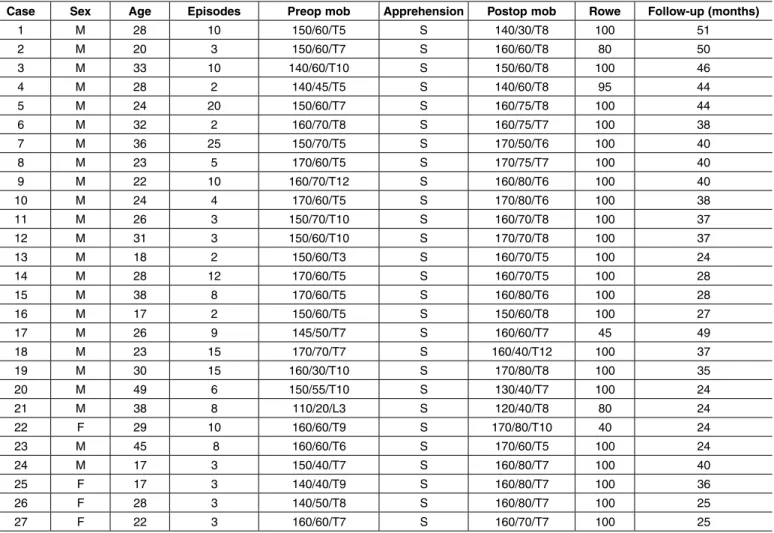
Table 3 – Patients operated using the technique of opening the anterior glenoid rim.
Case Sex Age Episodes Preop mob Apprehension Postop mob Rowe Follow-up (months)
1 M 28 10 150/60/T5 S 140/30/T8 100 51
2 M 20 3 150/60/T7 S 160/60/T8 80 50
3 M 33 10 140/60/T10 S 150/60/T8 100 46
4 M 28 2 140/45/T5 S 140/60/T8 95 44
5 M 24 20 150/60/T7 S 160/75/T8 100 44
6 M 32 2 160/70/T8 S 160/75/T7 100 38
7 M 36 25 150/70/T5 S 170/50/T6 100 40
8 M 23 5 170/60/T5 S 170/75/T7 100 40
9 M 22 10 160/70/T12 S 160/80/T6 100 40
10 M 24 4 170/60/T5 S 170/80/T6 100 38
11 M 26 3 150/70/T10 S 160/70/T8 100 37
12 M 31 3 150/60/T10 S 170/70/T8 100 37
13 M 18 2 150/60/T3 S 160/70/T5 100 24
14 M 28 12 170/60/T5 S 160/70/T5 100 28
15 M 38 8 170/60/T5 S 160/80/T6 100 28
16 M 17 2 150/60/T5 S 150/60/T8 100 27
17 M 26 9 145/50/T7 S 160/60/T7 45 49
18 M 23 15 170/70/T7 S 160/40/T12 100 37
19 M 30 15 160/30/T10 S 170/80/T8 100 35
20 M 49 6 150/55/T10 S 130/40/T7 100 24
21 M 38 8 110/20/L3 S 120/40/T8 80 24
22 F 29 10 160/60/T9 S 170/80/T10 40 24
23 M 45 8 160/60/T6 S 170/60/T5 100 24
24 M 17 3 150/40/T7 S 160/80/T7 100 40
25 F 17 3 140/40/T9 S 160/80/T7 100 36
26 F 28 3 140/50/T8 S 160/80/T7 100 25
27 F 22 3 160/60/T7 S 160/70/T7 100 25
Source: Medical files of Irmandade Santa Casa de Misericórdia de São Paulo.
matic anterior shoulder instability is Bankart lesion repair, with reinsertion of the anteroinferior labrum in the anterior glenoid rim or in the neck of the
sca-pula(10). Studies conducted more recently have
rejec-ted suturing of the lesion at the neck of the scapula and have recommended suturing at the glenoid rim, along a length of 2 to 3 mm on the anterior internal glenoid rim, in the belief that the barrier created through labral and capsular reinsertion might help to stabilize the shoulder, thereby functioning as a mechanical barrier(17).
Burkhart et al(18) described a technique for opening
the anterior joint margin of the glenoid cavity and ex-posing the subchondral bone, with capsule-ligament fixation using bioabsorbable anchors in this bed, whi-ch they believed would lead to better healing of the lesion. We share this opinion, since healing over the joint cartilage could be one of the contributory rea-sons for recurrence of dislocations.
Recent studies have demonstrated that the results from treatment with arthroscopic viewing are compa-rable with those from an open approach, with the ad-vantage of providing better postoperative comfort for the patients and the possibility of making an inventory of the joint cavity to seek diagnoses and treatments for
associated lesions(15,16,20), which was done frequently
among our patients.
From a review of the literature, Mohtadi et al(21)
demonstrated a recurrence rate of 10% when an-chors were used under arthroscopic viewing. Barber
et al(22) reported recurrence of 7% from using
bioab-sorbable anchors in 57 patients. From a
meta-analy-sis, Hobby et al(16) described a mean recurrence rate
of 8.9%. In our setting, Godinho et al(12) presented
results from arthroscopic surgical treatment after two years of follow-up with a recurrence rate of 8.9%. We found a recurrence rate of 7% after two years of follow-up, which is comparable with the best rates found in the literature.
Among our patients who suffered recurrence of instability, one was a professional Greco-Roman wrestler who went back to his sport without any symptoms of instability, but then presented a new episode of traumatic dislocation during a competi-tion, after two years of postoperative follow-up. From reviewing the literature, we noted that there was a
hi-could range from 15% to 25%(23). In the index
descri-bed by Boileau, young patients practicing sports at a high level had better results when the treatment was
done as open surgery(24), which has led us to think
again regarding indications for arthroscopic treatment among patients with this profile. The other of our pa-tients who presented recurrence of instability suffered an episode of dislocation after one and a half years of follow-up, also with traumatic etiology.
Two patients continued to complain of pain in the operated shoulder: one case with pain during intense physical activity practice and the other with pain even at rest. These two cases are still undergoing diagnostic investigation, without coming to any conclusion so far, because they do not present any signs of insta-bility on physical examination, or any abnormality suggestive of joint lesions on magnetic resonance imaging that would explain the pain.
There was an improvement in the range of motion in relation to the preoperative situation, albeit without statistical significance, consisting of seven degrees of elevation, 10 degrees of lateral rotation and one verte-bral level of medial rotation. These findings differed
from the results presented by Mazzocca et al(23), who
demonstrated that there was a slight decrease in lateral rotation after the operation, in contact sport players.
According to the Rowe scale, Fabbriciani et al(20)
obtained a mean score of 91 points for cases of ins-tability that were treated with arthroscopic viewing,
Barber et al(22) found a mean of 93 points, with a range
from 40 to 100 points, and Kim et al(11) found that
95% of the results were satisfactory. In our setting,
Godinho et al(12) obtained a mean of 92 points, with
a range from 25 to 100 points. In the same way, the mean result presented by our patients was 95 points, with a range from 45 to 100 points, which is com-parable with the best results found in the literature, with excellent results in 93% of the cases and poor results in 7%.
CONCLUSION
REFERENCES
1. Gartsman GM, Roddey TS, Hammerman SM. Arthroscopic treatment of ante-rior-inferior glenohumeral instability. Two to five-year follow-up. J Bone Joint Surg Am. 2000;82(7):991-1003.
2. Jakobsen BW, Johannsen HV, Suder P, Søjbjerg JO. Primary repair versus con-servative treatment of first-time traumatic anterior dislocation of the shoulder: a randomized study with 10-year follow-up. Arthroscopy. 2007;23(2):118-23. 3. Bigliani LU, Kurzweil PR, Schwartzbach CC, Wolfe IN, Flatow EL. Inferior
capsular shift procedure for anterior-inferior shoulder instability in athletes. Am J Sports Med. 1994;22(5):578-84.
4. Bankart A. The pathology and treatment of recurrent dislocation of the shoulder joint. Br J Surg 1938;26(1):23-9.
5. Rowe CR, Patel D, Southmayd WW. The Bankart procedure: a long-term end--result study. J Bone Joint Surg Am. 1978;60(1):1-16.
6. Karlsson J, Magnusson L, Ejerhed L, Hultenheim I, Lundin O, Kartus J. Compa-rison of open and arthroscopic stabilization for recurrent shoulder dislocation in patients with a Bankart lesion. Am J Sports Med. 2001;29(5):538-42. 7. Snyder SJ. Shoulder instability. In: Shoulder arthroscopy. New York:
McGraw--Hill; 1994. p. 179-213.
8. Johnson LL. Shoulder arthroscopy. In: Arthroscopic surgery: principals and practice. 3rd. St Louis: CV Mosby; 1986. p. 398-412.
9. Morgan CD, Bodenstab AB. Arthroscopic Bankart suture repair: technique and early results. Arthroscopy. 1987;3(2):111-22.
10. Wolf EM. Arthroscopic capsulolabral repair using suture anchors. Orthop Clin North Am. 1993;24(1):59-69.
11. Kim SH, Ha KI, Cho YB, Ryu BD, Oh I. Arthroscopic anterior stabilization of the shoulder: two to six-year follow-up. J Bone Joint Surg Am. 2003;85(8):1511-8. 12. Godinho GG, França FO, Freitas JMA, Menezes CM, Freire SG, Wanderley AL,
et al. Tratamento artroscópico da instabilidade anterior traumática do ombro: resultados a longo prazo e fatores de risco. Rev Bras Ortop. 2008;43(5):157-66. 13. Ozbaydar M, Elhassan B, Diller D, Massimini D, Higgins LD, Warner JJ. Results of arthroscopic capsulolabral repair: Bankart lesion versus an-terior labroligamentous periosteal sleeve avulsion lesion. Arthroscopy. 2008;24(11):1277-83.
14. Hantes ME, Venouziou AI, Liantsis AK, Dailiana ZH, Malizos KN. Arthroscopic repair for chronic anterior shoulder instability: a comparative study between patients with Bankart lesions and patients with combined Bankart and superior labral anterior posterior lesions. Am J Sports Med. 2009;37(6):1093-8. 15. Bottoni CR, Smith EL, Berkowitz MJ, Towle RB, Moore JH. Arthroscopic versus
open shoulder stabilization for recurrent anterior instability: a prospective ran-domized clinical trial. Am J Sports Med. 2006;34(11):1730-7.
16. Hobby J, Griffin D, Dunbar M, Boileau P. Is arthroscopic surgery for stabilisation of chronic shoulder instability as effective as open surgery? A systematic review and meta-analysis of 62 studies including 3044 arthroscopic operations. J Bone Joint Surg Br. 2007;89(9):1188-96.
17. Itoi E, Cofield RH, Steinmann SP. Does the “bumper” created during Bankart repair contribute to shoulder stability? In: 11th International Congress of Shoul-der and Elbow Surgery, Edinburgh, 2010.
18. Burkhart SS, Lo IKY, Brady PC. “Instability: arthroscopy Barkart repair”. In: Burkhart”s view of the shoulder: a cowboy”s guide to advanced shoulder ar-throscopy. Philadelphia: Lippincott Williams e Wilkins; 2006. p. 217-20. 19. American Academy of Orthopeadic Surgeons; Joint motion: method of
measu-ring and recording. Chicago, AAOS meeting, 1965. p. 10-43
20. Fabbriciani C, Milano G, Demontis A, Fadda S, Ziranu F, Mulas PD. Arthros-copic versus open treatment of Bankart lesion of the shoulder: a prospective randomized study. Arthroscopy. 2004;20(5):456-62.
21. Mohtadi NG, Bitar IJ, Sasyniuk TM, Hollinshead RM, Harper WP. Arthroscopic
versus open repair for traumatic anterior shoulder instability: a meta-analysis. Arthroscopy. 2005;21(6):652-8.
22. Barber FA, Snyder SJ, Abrams JS, Fanelli GC, Savoie FH 3rd. Arthroscopic Bankart reconstruction with a bioabsorbable anchor. J Shoulder Elbow Surg. 2003;12(6):535-8.
23. Mazzocca AD, Brown FM Jr, Carreira DS, Hayden J, Romeo AA. Arthroscopic anterior shoulder stabilization of collision and contact athletes. Am J Sports Med. 2005;33(1):52-60.