J of Evolution of Med and Dent Sci/ eISSN- 2278-4802, pISSN- 2278-4748/ Vol. 3/ Issue 49/Oct 02, 2014 Page 11698
MICROBIOLOGICAL STUDY OF EAR DISCHARGE AND THEIR ANTIBIOTIC
SENSITIVITY PATTERN IN CHRONIC SUPPURATIVE OTITIS MEDIA
V. Rama Chandra Rao1, K. Srilatha2, S. Visweswara Rao3, K. N. Manohar4
HOW TO CITE THIS ARTICLE:
V. Rama Chandra Rao, K. Srilatha, S. Visweswara Rao, K. N. Manohar. Microbiological Study of Ear Discharge and their Antibiotic Sensitivity Pattern in Chronic Suppurative Otitis Media. Journal of Evolution of Medical and Dental Sciences 2014; Vol. 3, Issue 49, October 02; Page: 11698-11705, DOI: 10.14260/jemds/2014/3537
ABSTRACT: INTRODUCTION: Chronic Suppurative otitis media (CSOM) is the most common condition encountered by otolaryngologists in day to day practice. The importance of chronic otitis
media lies in its dreaded complications and deafness. AIM: This study was under taken to identify the
microbiological isolates of the ear discharge in CSOM cases and their sensitivity to antibiotics.
SETTINGS AND DESIGN: Tertiary care hospital in north costal Andhra Pradesh. It was a Prospective
study. MATERIALS & METHODS: About 100 patients having ear discharge who attended ENT
outpatient department from July 2013 to Feb 2014 for a period of 8months were studied. Aural swabs were sent to microbiology lab for culture &sensitivity. RESULTS: Culture reports showed aerobic bacterial isolates in 85 cases, fungi in 7 cases and sterile in 8 cases. Of the 85 cases of aerobic bacteria, staphylococcus aureus was isolated in 34 cases (40%) followed by Pseudomonas aeruginosa in 29 cases (34%), klebsiella in 16 cases (18.8%), E.coli in 4 cases (4.7%) and proteus in 2 cases. Antibiotic sensitivity reports showed Staphylococcus was more susceptible to netilmycin (97%), amoxiclav (91.7%) and least sensitive to ceftazidime (64.7%). Pseudomonas was more sensitive to amikacin (96.5%), gentamycin (93.1%) and least sensitive to amoxyclav (79.3%), ampicillin +
sulbactum (82.75%). CONCLUSION: Mono microbial etiology, especially Staphylococcus species was
found to be the most common organism causing chronic otitis media. Knowledge of the prevailing flora and their susceptibility to antimicrobials will guide the clinicians for early and effective treatment thereby avoiding complications.
KEYWORDS: Chronic Suppurative otitis media, culture and sensitivity, Staphylococcus aureus
INTRODUCTION: Chronic Suppurative otitis media (CSOM) denotes chronic inflammation within the mucosa of middle ear and mastoid leading to production of ear discharge via tympanic membrane perforation.1 CSOM results from long term Eustachian tube dysfunction with poorly aerated middle
ear space, multiple bouts of acute otitis media and persistent middle ear infection.2 Risk factors
include mechanical obstruction of Eustachian tube due to adenoid hypertrophy, sinusitis, immunodeficiency and environmental factors such as lack of breast feeding in infancy, passive
exposure to smoking and low socio economic status.3
Major cause of ear infection are bacterial isolates predominantly aerobic gram negative bacteria such as Pseudomonas, E.coli, Proteus, klebsiella and gram positive bacteria Staphylococcus spps. Anaerobic bacteria include bacteroid spps4. Frequent upper respiratory tract infections and
poor socio economic status condition, overcrowded housing, poor hygiene, and poor nutrition may be
related to development of CSOM.5-9
J of Evolution of Med and Dent Sci/ eISSN- 2278-4802, pISSN- 2278-4748/ Vol. 3/ Issue 49/Oct 02, 2014 Page 11699 may lead to complications including septicemia, meningitis, brain abscess and facial palsy.16,17
Therefore, the microbial culture and sensitivity will help in appropriate management of otitis media and its complications and thus preventing the emergence of resistant bacterial strains.
Since no previous similar studies were done in north costal Andhra Pradesh, we undertook this study to identify the microorganisms causing CSOM and to detect antibiotic sensitivity of the isolates in north coastal Andhra Pradesh where prevalence of CSOM is high.
MATERIALS & METHODS: The study design is Prospective. Informed consent was taken from all the patients and the study was approved by institution ethics committee.100 patients with CSOM who presented to ENT outpatient department of our sub urban medical college hospital in north costal Andhra Pradesh from July 2013 to February 2014 were studied for a period of 8months. Ear discharge samples from the clinically diagnosed cases of CSOM who have not taken antibiotics 10 days prior were sent for culture and sensitivity to the microbiology laboratory. Two Sterile cotton swabs were used to collect the samples.
In the laboratory, the first swab was used to make a smear on clean grease-free glass slide for bacterial differentiation by gram stain examination and direct microscopy of specimen in KOH for
fungal examination. The second swab was used for the bacterial culture on blood agar, mac Conkey’s
agar and peptone water for 24hrs at 370C in incubator. Isolates were identified using colony
morphology and standard biochemical tests. After identifying isolates antimicrobial susceptibility test was performed using modified Kirby- Bauer disc diffusion method according to Clinical and Laboratory Standards Institute (CLSI) guidelines on nutrient agar.
Antibiotics tested for gram negative bacteria were cefotaxime (10mcg), amikacin (10mcg), ampicillin+sulbactum (15mcg), amoxiclav (10mcg), cefuroxime (30mcg), Ofloxacin (5mcg), gentamycin (30mcg), ceftriaxone (10mcg), tetracycline (10mcg) and for gram positive bacteria were cefazolin (30mcg), ampicillin + sulbactum (10mcg), ciprofloxacin (10mcg), vancomycin (10mcg), amoxiclav (10mcg), erythromycin (10mcg), ceftazidime (30mcg) and netilmicin (10mcg). A part of
the discharge was cultured on Sabouraud’s dextrose agar slant with Chloramphenicol . 5 and was
examined for gross and microscopic morphology of the fungi using lacto phenol cotton blue stain.
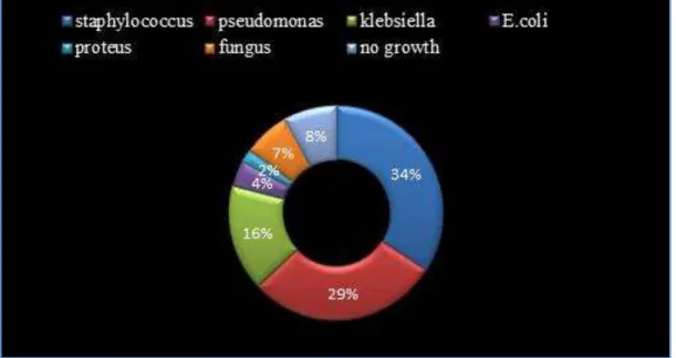
RESULTS: In the present study of 100 cases of ear discharge highest incidence of was observed in the age group of 11-20 years (28%) followed by 21-30yr (18%), 0-10yr(13%), 31-40yr(13%), 41-50yr(13%), 51-60yr(10%) and >60yr (5%). Analysis of sex incidence revealed that COM is more common in female population (54%) than males (46%). Out of 100 samples of ear discharge, direct examination revealed the presence of bacteria in 85 cases (85%), fungi in 7 cases (7%) and sterile in 8 cases (8%), as shown in table 1.
Out of 85 bacterial isolates, staphylococcus is isolated in 34 cases (40%) followed by pseudomonas in 29 cases (34%), klebsiella in 16 cases (18.8%), E. coli in 4 cases(4.7%), and proteus in 2 cases(2.3%) as shown in table 2 and figure 1. In the fungus isolates, aspergillus spp. was isolated in 7 cases (87.5%) of which aspergillus Niger is seen in 3 cases (42.85%) followed by aspergillus fumigatus in 2 cases(28.5%), aspergillus flavus in one case (14.28%) and candida albicans in one case (14.28%) as shown in table 3.
J of Evolution of Med and Dent Sci/ eISSN- 2278-4802, pISSN- 2278-4748/ Vol. 3/ Issue 49/Oct 02, 2014 Page 11700 34(88.2%), cefazolin (88.2%), ciprofloxacin (88.2%), vancomycin (88.2%) and showed lower sensitivity to ceftazidime 12out of 34 isolates (35%) as shown in table 4, figure 2.
Pseudomonas showed highest sensitivity to amikacin i.e. 28 of 29 isolates (96.5 %) followed by gentamycin 27 of 29(93.1%), ceftriaxone (93.1%), ofloxacin (89.65%), cefotaxime (72.41%), cefuroxime (65.5%) and showed lower sensitivity to tetracycline (10.3 %), ampicillin+sulbactum (17.2%) and amoxiclav (20.6 %).
Klebsiella showed higher sensitivity to ceftriaxone 16 out of 16(100%), gentamycin 16 out of 16(100%), amikacin (93.7%), ampicillin+sulbactum (87.5%), cefotaxime (81.25%), cefuroxime (81.25 %), ofloxacin (68.75%) and lower sensitivity to amoxiclav 7 of 16(43.7%), and tetracycline (43.7%). E.coli showed higher sensitivity to amikacin (75%), ampicillin+sulbactum (75%), cefuroxime (75%), ofloxacin (75%), gentamycin (75%) and showed 0% sensitivity to amoxiclav. Proteus showed sensitivity to amikacin, ofloxocin, gentamycin, ceftrioxone and resistance to ampicillin + sulbactum and amoxiclav.
Distribution of CSOM in north costal Andhra Pradesh showed the disease prevalence more in southern part of the area. Pseudomonas was more common in northern part and staphylococcus was more common in southern part. Analysis of socioeconomic status according to modified Kuppuswamy scale showed the disease more common in rural population and upper lower class.
DISCUSSION: Knowledge of the microorganisms causing CSOM and their antibiotic susceptibility pattern helped us to give appropriate medical treatment there by reducing complications and antibiotic resistance among patients and ultimately reducing the cost of the treatment. Deafness rate can be minimized there by reducing burden on the society.
Overall incidence of COM was found to be higher in females accounting for 54% and male 46%. Majority of cases were of age group 11-20 being 28%.This correlates with the study conducted
by, Baruah PC, Gulati et al.18,19 Analysis of socioeconomic status according to modified Kuppuswamy
scale showed the disease is more common in rural population and lower class.
Among the bacterial causes of COM, a single bacterium was found to be the cause. Most common bacteria isolated were staphylococcus aureus (34%) followed by pseudomonas 29%, klebsiella 16%, E.coli 4%, and proteus (2%). This correlates with the study conducted by Prakash R
etal.20-23 In contrary, this observation is different from other studies done by Yeo SG and Poorey VK
where Pseudomonas species were isolated commonly.24,25 Coagulase negative staphylococcus has
been excluded, they being the natural commensals of the body.26,27
Staphylococcus is universally harbored with in human nares and Pseudomonas is known to reside in the moist environment of ear canal. The proximity of these bacteria reflects the likelihood of their eventual presence in middle ear.28 In the fungus isolates aspergillus species was found to be
most common (85.7%) of which aspergillus Niger had the highest prevalence of 42.85% followed by aspergillus fumigatus, aspergillus flavus and candida albicans.29, 30
Unusual prevalence of fungi in this study could be explained by an excessive and uncontrolled
use of antibiotics as it was reported by Araiza J etal.30 Results showed no growth in 8 % of cases and
J of Evolution of Med and Dent Sci/ eISSN- 2278-4802, pISSN- 2278-4748/ Vol. 3/ Issue 49/Oct 02, 2014 Page 11701 Pseudomonas showed maximum sensitivity with amikacin (96.5%), gentamycin (93.1%), and ceftriaxone (93.1%) and resistant to ampicillin + sulbactum and amoxyclav. Now with the introduction of newer and higher antibiotics, the complications of otitis media have become less common. But due to increased and irrational use of broad spectrum antibiotics particularly by the registered/private medical practitioners in rural areas like ours, the resistance in the bacterial isolates has become very common.
CONCLUSION: Continuous and periodic evaluation of microbiological pattern and their antibiotic sensitivity pattern of CSOM in local area is necessary in prescribing appropriate antibiotics for successful treatment of otitis media and minimizing its complications and emergence of resistant strains. Mono microbial etiology, especially Staphylococcus species and Pseudomonas aeruginosa, was found to be the most common in our study. Staphylococcus species are highly resistant to ceftazidime and erythromycin. Pseudomonas aeruginosa is becoming less sensitive against commonly used antimicrobials viz. Ampicillin + sulbactum and amoxyclav. We believe that our data may contribute to an effective medical management of chronic supportive otitis media.
REFERENCES:
1. Browning GG, Merchant SN, Kelly G, Swan LRC, Canter R, Mc kerrow WS. Chronic otitis media.
)n: Gleeson M editor. Scott Brown’s otolaryngology.7th edition. London. Hodder Arnold; 2008; 3:p. 3395-3399.
2. Gopen Q. Pathology and clinical course of the inflammatory diseases of the middle ear. In: Gulya
AJ, Minor LB, Poe DS, editors. Glasscock - shambaugh surgery of the ear. 6th edition. Shelton, CT.
People’s medical publishing house; 2012. p. 425-429.
3. Chole RA, Nason R. Chronic otitis media and cholesteatoma.In: SnowJr JB, Wackym PA editors.
Ballenger’s otorhinolaryngology 7 head and neck surgery. Centennial edition. Shelton, CT. BC
Decker Inc; 2009. p. 217-226.
4. Young R. Chronic suppurative otitis media-mucosal disease. In: Ludman H, Wright T editors.
Diseases of the ear. 6th edition. London. Arnold publishers; 2002.p. 374-384.
5. Okafor B.C. The chronic discharging ear in Nigeria. J Laryngol Otol 1984; 98: 113-9.
6. Homoe P. Otitis media in Greenland. Studies on historical, epidemiological, microbiological, and
immunological aspects. Int J Circumpolar Health 2001; 60 suppl 2: 1–54.
7. Daly KA, Hunter LL, Levine SC, et al. Relationships between otitis media sequelae and age.
Laryngoscope 1998; 108: 1306–10.
8. Verhoeff M, Vander V, Rovers MM, Sanders EA, Schilder AG. Chronic suppurative otitis media: a
review. Int J Pediatr Otorhinolaryngol 2006; 70:1–12.
9. Vander Veen EL, Schilder AG, van Heerbeek N, Verhoeff M, Zielhuis GA, Rovers MM. Predictors
of chronic suppurative otitis media in children. Arch Otolaryngol Head Neck Surg 2006; 132: 1115-8.
10. Ogisi FO. Impedance screening for otitis media with effusion in Nigerian children. J.L.O 1988; 102: 986-8.
J of Evolution of Med and Dent Sci/ eISSN- 2278-4802, pISSN- 2278-4748/ Vol. 3/ Issue 49/Oct 02, 2014 Page 11702
12. Seely DR, Gloyd SS and Wright ADO. Hearing loss prevalence and risk factors among Sierra
Leonean children. Arch Otolaryngol Head Neck Surg 1995; 121: 853-8.
13. Berman S. Otitis media in developing countries.Paediatrics 1995; 96: 126-31.
14. Madana J, Yolmo D, Kalaiarasi R, Gopalakrishnan S, Sujatha S. Microbiological profile with
antibiotic sensitivity pattern of cholesteatomatous chronic suppurative otitis media among children. Int J Pediatr Otorhinolaryngol 2011; 75: 1104-8.
15. Kerschner JE. Otitis media. In: Kliegman RM, Jenson HB, Behrman RE, Stanton BF, editors.
Nelson text book of paediatrics. 18th edition. Philadelphia. W.B. Saunders; 1994. vol 2.p.
2632-2645.
16. Harker LA, Shelton C. Complications of Temporal bone infections. In: Cummings CW, Flint PW,
Harker LA, Haughey BH, Richardson MA, Robbins KT, Schuller DE et al. editors. Cummings
Otolaryngology Head & neck surgery.4th edition. Pennsylvania. Eslevier Mosby; 2005. P.
3013-16.
17. Yorgancilar E, Yildirim M, Gun R, Bakir S, Tekin R, Gocmez C, Meric F, Topcu I. Complications of
chronic suppurative otitis media: a retrospective review. Eur Arch Otorhinolaryngology 2013; 270: 69-76.
18. Gulati J, Tandon PL, Singh W, Study of bacterial flora in chronic suppurative otitis media. Indian Journal of Otolaryngology and Head & Neck Surgery 1969; 21p.198-202.
19. Baruah PC, Agarwal SC, Arora MML, Mehra YN, Clinical and micribiological studies in
suppurative otitis media in Chandigarh. Indian Journal of Otolaryngology 1972; 24 p.157-160. 20. Prakash R, Juyal D, Negi V, Pal S, Adekhandi S, Sharma M, Sharma N. Microbiology of chronic
suppurative otitis media in a tertiary care setup of uttarakhand state, India. N Am J Med Sci 2013; 5: 282-7.
21. Agrawal A, Kumar D, Goyal A, Goyal S, Singh N, Khandelwal G. Microbiological profile and their
antimicrobial sensitivity pattern in patients of otitis media with ear discharge. Indian J Otol 2013; 19:5-8.
22. Nandan S, Bhaskar R, Microbiological study of otitis media. Indian Journal of Otolaryngology
and Head & Neck Surgery 1972; 24 p.161-164.
23. Anifasi WB, Tumushime - Buturo CG. Bacteriology and drug sensitivity of chronic suppurative
otitis media in central hospital in Zimbabwe. Cent Afr J Med 1989; 35:481-3.
24. Yeo SG, Park DC, Hong SM, Cha CI, Kim MG. Bacteriology of chronic suppurative Otitis media-a
multicenter study. Acta Otolaryngol. 2007 Oct; 127(10): 1062-7.
25. Poorey VK, Lyer A. Study of bacterial flora in csom and its clinical significance. Indian Journal of Otolaryngology and Head and Neck Surgery2002; 54: 91-95.
26. Ananthanarayan R, Paniker CK. Staphylococcus. In: Arti kapil editor. Text book of Microbiology.
9th ed. Hyderabad. Universities Press; 2013.p.199-207.
27. Gupte S. Staphylococcus.In: Gupte S editor. The short textbook of medical microbiology. 9th ed.
New Delhi. Jaypee brothers; 2006. P.159-167.
28. Chole RA, Nason R. Chronic otitis media and cholesteatoma.In: SnowJr JB, Wackym PA editors.
J of Evolution of Med and Dent Sci/ eISSN- 2278-4802, pISSN- 2278-4748/ Vol. 3/ Issue 49/Oct 02, 2014 Page 11703
29. Hueso Gutierrez P, Jimenez Alvarez S, Gil-Carcedo Sanudo E, Gil-Carcedo Garcia LM, Ramos
Sanchez C, Vallejo Valdezate LA. Presumption diagnosis: otomycosis. A 451 patients study. Acta Otorhinolaryngol Esp. 2005; 56: 181-86.
30. Araiza J, Canseco P, Bonifaz A. Otomycosis: clinical and mycological study of 97 cases. Rev Laryngol Otol Rhinol 2006; 127: 251-4.
Total no. of cases 100
Bacterial isolates 85
Fungal isolates 07
Sterile 08
Table 1: Culture results of ear discharge in CSOM
S. NO. Bacteria isolated Number
1. Staphylococcus 34 (40%)
2. Pseudomonas aeruginosa 29 (34%)
3. Klebsiella 16
4. E. Coli 04
5. Proteus 02
Table 2: Incidence of various bacteria isolated in 85 cases of CSOM
Fungal isolate Number Percentage
Candida 01 14.28%
Aspergillus flavus 01 14.28%
Aspergillus fumigatus 02 28.5%
Aspergillus niger 03 42.85%
Total 07 100%
Table 3: Incidence of various Fungi isolated in cases of CSOM
Bacterial isolates
Total
no. CZ AS CIP VA AC E CAZ NT CTX AK CXM OF G CTR T
Staph. aureus 34 30 (88.2) 30 (88.2) 30 (88.2) 30 (88.2) 31 (91.1) 21 (61.7) 12 (35.2) 33
(97) - - - -
Pseudomonas
spp 29 -
5
(17.2) - - 6
(20.6) - - - 21 (72.4) 28 (96.5) 19 (65.5) 26 (89.6) 27 (93.1) 27 (93.1) 3 (10.3)
klebsiella 16 - 14
(87.5) - - 7
(43.7) - - - 13 (81.2) 15 (93.7) 13 (81.2) 11 (68.7) 16 (100) 16 (100) 7 (43.7)
E. coli 4 - 3
(75) - - 0
(0) - - - 2 (50) 3 (75) 3 (75) 3 (75) 3 (75) 2 (50) 2 (50)
Proteus 2 - 0
(0) - - 0
J of Evolution of Med and Dent Sci/ eISSN- 2278-4802, pISSN- 2278-4748/ Vol. 3/ Issue 49/Oct 02, 2014 Page 11704 Cefazolin (CZ), Ampicillin + sulbactum (AS), Ciprofloxacin (CIP) Vancomycin (VA), Amoxiclav (AC), Erythromycin (E), Ceftazidime (CAZ), Netilmycin (NT), Cefotaxime (CTX), Amikacin (AK) Cefuroxime (CXM), Ofloxocin (OF),Gentamycin (G), Ceftriaxone (CTR), Tetracycline (T).
Fig. 1: Incidence of various microorganisms isolated from 100 specimens in CSOM
J of Evolution of Med and Dent Sci/ eISSN- 2278-4802, pISSN- 2278-4748/ Vol. 3/ Issue 49/Oct 02, 2014 Page 11705
AUTHORS:
1. V. Rama Chandra Rao 2. K. Srilatha
3. S. Visweswara Rao 4. K. N. Manohar
PARTICULARS OF CONTRIBUTORS:
1. Associate Professor, Department of ENT, Maharajah Institute of Medical Sciences. 2. Junior Resident, Department of ENT,
Maharajah Institute of Medical Sciences. 3. Associate Professor, Department of ENT,
Maharajah Institute of Medical Sciences. 4. Assistant Professor, Department of ENT, Maharajah Institute of Medical Sciences.
NAME ADDRESS EMAIL ID OF THE CORRESPONDING AUTHOR:
Dr. V. Rama Chandra Rao, Associate Professor, Department of ENT,
Maharajah Institute of Medical Sciences, Vizianagaram-535217.
Andhra Pradesh, India.
Email: [email protected]