Microspines in the pylorus of Pseudonannolene tricolor and
Rhinocricus padbergi (Arthropoda, Diplopoda)
Akio R. Miyoshi, Vagner A. Gabriel, Evandro R. Fantazzini & Carmem S. Fontanetti
ABSTRACT. The aim of this paper is to report the morphology and distribution of microspines in diplopods pylorus, as these are important structures present along the alimentary tract of arthropods. The morphology of the internal surface of the pylorus of
Pseudonannolene tricolor Brolemann, 1901 and Rhinocricus padbergi Verhoeff, 1938 was analyzed by SEM. Pseudonannolene tricolor
presents two morphologically distinct pyloric regions: anterior and posterior. The first region is characterized by the presence of thin microspines that increase in number and size towards the posterior portion; the second region presents smaller and triangular-shaped microspines distributed throughout small plates. The pylorus of R. padbergi does not present differentiated regions; the anterior portion is characterized by microspines grouped in plates that decrease in number and increase in size towards the ileum.
KEYWORDS. Alimentary canal, hindgut, morphology, scanning microscopy.
Departamento de Biologia, Instituto de Biociências, Universidade Estadual Paulista, Av. 24A, 1515, 13506-900 Rio Claro, SP, Brazil. (fontanet@rc.unesp.br)
RESUMO. Microespinhos no piloro de Pseudonannolene tricolor e Rhinocricus padbergi(Arthropoda, Diplopoda). Este trabalho relata a morfologia e distribuição de microespinhos no piloro de diplópodos, uma vez que estas são estruturas importantes presentes ao longo do trato digestório dos artrópodos. A morfologia da superfície interna do piloro de Pseudonannolene tricolor Brolemann, 1901 e
Rhinocricus padbergi Verhoeff, 1938 foi analisada por meio de MEV. Pseudonannolene tricolor apresenta duas regiões pilóricas morfologicamente distintas: anterior e posterior. A primeira região caracteriza-se pela presença de finos microespinhos que aumentam em número e tamanho em direção a porção posterior; a segunda região apresenta microespinhos pequenos, de formato triangular, distribuído em pequenas placas. O piloro de R. padbergi não apresenta regiões diferenciadas; a porção anterior caracteriza-se por microespinhos agrupados em placas, os quais decrescem em número e aumentam em tamanho, em direção ao íleo.
PALAVRAS-CHAVE. Canal alimentar, intestino posterior, morfologia, microscopia eletrônica de varredura.
Diplopoda are known for their role in the decomposition of vegetal organic matter and in soil aeration and enrichment (ROMELL, 1935). Despite their
ecological importance, papers on the morphology of their alimentary tract are scarce and usually related to European and North American species (HEFNER, 1929; HOPKINS &
READ, 1992; MILEY, 1930; NUNEZ & CRAWFORD, 1977).
Recently some studies regarding two Neotropical species, Plusioporus setiger (Brolemann, 1901) andRhinocricus padbergi Verhoeff, 1938 were conducted (FANTAZZINIet
al., 1998, 2002; FONTANETTI & CAMARGO-MATHIAS, 1997;
FONTANETTIet al., 2001).
The alimentary canal of arthropods is basically composed of a tube through their bodies divided into three regions: foregut, midgut and hindgut. Both the foregut and hindgut are epidermal invaginations, whose cells secret a layer of cuticle similar to that of the external surface of the body. This cuticle, called intima, presents protuberances referred to as denticles, acanthae, spicules, microspines or setae (ELZINGA, 1998).
In diplopods, the hindgut arises from a pyloric valve, whose internal face may present cuticle projections in the intima (ELZINGA, 1998; HOPKINS & READ, 1992).
According to ELZINGA (1998), the microspines
found along the alimentary tract of arthropods seem to be symplesiomorphic and may act in the filtering of the digested food, in the propelling of the continuous food movement through the alimentary tract avoiding reflux, in the retention of symbiont organisms, and in the protection of cellular layers of the ingested particles.
This paper describes the morphology and
distribution of microspines in the pylorus of Pseudonannolene tricolor Brolemann, 1901 and R. padbergi by means of the Scanning Electronic Microscopy.
These species are very common in the State of São Paulo and belong to distinct families in the group.
MATERIAL AND METHODS
Males and females of P. tricolor were collected in Descalvado, São Paulo, Brazil (21o
54’ S - 47o
36’ W) and Corumbataí, SP, Brazil (22o
14’ S - 47o
41’ W), on III.11.2002 and XI.03.2002, respectively.
Males and females of R. padbergi were collected in different seasons of the year at the Campus of UNESP, in Rio Claro, SP, Brazil (22o
23’ S - 47o
32’ W).
The pylorus was removed and placed in physiologic solution, fixed in KARNOVSKY (1965) fixative
and dehydrated in a series of progressive acetone concentrations. After drying to a critical point, the pieces were mounted on aluminum stubs with adhesive tape, sputter coated with gold and examined with a Philips Scanning Electronic Microscope.
RESULTS
The pylorus of diplopods is a tubular structure that indicates the beginning of the hindgut, where the Malpighian tubes are inserted (fig. 1). The length of this structure differs depending on the species (FANTAZZINIet
As a general characteristic of the arthropods, a cuticle that may present different structures internally covers the pylorus. The diplopods species herein analyzed, although presenting small differences in their general constitution, are both provided with microspines in their pyloric cuticle.
Pseudonannolene tricolor presents two distinct regions in the pylorus, which will be called the anterior and the posterior pyloric regions (fig. 2).
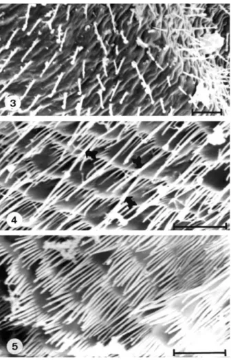
The anterior portion is characterized by the presence of microspines along the structure, which
resemble thin setae and are heterogeneous in size, number and shape. Initially, few and dispersed microspines can be seen (fig. 3), which progressively increase in number and size (figs. 4, 5). The intermediate portion of the anterior region (fig. 4) presents microspines of several sizes, distributed on plate structures; some of the larger ones repeated from space to space, are also observed intermixed with smaller and thinner microspines (arrows in fig. 4). In the final portion, no differences between microspines in spite of their large number are observed (fig. 5).
The transition between the anterior and posterior pyloric portions is remarkable (fig. 6); the long and numerous microspines disappear and other microspines of smaller size, triangular-shaped and uniformly distributed on small plates as multispinose can be seen (fig. 7).
Rhinocricus padbergi does not present the same differentiation in the pylorus (fig. 8) as observed in P. tricolor. Several filamentous structures are observed in this species, especially in the midgut/pylorus and pylorus/ ileum transitions (arrows in fig. 8). Most of these filamentous structures present their bases on the cuticular intima (arrow in fig. 9).
Microspines in multispinose are observed at the beginning of the pylorus (fig. 10), resulting in a scale-like aspect. Progressively, these microspines decrease in number and increase in size, thus characterizing the unispinose (figs. 11, 12).
Fig. 1. Scheme of a Diplopoda pyloric region (hg, hindgut; i, ileum; mg, midgut; Mt, Malpighian tube; p, pylorus).
Fig. 2. Scheme of the pylorus internal aspect of Pseudonannolene tricolor (ap, anterior pyloric region; i, ileum; mg, midgut; p, pylorus; pp, posterior pyloric region).
In the direction of the pylorus/ileum transition, the same organization seen in the beginning of the pylorus is observed, with the cuticle presenting microspines in multispinose (fig. 13). At the pylorus/ileum transition point, cuticular expansions larger than the denticles are observed (arrow in fig. 14).
DISCUSSION
The alimentary tract of millipedes is basically composed of a tube that extends from the mouth to the anus, divided into foregut, midgut and hindgut. The foregut and the hindgut have ectodermal origin and their cells secrete cuticle similar to that, which covers the external surface of the body. This cuticle, known as the intima, does not appear in the midgut where the digestion occurs. This cuticle lining can show different configurations in the animal groups depending basically on the alimentary diet.
Similarly to other arthropods, cuticular expansions are observed at the cuticular intima of the alimentary tract of diplopods, called spines by HOPKINS & READ (1992)
and in this group, these spines only occur in the hindgut. In both species studied here, these spines are restricted to the pylorus, the first portion of the hindgut.
Here the word microspine is used as suggested by ELZINGA (1998), to name a tiny structure presenting no
articulated basis. Microspines have also been observed in other species of diplopods; SCHLÜTER (1980) studying
Tachypodoiulus niger (Leach, 1815) and Polydesmus angustus Latzel, 1884 observed in T. niger that the spines are superficial, not attached to the epithelium, with the function of preventing the anterior movement of the fecal bolus by holding the leftovers in the perithrophic membrane. Our results suggest the same as observed in histological sections; the perithrophic membrane marked with microspines. These microspines are formed by simple cuticular expansions projected towards the intestine lumen without attaching to the epithelium. In Polydesmus angustus, the microspines are attached to the epithelium and each one is also attached to a longitudinal muscle provided with movement. SCHLÜTER (1980) suggested that this type
of spine is able to break the perithrophic membrane, allowing higher exchange of ions and fluids with the fecal material. In that paper, the author did not mention the region of the hindgut in which these structures appear in both studied species.
ELZINGA (1998) also reported microspines only in
the pylorus of Spirobolus sp. and Orthoporus sp.; these species present short microspines (1 to 2 µm) at the beginning of the pylorus that increase, reaching up to 50-60 µm with no other morphological alteration. The microspines seem to be isolated, in other words, as a microspine in unispinose.
The species studied here also present a progressive increase in size of microspines along the pylorus, especially in P. tricolor, but other morphological Figs. 8-10. SEM of pylorus of Rhinocricus padbergi. 8, general view; 9, detail of the midgut/pylorus transition; 10, detail of microspines at the beginning of the pylorus (i, ileum; mg, midgut; p, pylorus; arrows = filamentous structures). Bars, 100 µm, fig. 8; 50 µm, fig. 9; 2,5 µm, fig. 10.
alterations are also observed. The microspines appear as multispinose in both species.
The presence of microspines with multispinose aspects as observed in P. tricolor and R. padbergi, is also found in other groups. This has been reported in other parts of the alimentary tract including the pylorus, in several families of Orthoptera (BENTOS-PEREIRA &
LORIER, 1995; ELZINGA, 1996), in some species of Blattaria
(ELZINGA & HOPKINS, 1995), and in the foregut of
Scorpionida and nymphs of Odonata (ELZINGA, 1998).
Comparing our data to those found in other groups of Arthropoda, with regard to the region of the digestive tube where the microspines appear, similarities with data from Insecta were observed; as in Diplopoda, insects also present microspines in the pylorus, but such a presence was not observed in Pycnogonida, Merostomata, Arachnida, Crustacea and Chilopoda (ELZINGA, 1998).
ELZINGA (1998), in a phylogenetic analysis based
on molecular data combined with morphological characters of the alimentary canal of Arthropoda,
Onychophora and Annelida, reported that the presence of microspines in the phylogeny was firstly unispinose, being small and isolated within the foregut and hindgut of Chelicerata and Mandibulata. The presence of multispinose microspines occurred in different numbers. ELZINGA (1998) also explains that the occurrence of
microspines and other cuticular modifications may be correlated with the presence of well defined hindgut subdivisions and the presence of valves between these sections in Mandibulata. This condition seems to be a modification for the ingestion of particulate food. Such subdivisions (chambers) permit better regulation of food movement and also the retention of the remnants of the digested food to be acted upon by symbionts. This last hypothesis also seems to be valid for diplopods, as the pylorus is the only portion of the digestive system presenting microspines and the presence of symbionts in the digestive system is large and diversified (CRAWORD
et al., 1983; HOPKINS & READ, 1992; MOSS & TAYLOR, 1996).
Filamentous structures similar to those observed here were also found in the cuticle of the hindgut of Ophyiulus pilosus (Newport, 1843) and Cylindroiulus punctatus (Leach, 1815) (MOSS & TAYLOR, 1996).
According to those authors, they are related to microbionts of the order Eccrinales (Class Trichomycetes), frequently collected in millipedes of tropical, subtropical and temperate regions. It seems to be a cosmopolitan and ubiquitous infestation; however, the definition of these species is not easy as they have not been reared and besides, the host specificity and the influence that the intestinal environment may have on the morphology of Eccrinales is so far unknown. It is known that inhabitants of millipedes belong to the family Eccrinaceae; in England all species of diplopods analyzed belong to genusEnterobryus (Leidy, 1849) and the filaments are attached to the intestine cuticle, but they do not penetrate into it (MOSS & TAYLOR, 1996).
The cuticular structures of the stomodeum, such as microspines, have been found to be of value as systematic characters in some insect groups (BENTOS
-PEREIRA & LORIER, 1995), but in the hindgut it is less
discussed. In diplopods these structures do not appear in the foregut and because of the scarce studies (only four species) this question is not discussed.
With regard to these findings, a comparative study of the morphology of microspines in the digestive system of different diplopods species would be interesting, as a great diversity was found in the few species that were studied. Studies in different families and/or orders could help in the taxonomic problems and the phylogenetic correlations in the group.
Acknowledgments. We would like to thank Guilherme Guindolin Galassi, Izabela Braggião Calligaris and Rogilene Aparecida Prado (UNESP, Rio Claro, SP) for collecting samples ofP. tricolor; Antônio Teruyoshi Yabuki, Cristiane Márcia Miléo and Monika Iamonte (UNESP, Rio Claro, SP) for the technical support; Jonathan Burgess and Cybel Burgess for the English review and CNPq for the financial support.
REFERENCES
BENTOS-PEREIRA, A. & LORIER, E. 1995. Taxonomic value of the
cuticular structure of the stomodeum in Acridomorpha Figs. 11-14. SEM of pylorus of Rhinocricus padbergi: 11, 12,
(Orthoptera). Journal of Orthoptera Research 4:185-195. CRAWFORD, C. S.; MINION, G. P. & BOYERS, M. D. 1983. Intima morphology, bacterial morphotypes, and effects of annual molt on microflora in the hindgut of the desert millipede
Orthoporus ornatus (Girard) (Diplopoda: Spirostreptidae).
International Journal of Insect Morphology and Embryology 12(5/6):301-312.
ELZINGA, R. J. 1996. A comparative study of microspines in the alimentary canal of five families of Orthoptera (Saltatoria).
International Journal of Insect Morphology and Embryology 25(3):249-260.
___. 1998. Microspines in the alimentary canal of Arthropoda, Onycophora, Annelida. International Journal of Insect Morphology and Embryology 27(3):341-349.
ELZINGA, R. J. & HOPKINS, T. L. 1995. Microspine variation in
hindgut regions of four families of cockroaches (Blattaria).
International Journal of Insect Morphology and Embryology 24(2):203-211.
FANTAZZINI, E. R.; FONTANETTI, C. S. & CAMARGO-MATHIAS, M. I. 1998.
Anatomy of the digestive tube, histology and histochemistry of the foregut and salivary glands of Rhinocricus padbergi
Verhoeff. (Diplopoda: Spirobolida: Rhinocricidae). Arthropoda Selecta 7(4):257-264.
___. 2002. Midgut of the millipede, “Rhinocricus” padbergi
Verhoeff, 1938 (Diplopoda: Spirobolida): histology and histochemistry. Arthropoda Selecta 11(2):135-142. FONTANETTI, C. S. & CAMARGO-MATHIAS, M. I. 1997. Histoanatomy
of the digestive tract in Plusioporus setiger diplopod
Recebido em outubro de 2004. Aceito em maio de 2005. ISSN 0073-4721
Brolemann, 1901 (Spirostreptida, Spirostreptidae). Brazilian Journal of Morphological Sciences 14(2):205-211. FONTANETTI, C. S.; CAMARGO-MATHIAS, M. I. & CAETANO, F. H. 2001.
Apocrine secretion in the midgut of Plusioporus setiger
(Brolemann, 1901) (Diplopoda, Spirostreptidae). Naturalia 26:35-42.
HEFNER, R. A. 1929. The micro–anatomy of the alimentary canal
of Parajulus impressus Say. Transactions of the American Microscopical Society 48(4):321–351.
HOPKINS, S. P. & READ, H. J. 1992. The Biology of Millipedes. Oxford, Oxford University Press. 233p.
KARNOVSKY, M. J. 1965. A formaldehyde-glutaraldehyde fixative at high osmolarity for use in electron microscopy. The Journal of Cell Biology 27:137-138.
MILEY, H. H. 1930. Internal anatomy of Euryurus erythropygus
(Brant) (Diplopoda). Ohio Journal of Science 30 (4):229-254.
MOSS, S. T. & TAYLOR, J. 1996. Mycobionts in the gut of millipedes – The Eccrinales. The Mycologist 10(3):121-124. NUNEZ, F. S. & CRAWFORD, C. S. 1977. Anatomy and histology of
the alimentary tract of the desert millipede Orthoporus ornatus
(Girard) (Diplopoda: Spirostreptidae). Journal of Morphology 151(1):121-130.
ROMELL, L. S. 1935. An example of Myriapods as mull formers.
Ecology 16(1):67-71.
SCHLÜTER, U. 1980. Ultrastruktur der Pyloruszähnchen zweier Tausendfüssler (Tachypodoiulus niger, Polydesmus angustus).
Acta Zoologica 61:171-178.