INTRODUCTION
The order Gymnotiformes is a monophyletic group of electrogenic freshwater fish. It comprises six families of which the family Gymnotidae is the most widely dis-tributed in neotropical regions (Mago-Leccia, 1994). Gym-notus is the only genus of Gymnotidae and, until now, en-compasses about 15 nominal species, although many undes-cribed species probably still exist (Nelson, 1994). In Bra-zil Gymnotus species are usually known as “sarapó” (pro-nounced “säräpô”).
The species diversity of Gymnotus is well known in the Amazon basin where nine species are recognized (Albert
et al., 1999), but few data are available about species di-versity, distribution and population structure of this genus in other neotropical basins. In the southeastern Brazilian basins, four morphologically distinct Gymnotus species have been detected (Fernandes-Matioli et al., 1998). Cy-togenetic and electric organ discharge (EOD) analyses of these species have been carried out (Almeida-Toledo, 1978, Foresti et al., 1984, Foresti, 1987, Fernandes-Matioli, 1996; Fernandes-Matioli et al., 1997, 1998) but were in-conclusive for species identification since some of these species present identical karyotypes and/or EOD patterns. A very good specific molecular marker, obtained by the single primer amplification reaction (SPAR) technique, was described by Gupta et al. (1994). This technique is based on polymerase chain reaction (PCR) amplification of DNA markers using single primers of simple sequence repeats (SSR) or microsatellites.
The aim of the present work was the characterization of species and populations of Gymnotus from southeast-ern Brazilian basins using the SPAR technique to analyze species diversity and geographic distribution.
MATERIAL AND METHODS
Specimens
Four species, Gymnotus carapo (94 specimens), G. pantherinus (42 specimens), G. sylvius (58 specimens), and G. inaequilabiatus (four specimens) from eight south-eastern Brazilian river basins were analyzed using the SPAR technique (Table I and Figure 1). The 58 G. sylvius speci-mens were also analyzed based on polymorphic micro11 patterns. All specimens were obtained from wild popula-tions. Unless they were to be preserved for further analy-ses, the individuals were released at the collection site. Two other fish genera, Eigenmannia (Sternopygidae, Gymno-tiformes) and Pseudostegophilus (Trichomycteridae, Siluriformes), from neotropical basins were included in the experiment to verify the efficacy of micro11. Tissue samples of specimens from these two species were kindly provided by Dr. D. Calcagnotto and Dr. M. de Pinna, re-spectively.
Total DNA
DNA was extracted from blood, fin and/or scales and was isolated by the standard phenol:chloroform protocol (Sambrook et al., 1989).
Primers
Tetranucleotide repeat primers (16 bp) were em-ployed. Pilot experiments were carried out using 36 com-binations of the following oligonucleotides: (GGGT)4, (CACT)4, (GGAT)4, (AACC)4, (GACA)4, (GGAC)4, (TAGG)4 and (AAGC)4. Tetranucleotide primers were used because
Species diversity and geographic distribution of
Gymnotus
(Pisces: Gymnotiformes) by nuclear (GGAC)
nmicrosatellite analysis
F.M.C. Fernandes-Matioli, S.R. Matioli and L.F. Almeida-Toledo
Abstract
Patterns of amplified DNA fragments flanked by (GGAC)n microsatellites, obtained by single primer amplification reaction (SPAR), from 198 Gymnotus specimens (Pisces: Gymnotiformes) sampled from 8 southeastern Brazilian river basins were analyzed. The species stud-ied were Gymnotuscarapo, G. pantherinus, G. inaequilabiatus, and G. sylvius. The indirectly obtained patterns reflected the distribu-tion of simple sequence repeats in the nuclear genome of the specimens. Species-specific patterns of DNA amplificadistribu-tion were found and were useful for the analysis of the geographic distribution of Gymnotus species. Monomorphic patterns were found in G. carapo, G. pantherinus, and G. inaequilabiatus. Three polymorphic patterns were found in G. sylvius populations. The SPAR technique could be a useful molecular tool in conservation programs involving communities of neotropical freshwater fish.
shorter or longer repeats produce inferior PCR products (Gupta et al., 1994). Other possible repeats were not used to avoid primer-dimers.
PCR conditions
One nanogram of DNA was amplified in a final vol-ume of 30 µl containing 10 mM Tris-HCl, pH 8.4, 0.5% nonidet P-40, 50 mM KCl, 2.5 mM MgCl2, 100 µM each of dNTP, 5 pmol primers, and 1.25 units of Taq DNA poly-merase (Life Technologies). Amplifications were per-formed in a Perkin Elmer TC1 thermocycler for 35 cycles. The cycles consisted of 45 s at 94°C, 60 s at 53°C and 60 s at 72°C. All products were analyzed on 1.4% agarose gels stained with ethidium bromide. The molecular weight mark-ers used were λ digests with HindIII and EcoR, pBR328 digests with BamHI, BglI and HinfI, and a 123-bp DNA lad-der (Gibco BRL).
This work was conducted in three major steps. 1) We carried out PCR amplifications using different combina-tions of SSR primers in species previously identified by morphological criteria in a sample of 16 specimens. 2) After the characterization of a species-specific molecular marker we analyzed the geographic distribution of the 198 specimens sampled. 3) We analyzed the geographic distri-bution of the micro11 polymorphic patterns detected in G.
sylvius.
RESULTS AND DISCUSSION
Thirty-six combinations of primers were tested in some specimens previously identified by morphological analysis (Britsk, H.A., personal communication; Fernandes-Matioli et al., 1998). The primer combinations (TAGG)4 + (GGAT)4 and (GACA)4 + (AAGC)4, and by SPAR, (AAGC)4 and (CACT)4, albeit presenting positive amplification, pro-duced very faint bands and were not used. (GACA)4 gave monomorphic interspecific patterns.
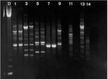
The most informative results were those obtained by SPAR using the primer (GGAC)4 and this molecular marker was named micro11; species-specific amplification patterns were only observed with this marker (Figures 2). Amplifi-cation was obtained with micro11 in two other fish genera,
Eigenmannia and Pseudostegophilus (Figure 2). The posi-tive amplification showed that this marker may be useful for other fish groups.
The results obtained by the amplification of regions flanked by (GGAC)n microsatellite loci in the four species studied revealed great intraspecific conservation of the amplification patterns. The intraspecific conservation of this molecular marker in Gymnotus species may be used for species characterization and was recently included as a tool in the description of the species G. sylvius (Albert et al., 1999).
In the second step of our work we tested specimens from different populations. All samples were successfully
Table I - Samples of Gymnotus species analyzed. Basin (number in Figure 1) Species Sample size Paranapanema River (1) G. carapo 34
G. carapo 28
Tietê River (2) G. sylvius 1
G. inaequilabiatus 4 Ribeira de Iguape River (3) G. sylvius 18 Pantanal do Miranda (4) G. carapo 19
G. sylvius 4
Tietê River (headwaters) (5) G. pantherinus 20 Coastal rivers (6) G. pantherinus 22 Paraíba do Sul River (7) G. carapo 1
G. sylvius 26
Mogi Guaçu River (8) G. carapo 12
G. sylvius 9
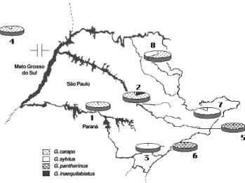
Figure 1 - Geographic localities of Gymnotus collection sites. 1, Parana-panema River (sample size: 34); 2, Tietê River (33); 3, Ribeira de Iguape River (18); 4, Pantanal do Miranda (23); 5, Tietê River (headwater) (20); 6, Coastal rivers (22); 7, Paraíba do Sul River (27); 8, Mogi Guaçu River (21). *, Sample previously studied by morphological analysis.
South America
Mato Grosso
do Sul São Paulo
Paraná
analyzed. The use of the micro11 patterns in combination with morphological analysis allowed the identification of the geographic distribution of Gymnotus species in the southeastern Brazilian basins (Figure 3) and analysis using
micro11 made it possible to quickly and easily identify a large number of specimens. All analyses were performed at least twice, with identical results. The species identifi-cation first obtained with micro11 in 64 specimens (20 G.
carapo, 20 G. pantherinus, 20 G. sylvius and 4 G. inaequilabiatus) was subsequently confirmed by morpho-logical analysis (Britsky, H., personal communication).
The total congruence between the morphological and molecular analyses is very useful because it would no longer be necessary to sacrifice numerous individuals or modify wild populations for analysis, permitting studies that do not interfere with biological conservation.
G. carapo was the most widely distributed of the spe-cies analyzed, being found in most of the small tributaries in southeastern Brazilian basins (Figure 3). G. pantherinus
was only detected in the coastal river basins of the State of São Paulo (sample 6, Figure 1), and in the headwaters of the Tietê River (sample 5, Figure 1). G. inaequilabiatus
was only found in tributaries of the Tietê River (sample 2, Figure 3). The populations of each of these three species presented a geographic distribution that was continuous in all the sampled basins. Populations of G. sylvius, on the other hand, were patchily assigned to five basins (samples 2, 3, 4, 7 and 8, Figure 3).
In some basins, Gymnotus populations of different species were found in sympatry, but in all cases one of the species was present at a significantly lower frequency. This was the case of the Paraíba do Sul River basin (sample 7, Figure 1), where 26 G. sylvius and one G. carapo were found sympatrically in a sample of 27 individuals (Figure 3). Sym-patry involving G. carapo (N = 28) and G. inaequilabiatus
(N = 4) was also observed in 32 specimens from the Tietê River basin (sample 2, Figure 3). However, there was no evi-dence of hybridization or introgression between the
Gymnotus species found in sympatry. Furthermore, the spe-cies found in sympatry had very different chromosome num-bers (Fernandes-Matioli et al., 1998), a factor that usually precludes introgression or hybridization on a large scale.
Sympatry was also observed in some parts of the Mogi Guaçu River, Tietê River, and Pantanal do Miranda basins (samples 8, 2, 4, respectively, Figure 3). It is known that in these areas Gymnotus specimens are often sold as bait for fishing, so this species mixture might be attributable to human interference.
In the third step of the work, we analyzed the intraspe-cific micro11 pattern variation in 58 individuals of G. sylvius from different populations (see Table I). Three dis-tinct patterns were found (A, B, C) (Figures 4b and 5). In the Paraíba do Sul River basin only the A pattern was found. The A and C patterns were found in the Ribeira do Iguape River basin, and all three patterns were found in the Mogi Guaçu River basin.
Figure 4 - Schematic drawing of micro11 patterns. a) Species-specific pat-terns: D, 123-bp DNA ladder; 1, Gymnotus carapo; 2, G. inaequilabiatus; 3, G. pantherinus. b) The three micro11 patterns obtained for G. sylvius.
D 1 2 3 A B C
a b
G. sylvius São Paulo
micro11 patterns
Figure 5 - Geographic distribution of micro11 patterns (see Figure 4) of three Gymnotus sylvius populations.
Mato Grosso do Sul
São Paulo
Paraná
G. carapo G. sylvius G. pantherinus G. inaequilabiatus
Since the data did not allow a classical analytical ap-proach based on allele frequencies, a comparative study of intraspecific polymorphisms based on the micro11 marker was adopted. The variant patterns may have been due to spu-rious PCR products, making the variation entirely arte-factual; however, taking into account the highly stringent conditions under which the experiments were performed, the micro11 intraspecific pattern variation could indeed be reflecting polymorphism within species. If so, the varia-tion might be generated by inverted sequence blocks or might reside within some larger repeat structure, such as satellite sequences or ribosomal internal transcriber spacer regions. Alternatively, this variation might be associated with some kind of dispersed repeats, such as SINEs (short interspersed repeated sequences).
After testing different PCR conditions (e.g., anneal-ing temperatures (50°, 51°, 52° and 53°C) and Mg2+
con-centrations (2.0, 2.5, and 3.0 mM)), we concluded that G. sylvius has at least three polymorphic micro11 patterns (Figure 4b).
The presence of the A (N = 7) and C (N = 11) patterns in the Ribeira de Iguape River basin (sample 3; Figure 5) and only pattern A in the Paraíba do Sul River basin (sample 7; Figure 5) may indicate an ancestral polymorphism (Fig-ure 5). The occurrence of a few G. sylvius specimens in the Tietê River basin (one specimen, sample 2; Figure 3) and in the Pantanal do Miranda basin (two individuals, sample 4; Figure 3) presenting the C and A patterns, respec-tively, was considered as evidence of species introduction because these patterns seem to be characteristic of other basins (see Ribeira de Iguape basin, sample 3, and Paraíba do Sul basin, sample 7; Figure 5). All the three micro11 patterns observed in G. sylvius were found in the Mogi Guaçu River basin (sample 8; Figure 5) (A, N = 7; B, N = 11, and C, N = 3, in a sample of 21 fish). The occurrence of pattern B only in the Mogi Guaçu River basin may indicate, in spite of the commercial profile of this site, that natural populations of G. sylvius might actually occur in this re-gion since this was the only place where this pattern was detected. On the other hand, the presence of patterns A and C in the Mogi Guaçu River basin could be evidence of speci-men introduction since they are characteristic of very dis-tant populations.
Other studies involving G. sylvius (including the analy-sis of individuals that presented A, B, and C micro11 pat-terns) showed homogeneity in chromosome number (2n = 40, Fernandes-Matioli et al., 1998), and morphological and bioelectrical traits (Albert et al., 1999). According to these studies, the hypothesis of sibling species living in sympa-try could be discarded.
The presence of only one micro11 pattern in the G. sylvius population from the Paraíba do Sul basin (sample 7; Figure 5) may be indicative of the loss of genetic vari-ability due to genetic drift. This way, this population had probably originated from colonization events associated with the founder effect. In fact, this is very plausible
be-cause of the generally small size of the populations that migrate from one basin to another. This has already been observed in cichlid fish (Agnèse et al., 1997).
The present work outlines the first steps of an appro-priate application of a novel molecular technique to im-prove the understanding of species diversity and geographic distribution of Gymnotus species, an approach which may be very useful in conservation programs involving com-munities of neotropical freshwater fish.
ACKNOWLEDGMENTS
We most gratefully thank Dr. Joan Strassman for sugges-tions of microsatellites analysis and Dr. Anita Wajntal for helping in the lab work. We also thank Dr. H.A. Britski, Museu de Zoologia da Universidade de São Paulo, for the identification of species, and Dr. M. de Pinna for providing some tissue samples. This manu-script was improved by comments from Dr. Silvio A. Toledo-Filho, Dr. Daniela Calcagnotto and Ms. Cinthia B. Moysés. We would like to thank the anonymous reviewers whose comments improved the earlier version of the manuscript. This study was supported by grants from the CNPq, CAPES, FINEP and FAPESP (No.92/ 4969-8). Publication supported by FAPESP.
RESUMO
No presente estudo foram analisados os padrões de ampli-ficação de fragmentos de DNA nuclear flanqueados por micros-satélites (GGAC)n obtidos a partir de 198 exemplares do gênero
Gymnotus (Pisces: Gymnotiformes) amostrados em 8 bacias hidrográficas do sudeste brasileiro. As espécies analisadas foram Gymnotuscarapo, G. pantherinus, G. inaequilabiatus e G. syl-vius. Os padrões de amplificação foram obtidos através da técnica de SPAR (single primer amplification reaction) e refletem, indiretamente, a distribuição de seqüências repetitivas simples no genoma nuclear dos espécimens. Foram encontrados padrões de amplificação espécie-específicos, os quais foram utilizados como potentes ferramentas na análise da distribuição geográfica e diversidade de espécies de Gymnotus. Padrões monomórficos foram observados em G. carapo, G. pantherinus e G. inaequi-labiatus. Três padrões polimórficos foram verificados em G. syl-vius. Os resultados obtidos através da técnica SPAR indicam que esta é uma abordagem promissora como ferramenta molecular em programas de conservação de comunidades de peixes de água doce neotropicais.
REFERENCES
Agnèse, J.F., Adépo-Gourène, B., Abban, E.K. and Fermon, Y. (1997). Ge-netic differentiation among natural populations of the Nile tilapia Oreo-chromis niloticus (Teleostei, Cichlidae). Heredity 79: 88-96. Albert, J., Fernandes-Matioli, F.M.C. and Almeida-Toledo, L.F. (1999). A
new species of Gymnotus (Gymnotiformes, Teleostei) from Southeast-ern Brazil: towards the deconstruction of Gymnotus carapo. Copeia 2: 410-421.
Almeida-Toledo, L.F. (1978). Contribuição à citogenética de Gymnotiformes (Pisces, Ostariophysi). Doctoral thesis, IB, Universidade de São Paulo, Brasil.
Fernandes-Matioli, F.M.C., Almeida-Toledo, L.F. and Toledo-Filho, S.A. (1997). Extensive nucleolus organizer region polymorphism in Gymnotus carapo (Gymnotoidei, Gymnotidae). Cytogenet. Cell Genet. 78: 236-239. Fernandes-Matioli, F.M.C., Marchetto, M.C.N., Almeida-Toledo, L.A. and Toledo-Filho, S.A. (1998). High intraspecific karyologycal conserva-tion in four species of Gymnotus (Pisces: Gymnotiformes) from South-eastern Brazilian basins. Caryologia 51: 221-234.
Foresti, F. (1987). Estudos cromossômicos em Gymnotiformes (Pisces, Ostariophysi). Livre Docência thesis, Instituto Básico de Biologia Mé-dica e Agrícola, Universidade Estadual “Júlio de Mesquita”, Botucatu, Brasil.
Foresti, F., Almeida-Toledo, L.F. and Toledo-Filho, S.A. (1984). Chromo-some studies in Gymnotus carapo and Gymnotus sp. (Pisces, Gymno-tidae). Caryologia 37: 141-146.
Gupta, M., Chyi, Y.-S., Romero-Severson, J. and Owen, J.L. (1994). Ampli-fication of DNA markers from evolutionarily diverse genomes using single primers of simple-sequence repeats. Theor. Appl. Genet. 89: 998-1006.
Mago-Leccia, F. (1994). Electric Fishes of the Continental Waters of America. Biblioteca de la Academia de Ciencias Fisicas, Matematicas, y Naturales. Caracas, Venezuela, 29: 1-206.
Nelson, J.S. (1994). Fishes of the World. 3rd edn. John Wiley & Sons, Inc., New York, pp. 172-175.
Sambrook, J., Fritsch, E.F. and Maniats, T. (Eds.) (1989). Molecular Clon-ing, A Laboratory Manual. 2nd edn. Cold Spring Harbor Laboratory, Cold Spring Harbor, NY.