Full paper published online: May 30, 2009 ISSN 1678-9199.
ALT-C, A DISINTEGRIN-LIKE CYS-RICH PROTEIN FROM Bothrops alternatus,
INCREASES SKELETAL MYOBLAST VIABILITY
Mesquita-Ferrari RA (1), de Moraes CK (2), Micocci KC (2), Selistre-de-Araújo HS (2)
(1) Department of Physiotherapy, Nove de Julho University, UNINOVE, São Paulo, São Paulo State, Brazil; (2) Department of Physiological Science, Federal University of São Carlos, São Carlos, São Paulo State, Brazil.
ABSTRACT: ALT-C, an ECD motif (glutamic acid, cysteine, aspartic acid) disintegrin from Bothrops alternatus snake venom, induces α2β1 integrin-mediated signaling and neutrophil chemotaxis. In vitro, in human umbilical vein endothelial cells (HUVEC), ALT-C induces cell proliferation, thus showing an interesting potential for tissue regeneration studies. This work aimed to evaluate the influence of ALT-C in myoblast viability and differentiation. Myoblasts were obtained from hind limb muscles of 3 to 4-day old Wistar rats. The cells were incubated with ALT-C at different concentrations and incubation periods were followed by total RNA isolation. cDNA synthesis and real time polymerase chain reaction (PCR) were performed with primers of myoD as well as of both (slow and fast) myosin heavy chain isoforms (MHC). ECD-disintegrin increased myoblast viability in a dose-dependent way, mostly with 50 to 100 nM concentrations, and such effect was more prevalent after 48 hours. No changes in gene expression of both MHC isoforms were observed in ALT-C-treated cells. MyoD expression was not detected, which suggests that myoblasts were in mature stages. Protease activity and cytokine array tested in a medium of 50 nM ALT-C-treated cells after 48 hours were not different from controls. In conclusion, it was shown that myoblats are sensitive to ALT-C indicating an integrin-mediated intracellular signaling that increases cell viability.
KEY WORDS: viability, myosin heavy chain, myoblast, disintegrin, skeletal muscle,
Bothrops alternatus.
CONFLICTS OF INTEREST: There is no conflict.
FINANCIAL SOURCE: FAPESP and CNPq.
CORRESPONDENCE TO:
RAQUEL AGNELLI MESQUITA-FERRARI, Departamento de Fisioterapia, Mestrado
em Ciências da Reabilitação, Universidade Nove de Julho, UNINOVE, Centro de
Pós-graduação, Av. Francisco Matarazzo, 612, São Paulo, SP, 05001-100, Brasil.
INTRODUCTION
Function and maintenance of tissue integrity depend on specific interactions of cells
with the surrounding extracellular matrix (ECM). Transmembrane receptors are
involved in the polymerization and assembly of the matrix as well as in providing a
mechanical connection with the cytoskeleton and a way of transducing signals from
the extracellular matrix to the nucleus (1).
Integrins form a family of cell surface adhesion receptors, mediating both cell-to-cell
and cell-to-matrix interactions. They are heterodimeric transmembrane glycoproteins
that consist of α and β chains, non-covalently associated (2, 3). At least 18 α and
eight β chains have already been identified, which differently combined form about 24
distinct dimers (1, 4).
Studies carried out with birds have indicated that β1 integrins are involved in cell
migration from the somite and terminal differentiation of myoblasts into myotubes,
and that particular integrins are expressed in different stages of muscular
development and differentiation (5). The integrins α5β1 (fibronectin receptor), α6β1
and α7β1 (laminin receptor) are widely expressed during development and
down-regulated after myotube formation, whereas α7β1 is mainly restricted to skeletal and
cardiac muscles and is strongly up-regulated during myoblast fusion. The role of
α5β1 and α6β1 in muscle development and the reason they coexist at the myoblast
stage as ligand-opposing receptors have not bell well characterized yet (1).
A family of myogenic transcription factors, expressed at precise steps during
development, coordinates the process of myogenic differentiation. Myoblasts start to
express early specific markers known as muscle regulatory factors (MRF) including
desmin, Myf-5 and MyoD while the expression of myogenin, muscle regulatory factor
4 (MRF4) and myosin occurs later (6-9). However, the expression of these factors is
also regulated by extracellular signaling molecules from the ECM and other elements
involved in cell-to-cell contact (8).
Although myosin heavy chain (MHC) is known to be closely related to muscle
function, it consists of several isoforms (10, 11). Innervation or cellular signaling
affects myosin expression; however, fibers can express the slow myosin in early
phases independently of innervation (12, 13). Innervation-independent variation in
muscle fiber phenotype based on individual myoblasts gave rise to the concept that
contractile protein genes in fiber type specific patterns (10). Furthermore, when
skeletal muscle myoblasts were mechanically stretched, cell growth was facilitated
and mRNA expression in MHC 2b, 2d and 2a increased with rapid, medium and
slow stretching, respectively (10, 11).
Disintegrins were first described in 1989 as a protein group with low molecular weight
(5 to 9 kDa), that interact with integrin receptors on the cell surface (14-16). These
peptides represent a family of cysteine-rich proteins, isolated from snake venoms,
and are known to inhibit cell-to-matrix and cell-to-cell interactions mediated by
integrins (2, 17). Most disintegrins contain a RGD/KGD sequence within a hairpin
loop maintained by disulfide bonds and are very potent inhibitors of αIIbβ3
integrin-dependent platelet aggregation as well as cell-to-matrix interactions involved in tumor
cell metastasis and angiogenesis (2, 18, 19).
A different class of disintegrins, called disintegrin-like, is also found in snake venoms
and does not contain the RGD motif. These proteins are larger than RGD disintegrins
(about 30 kDa) and have an extra C-terminal, cysteine-rich domain. Additionally, they
do not bind to αIIbβ3, α5β1 or αvβ3 integrins, but interact with the collagen receptor,
α2β1 integrin, therefore inhibiting cell adhesion to collagen I. The D/ECD sequence
replaces the RGD motif, and it has been suggested that this sequence is involved in
integrin binding (20).
ALT-C, an ECD-disintegrin isolated from Bothrops alternatus venom, is synthesized
as a precursor form with a metalloproteinase domain from which it is released after
proteolytic processing, yielding a form with both disintegrin and cysteine rich
domains. ALT-C induces integrin-mediated signaling and chemotaxis in neutrophils
and also strongly leads to human vein endothelial cells (HUVEC) proliferation in vitro
by up-regulating the expression of some growth factors, including vascular
endothelial growth factor (VEGF) (2, 18, 20, 21).
There is considerable interest in skeletal muscle regeneration studies to improve
efficiency of repair in sports medicine, after severe injuries or muscle transplantation,
in muscular dystrophies and for recovery strength in disuse or space flight atrophy
(22). The use of integrin-binding molecules that up-regulate the expression of growth
factors may be useful in these studies. This is the first demonstration of the influence
MATERIALS AND METHODS
Protein Purification
ALT-C was purified from Bothrops alternatus venom by two steps of gel filtration
followed by anion exchange chromatography (16). The molecular mass of the
purified protein was estimated in 29 kDa by sodium dodecyl sulfate polyacrylamide
gel electrophoresis (SDS-PAGE). Protein concentration was estimated according to
the method of Bradford (23).
Cell Culture
Myoblasts were obtained from limb muscles of 3 to 4-day old Wistar rats, according
to Horn and Brodwick (24). Rats were killed and limb muscles were removed, cut into
small pieces and homogenized by trituration for 10 minutes with shears. After that,
tissue was submitted to an enzymatic digestion with tripsin (0.025% in PBS buffer)
and collagenase V (0.1% in PBS buffer) for 40 minutes at 37°C. Then, cells were
cultivated in a 10 cm plastic dish with Dubelcco’s minimum medium (DMEM –
Cultilab, Brazil) supplemented with 10% fetal bovine serum, 1% L-glutamine, 1%
antibiotic-antimycotic solution(Cultilab, Brazil) at 37°C, with 5% CO2 for 20 minutes to
remove other cell types and proteins such as fibroblasts, endothelial cells and
ptoteoglicans. Afterwards, cells were transferred to a culture flask containing DMEM
supplemented as described before.
Cell Viability Assay
Myoblasts (105 cells/well) were incubated in a 96-well plate in 200 μL of DMEM with
ALT-C at different concentrations at 37°C and 5% CO2 for 24, 48 and 72 hours. After
incubation time, media from cultures were saved for latter protein analysis. Cell
concentration was measured by addition of a tetrazolium salt – MTT
[1-(4,5-dimethylthiazol-2-yl)-3,5-diphenyl formazan, Sigma, USA], 0.5 mg/mL final
concentration – followed by incubation at 37oC and 5% CO2 for three hours. The
bound stain was dissolved in isopropyl alcohol and measured spectrophotometrically
at 595 nm. All experiments were repeated three times, and each sample was made
in triplicate. Some experiments were performed in collagen I-coated plates (1
employed for data analysis. Statistical significance was assessed by the Dunnett’s
test and acceptable p levels were less than 0.05 (25).
Detachment Assay
Myoblasts (105 cells/well) were incubated in collagen I-coated 96-well plates in 200
μL of adhesion buffer [20 mM Hepes (Sigma, USA), 150 mM NaCl, 5 mM MgCl2,
0.25 mM MnCl2,pH 7.4] at 37°C for four hours. Subsequent to incubation, cells were
washed five times with adhesion buffer, then ALT-C in adhesion buffer was added at
different concentrations and cells were incubated at 37°C and 5% CO2 for 48 hours.
Medium from cultures was removed, cells were washed again and cell concentration
was measured by adding MTT (0.5 mg/mL, final concentration) followed by
incubation at the same conditions for three hours. Data analysis was performed as
previously described.
Isolation of Total RNA
Cells (106 cells/well) were incubated in 10-cm plastic dishes in DMEM medium with
or without 50 nM ALT-C at 37°C and 5% CO2 for 24, 48 and 72 hours. Culture
medium was removed and cells were lysed with cold TRIzol® reagent (Invitrogen
Carlsbad, USA) for total RNA isolation, according to the manufacturer instructions.
Total RNA was quantified by spectrophotometry and RNA samples were treated with
DNAse (Invitrogen Carlsbad, USA) to avoid contamination with genomic DNA. All
solutions were prepared with 0.01% diethyl pyrocarbonate-treated water (DEPC –
Sigma, USA), while glassware and plasticware were treated against RNase using
standard procedures.
cDNA Synthesis and Real Time PCR
cDNA synthesis was performed using the AMV reverse transcriptase (Access Quick
RT-PCR System® (Promega, USA) and real time PCR was accomplished using the
SYBR Green PCR® Master Mix (Applied Biosystems, USA) in a Rotor-Gene RG
Oligonucleotide Primers
Specific primers for MHC I isoform (GenBank, accession n. X15939) and MHC IIa
were used for real time PCR (26). The primer sequences were:
• MHC I forward: 5’ AGA GAA TGG CAA GAC GGT GAC T; reverse: 5’ CAT GTC CTC GAT CTT GTC GAA CT (82 bp amplicon);
• MHC IIa foward: 5’ TAT CCT CAG GCT TCA AGA TTT G; reverse: 5’ TAA ATA GAA TCA CAT GGG GAC A (310 bp amplicon);
• MyoD forward: 5’ GGA GAC ATC CTC AAG CGA TGC; reverse: 5’ AGC ACC TGG TAA ATC GGA TTG.
A constitutive gene, β-actin, was emplyed to normalize data using the same amount
of cDNA. β-actin primers were forward 5’ CGTGGGCCGCCCTAGGCACCAGGG and
reverse 5’ CGGAGGAAGAGGATGCGGCAGTGG (604 bp amplicon).
To normalize data among control and treated groups, arbitrary units were calculated
as follows, Arbitrary Unit = 2 –ΔΔTC, and ΔΔTC = sample ΔTC – control ΔTC (TC,
threshold cycle).
Protease Activity and VEGF Expression
After incubation time, the cell medium was removed and tested for protease activity
by zymography, carried out as described by Allen et al (27) with small modifications.
For enzymatic assays, SDS-gels (15%) were prepared with gelatin (1 mg/mL) and 30
μg of protein was loaded per lane in sample buffer without reduction. After
electrophoresis, gels were washed twice for 15 minutes each with 2.5% Triton
X-100® (Sigma, USA) to eliminate SDS. Gels were then incubated overnight at 37°C in
substrate buffer (50 mM Tris-HCl, pH 8.5, 5 mM CaCl2, 0.02% NaN3). After this, gels
were stained for 30 minutes in 0.05% Coomassie Brilliant Blue R-250® (Sigma, USA)
in acetic acid:methanol:water (1:4:5) and destained in the same solution. All gels
were prepared and run at the same time. The bands were quantified by densitometry
using the Image Pro-plus software (Media Cybernetics, USA). ELISA assays for
VEGF detection were performed using the Quantikine® immunoassay kit for human
VEGF (R&D Systems, USA) according manufacturer instructions. Briefly, 200 μL of
conditioned media were added to each well, previously coated with human
and incubated with an enzyme-linked polyclonal anti-VEGF antibody. Following
another washing, substrate solution was added to wells and color developed in
proportion to the amount of VEGF bound in the initial step. The plate was read on a
Dynex plate reader (USA) at 450 nm.
Cytokine Array
The expression of cytokines was analyzed using the incubation medium of control
cells (without Alt-C) and treated cells (with Alt-C 50 nM), after 48 hours, using the
RayBio® human cytokine antibody array kit (R&D Systems, USA). The analyzed
cytokines were interleukins (IL-1α, IL-2, IL-3, IL-5, IL-6, IL-7, IL-8, IL-10, IL-13, IL-15);
tumor growth factor (TGF-β1); tumor necrosis factors (TNF-α, TNF-β); interferons
(IFN-γ, IFN-α, IFN-β); granulocyte colony-stimulating factor (GCSF);
granulocyte-monocyte colony-stimulating factor (GM-CSF); genes GRO and GRO-a; regulated
upon activation, normal T-cell expressed and secreted (RANTES); monokine induced
by interferon-gamma (MIG); macrophage/monocyte chemotactic proteins (MCP-1,
MCP-2, MCP-3).
RESULTS
ALT-C Induces Myoblast Viability
There was no significant difference of cell number in ALT-C treated myoblasts when
compared with control values 24 hours after incubation in the tested concentrations
(Figure 1). However, ALT-C induced a significant increase in viability of myoblasts at
50 nM concentration, 48 and 72 hours after incubation. Myoblast viability was also
induced by ALT-C at 100 nM, but only after 72 hours. These effects were not
observed with higher concentrations, such as 200 nM, which presented values not
significantly different from controls (Figure 1).
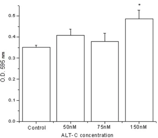
For cell growing on collagen-coated plates, similar results were found. ALT-C also
improved myoblast viability; however, higher concentrations, around 100 and 150
nM, were required (Figure 2). Furthermore, ALT-C did not provoke myoblast
detachment when it was added to a culture after myoblast adhesion (results not
Figure 1. Myoblast viability in absence (control) and presence of ALT-C at different
concentrations 24, 48 and 72 hours after incubation, performed in a non-coated
plastic plate (*p ≤ 0.05; ANOVA/Dunett).
Figure 2. Viability of myoblasts in absence (control) and presence of ALT-C at
different concentrations 48 hours after incubation in collagen I-coated wells (*p ≤
Influence of ALT-C on Myosin Heavy Chain Expression
Both myosin slow (MHCI, Figure 3 – A) and fast (MHCIIa, Figure 3 – B) isoforms
were expressed in all conditions: absence (control) and presence of ALT-C in all
incubation periods and no differences were observed when ALT-C was used.
Figure 3. Myosin heavy chain gene expression by myoblasts in absence (control)
and presence of ALT-C (50 nM) – 24, 48 and 72 hours after incubation – by real time
PCR. (A) Slow isoform (MHC I) and (B) fast isoform (MHCIIa) (*p ≤ 0.05).
Protease, VEGF and Cytokine Expression
Protease activity was detected in the culture medium of myoblast as a single ~72
kDa lytic band (Figure 4). This activity was completely abolished when gels were
incubated in substrate buffer with 15 mM EDTA (not shown), suggesting the
ALT-C-treated samples and controls. VEGF was not detected in the medium culture of both
control and treated cells (results not shown). There was no difference in cytokine
expression between control and treated cells (Figure 5). After membrane incubation
in the culture medium, only positive controls were marked.
Figure 4. Protease expression detected by zymography, using a culture medium
from myoblasts treated with ALT-C and from control (without ALT-C), at different
concentrations, 48 hours after incubation. The arrow indicates a band of ~72 kDa
with protease activity.
Figure 5. Cytokine expression in (A) absence (control) and (B) presence of ALT-C
(50 nM). Membranes were incubated in medium from myoblast culture treated for 48
DISCUSSION
It was previously demonstrated that ALT-C, an α2β1-integrin ligand, induces VEGF
expression in fibroblasts and endothelial cell viability in doses as low as 10 nM (19).
Therefore, we found interesting to study the effect of this disintegrin on other cell
types. A striking observation is that ALT-C induced myoblast viability in a
dose-dependent way, mostly in concentrations ranging from 50 to 100 nM. Interestingly, in
the presence of collagen I, increased protein concentrations were require to provoke
similar results (150 nM). In addition, this effect was also influenced by incubation time
and was more consistent after 48 hours. These results suggest that α2β1 integrin
may be relevant for muscle cell viability. Moreover, ALT-C did not induce myoblast
detachment as similarly observed in fibroblasts and HUVEC (19).
Cells express distinct sets of muscle-specific proteins at different stages of the
myogenic pathway. Desmin, Myf-5 and MyoD are expressed at relatively early
stages, whereas myogenin, myogenin regulatory factor 4 (MRF4) and myosin are
expressed at later stages. The cells utilized in the current study expressed both MHC
I and IIa isoforms of myosin, but not MyoD. These characteristics allow us to
conclude that they belong to later differentiation stages (6-9, 28, 29).
It has been reported that myoblasts in their mature stage, as myotube, express
mostly slow myosin, while the fast form can be found especially after innervation or
under signaling conditions (10, 11). The present data showed that myosin expression
occurred in myoblast cells and both isoforms, slow and fast, were expressed in
absence of innervation. Furthermore, there was no evidence of ALT-C influence in
the expression of these isoforms, since their values were similar in the presence and
absence of this disintegrin in all periods of cell incubation.
ALT-C was reported to induce in vitro VEGF expression by fibroblasts, but not by
HUVEC (18). However, in the present study VEGF expression was not detected
either at mRNA or protein level, suggesting that this effect may be cell-specific. It
also implies that the primary cultures were not contaminated with fibroblasts. We
could not determine, by the assays accomplished, which factors or pathways could
be involved in myoblast intracellular signaling induced by ALT-C. More studies, such
as cDNA arrays, are necessary to address these relevant questions for a better
understanding of the action mechanism of this desintegrin.
Disintegrin-related proteins (ADAM – a disintegrin and metalloproteinase), with a
organisms, in which they are involved in numerous physiological processeses such
as fertilization, cell differentiation and shedding of receptors (30). It has been
demonstrated that ADAM disintegrin and Cys-rich domains may also trigger
integrin-mediated intracellular signaling events and cell responses (31). Given the homology
between disintegrins from snake venoms and ADAM domains, it is reasonable to
suggest that the disintegrin-like domain of some ADAM may have a role in myoblast
viability.
In conclusion, our results demonstrated that ALT-C did not modify myoblast MHC
expression but induced an increase in cell viability. Therefore, ALT-C may be very
valuable for myogenesis and skeletal muscle regeneration studies.
ACKNOWLEDGEMENTS
This work was supported by grants from FAPESP (98/14138-2; 04/09671-6) and
CNPq (350111/1998-7), Brazil.
REFERENCES
1. Mayer U. Integrins: redundant or important players in skeletal muscle? J Biol
Chem. 2003;278(17):14587-90.
2. Mariano-Oliveira A, Coelho AL, Terruggi CH, Selistre-de-Araújo HS, Barja-Fidalgo
C, De Freitas MS. Alternagin-C, a non RGD-disintegrin, induces neutrophil migration
via integrin signaling. Eur J Biochem. 2003;270(24):4799-808.
3. Niewiarowski S, McLane MA, Kloczewiak M, Stewart GJ. Disintegrins and other
naturally occurring antagonists of platelet fibrinogen receptors. Semin Hematol.
1994;31(4):289-300.
4. Humphries JD, Byron A, Humphries MJ. Integrin ligands at a glance. J Cell Sci.
2006;119(Pt 19):3901-3.
5. Velleman SG, McFarland DC. β1 integrin mediation of myogenic differentiation:
implications for satellite cell differentiation. Poult Sci. 2004;83(3):245-52.
6. Cabane C, Englaro W, Yeow K, Ragno M, Dérijard B. Regulation of C2C12
myogenic terminal differentiation by MKK3/p38α pathway. Am J Physiol Cell Physiol.
2003;284:658-66.
7. Dominov JA, Dunn JJ, Miller JB. Bcl-2 expression identifies an early stage of
myogenesis and promotes clonal expansion of muscle cells. J Cell Biol.
8. Krauss RS, Cole F, Gaio U, Takaesu G, Zang W, Kang JS. Close encounters:
regulation of vertebrate skeletal myogenesis by cell-cell contact. J Cell Sci.
2005;118:2355-62.
9. Tannu NS, Rao VK, Chaudhary RM, Giorgianni F, Saeed AE, Gao Y, Raghow R.
Comparative proteomes of the proliferating C(2)C(12) myoblasts and fully
differentiated myotubes reveal the complexity of the skeletal muscle differentiation
program. Mol Cell Proteomics. 2004;3(11):1065-82.
10. Kurokawa K, Abe S, Sakiyama K, Takeda T, Ide Y, Ishigami K. Effects of
stretching stimulation with different rates on the expression of MyHC mRNA in mouse
cultured myoblasts. Biomed Res. 2007;28(1):25-31.
11. Sakiyama K, Abe S, Tamatsu Y, Ide Y. Effects of stretching stress on the muscle
contraction proteins of skeletal muscle myoblasts. Biomed Res. 2005;26(2):61-8.
12. Jordan T, Jiang H, Li H, DiMario JX. Inhibition of ryanodine receptor 1 in fast
skeletal muscle fibers induces a fast-to-slow muscle fiber type transition. J Cell Sci.
2004;117(Pt 25):6175-83.
13. Miller JB, Crow MT, Stockdale FE. Slow and fast myosin heavy chain content
defines three types of myotubes in early muscle cell cultures. J Cell Biol. 1985;101(5
Pt 1):1643-50.
14. Beviglia L, Stewart GJ, Niewiarowski S. Effect of four disintegrins on the adhesive
and metastatic properties of B16F10 melanoma cells in a murine model. Oncol Res.
1995;7(1):7-20.
15. Gould RJ, Polokoff MA, Friedman PA, Huang TF, Holt JC, Cook JJ, Niewiarowski
S. Disintegrins: a family of integrin inhibitory proteins from viper venoms. Proc Soc
Exp Biol Med. 1990;195(2):168-71.
16. Souza DHF, Iemma MRC, Ferreira LL, Faria JP, Oliva MLV, Zingali RB,
Niewiarowski S, Selistre-de-Araújo HS. The disintegrin-like domain of the snake
venom metalloprotease alternagin inhibits α2β1 integrin-mediated cell adhesion. Arch
Biochem Biophys. 2000;384(2):341-50.
17. Calvete JJ, Moreno-Murciano MP, Theakston RD, Kisiel DG, Marcinkiewicz C.
Snake venom disintegrins: novel dimeric disintegrins and structural diversification by
18. Cominetti MR, Terruggi CH, Ramos OH, Fox JW, Mariano-Oliveira A, De Freitas
MS, Figueiredo CC, Morandi V, Selistre-de-Araujo HS. Alternagin-C, a disintegrin-like
protein, induces vascular endothelial cell growth factor (VEGF) expression and
endothelial cell proliferation in vitro. J Biol Chem. 2004;279(18):18247-55.
19. Kamiguti AS, Hay CRM, Zuzel M. Inhibition of collagen-induced platelet
aggregation as the result of cleavage of α2β1 integrin by the snake venom
metalloproteinase jararhagin. Biochem J. 1996;320(pt2):635-41.
20. Selistre-de-Araújo HS, Cominetti MR, Terruggi CHB, Mariano-Oliveira A, De
Freitas MS, Crepin M, Figueiredo CC, Morandi V. Alternagin-C, a disintegrin-like
protein from the venom of Bothrops alternatus, modulates α2β1 integrin-mediated
cell adhesion, migration and proliferation. Braz J Med Biol Res. 2005;38(10):1505-11.
21. Ramos OH, Selistre-de-Araujo HS. Snake venom metalloproteases – structure
and function of catalytic and disintegrin domains. Comp Biochem Physiol C Toxicol
Pharmacol. 2006;142(3-4):328-46.
22. Grounds MD, White JD, Rosenthal N, Bogoyevitch MA. The role of stem cells in
skeletal and cardiac muscle repair. J Histochem Cytochem. 2002;50(5):589-610.
23. Bradford MM. A rapid and sensitive method for the quantization of microgram
quantities of protein utilizing the principle of protein-dye binding. Anal Biochem.
1976;72:248-54.
24. Horn R, Brodwick MS. Acetylcholine-induced current in perfused rat myoballs. J
Gen Physiol. 1980;75(3):297-321.
25. Löster K, Horstkorte R. Enzymatic quantification of cell-matrix and cell-cell
adhesion. Micron. 2000;31(1):41-53.
26. Jaschinski F, Schuler M, Peuker H, Pette D. Changes in myosin heavy chain
mRNA and protein isoforms of rat muscle during forced contractile activity. Am J
Physiol Cell Physiol. 1998;43(2):365-70.
27. Allen DL, Teitelbaum DH, Kurachi K. Growth factor stimulation of matrix
metalloproteinase expression and myoblast migration and invasion in vitro. Am J
Physiol Cell Physiol. 2003;284:805-15.
28. Di Carlo A, De Mori R, Martelli F, Pompilio G, Capogrossi MC, Germani A.
Hypoxia inhibits myogenic differentiation through accelerated MyoD degradation. J
29. Dogra C, Hall SL, Wedhas N, Linkhart TA, Kumar A. Fibroblast growth factor
inducible 14 (Fn14) is required for the expression of myogenic regulatory factors and
differentiation of myoblasts into myotubes. Evidence for TWEAK-independent
functions of Fn14 during myogenesis. J Biol Chem. 2007;282(20):15000-10.
30. Blobel CP. ADAMs: key components in EGFR signaling and development. Nat
Rev Mol Cell Biol. 2005;6(1):32-43.
31. Eto K, Huet C, Tarui T, Kupriyanov S, Liu HZ, Puzon-McLaughlin W, Zhang XP,
Sheppard D, Engvall E, Takada Y. Functional classification of ADAMs based on a
conserved motif for binding to integrin α9β1: implications for sperm-egg binding and