Brazilian Journal of Microbiology (2009) 40: 795-807 ISSN 1517-8382
PURIFICATION AND CHARACTERISATION OF AN EXTRACELLULAR PHYTASE FROM ASPERGILLUS NIGER 11T53A9
Ralf Greiner1*, Lucineia Gomes da Silva2,3, Sonia Couri2
1
Department of Food and Bio Process Engineering, Max Rubner-Institute, Federal Research Institute of Nutrition and Food,
Haid-und-Neu-Straße 9, D-76131 Karlsruhe, Germany; 2CEFET, Química de Nilópolis, Maracanã, RJ, Brasil; 3
Empresa Brasileira de
Pesquisa Agropecuária, Tecnologia de Alimentos, Guaratiba, RJ, Brasil.Submitted: November 07, 2008; Returned to authors for corrections: February 10, 2009; Approved: June 28, 2009.
ABSTRACT
An extracellular phytase from Aspergillus niger 11T53A9 was purified about 51-fold to apparent homogeneity with a recovery of 20.3% referred to the phytase activity in the crude extract. Purification was achieved by
ammonium sulphate precipitation, ion chromataography and gel filtration. The purified enzyme behaved as a
monomeric protein with a molecular mass of about 85 kDa and exhibited maximal phytate-degrading activity
at pH 5.0. Optimum temperature for the degradation of phytate was 55°C. The kinetic parameters for the
hydrolysis of sodium phytate were determined to be KM = 54 µmol l-1 and kcat = 190 sec-1 at pH 5.0 and 37°C.
The purified enzyme was rather specific for phytate dephosphorylation. It was shown that the phytase
preferably dephosphorylates myo-inositol hexakisphosphate in a stereospecific way by sequential removal of phosphate groups via D-Ins(1,2,4,5,6)P5, D-Ins(1,2,5,6)P4, D-Ins(1,2,6)P3, D-Ins(1,2)P2 to finally Ins(2)P.
Key words: Aspergillus niger; phytate-degrading enzyme; phytate; phytase
INTRODUCTION
Much scientific information has been reported in the last
few years linking diet, specific foods, or individual food
components with the maintenance of human health and the
prevention of chronic diseases such as coronary heart disease,
cancer or osteoporosis. Individual myo-inositol phosphate esters have been shown to have important physiological
functions in man (22). Some of these compounds, in
particular D-Ins(1,4,5)P3 and D-Ins(1,3,4,5)P4, have been
demonstrated to play an important role as intracellular second
messengers (22), and several isomers of myo-inositol phosphates have shown important pharmacological effects,
such as prevention of diabetes complications and
anti-inflammatory effects (2, 5) as well as antiangiogenic and
antitumour effects (17). In addition, dietary myo-inositol phosphates have been suggested to bring about benefits for
human health, such as amelioration of heart disease
conditions by controlling hypercholesterolemia and
atheriosclerosis (14), prevention of renal stone formation (8),
and protection against a variety of cancers, in particular colon
cancer (31).
*Corresponding Author. Mailing address: Max Rubner-Institute, Federal Research Institute of for Nutrition and Food Department of Food and Bio Process Engineering Haid-und-Neu-Straße 9 - 76131 Karlsruhe (Germany).; Phone: +49 (0) 721 / 6625 300 Fax: +49 (0) 721 / 6625 303.; E-mail:
During food processing and digestion, phytate can be
partially dephosphorylated by phytate-degrading enzymes
(phytases) to yield a large number of positional isomers of
myo-inositol pentakis-, tetrakis-, tris-, bis-, and monophosphates. The number and distribution of the
phosphate residues on the myo-inositol ring determines the metabolic effects triggered by the individual myo-inositol phosphate isomer. Different phytases [myo -inositol(1,2,3,4,5,6)hexakisphosphate phosphohydrolases]
may exhibit different phytate degradation pathways and
therefore lead to the generation and accumulation of different
myo-inositol phosphate intermediates. Phytate-degrading activity has been detected in plants, micro-organisms, and in
some animal tissues (15, 30) and phytases from several plant
and microbial species have been purified and characterised.
Furthermore, the complete pathway of enzymatic phytate
dephosphorylation was elucidated for several phytases. From
these studies it was concluded that the phosphate residue at
position C-2 of the myo-inositol is resistant to dephosphorylation by phytases (15). There was only one
exception reported so far in the scientific literature. For the
phytase of Aspergillus niger var. ficuum a dual phytate dephosphorylation pathway resulting in the generation of two
myo-inositol trisphosphates was reported (4). The appearance of D-Ins(1,5,6)P3 as one of the myo-inositol trisphosphate
intermediates requires removal of the phosphate residue at
position 2 of the myo-inositol ring. To confirm or disprove these results, a phytase from Aspergillus niger 11T53A9 was purified and its enzymatic properties including the pathway of phytate
dephosphorylation were determined.
MATERIALS AND METHODS
Chemicals
The Aspergillus niger strain 11T53A9 was obtained from the culture collection of Embrapa Agroindústria de Alimentos,
Rio de Janerio. Most of the enzyme substrates were purchased
from E. Merck (Darmstadt, Germany). Phytic acid, as a
dodecasodium salt, was obtained from Aldrich (Steinheim,
Germany) and CM Sepharose CL 6B, DEAE Sepharose CL 6B,
High load 16/60 Sephacryl S-200 HR, and Mono S HR 5/5
from Pharmacia (Freiburg, Germany). All reagents were of
analytical grade. Ultrasep ES 100 RP18 was purchased from
Bischoff (Leonberg, Germany) and HPIC column Carbo-Pac
PA-100 was from Dionex (Sunnyvale, CA, USA). AG1 X-4,
100-200 mesh resin was obtained from Bio-Rad (München,
Germany). The source of the myo-inositol phosphate standards were as indicated by Skoglund et al. (1998). The Pantoea agglomerans and Saccharomyces cerevisiae phytases were purified as described previously (9, 12).
Growth of the fungus
Aspergillus niger 11T53A9 was grown on wheat bran in Erlenmeyer flasks for 96 h at 30°C. The initial moisture was
adjusted to 60% and thereafter the medium was sterilized at
121°C for 15 min, cooled and inoculated with 107 spores per gram of fermentation medium.
Preparation of the crude extract
Enzyme extraction was carried out on a rotary shaker at
180 rpm and 30°C for 1 h using 150 ml of 0.1 M acetate buffer,
pH 5.0 per 40 g of fermentation medium. Insoluble material
was removed by filtration and the clear extract was submitted to
precipitation with 0-90% ammonium sulfate and centrifuged at
5000 rpm for 30 min at 4°C.
Protein determination
Total protein concentration was determined by the
Coomassie blue G-250 dye-binding assay using bovine serum
albumin as a standard (1).
Standard phytase assay
Phytase activity was determined at 37°C in 350 µl of 100
mM sodium acetate buffer, pH 5.0 containing 875 nmol sodium
phytate. The enzymatic reaction was started by adding 10 µl of
enzyme solution to the assay mixture. After incubating for 30
min at 37°C, the liberated phosphate was measured according to
A. niger phytase
modifications. Blanks were run by addition of the ammonium
molybdate solution prior to addition of the enzyme solution to
the assay mixture. One unit of enzyme activity was defined as
the amount of enzyme that liberates 1 µmol of inorganic
phosphate per minute under the assay conditions.
To determine the substrate selectivity of the purified
phytase, several phosphorylated compounds in addition to
phytate were used for KM and vmax estimation. The incubation
mixture consisted of 350 µL 0.1 M sodium acetate buffer, pH
5.0, containing the phosphorylated compound in a serial
dilution of a concentrated stock solution (10 mM). The
enzymatic reactions were started by adding 10 µl of enzyme to
the assay mixtures. The rate of reaction was linear for the 30
min incubation time (data not shown). The kinetic constants
(KM, vmax) were calculated from Lineweaver-Burk plots of the
data.
To study the pH-optimum and the pH-stability of the
purified phytase, the following buffers were used in the above
described standard assay: pH 1.0-3.5, 0.1 M glycine-HCl; pH
3.5-6.0, 0.1 M sodium acetate-HCl; pH 6.0-7.0, 0.1 M
Tris-acetic acid; pH 7.0-9.0, 0.1 M Tris-HCl; pH 9.0-10.0, 0.1 M
glycine-NaOH.
Effect of temperature on enzyme activity
The temperature profile of the purified phytase was
determined in the temperature range from 10 to 80°C using the
standard phytase assay. To check thermal stability, the purified
enzyme was incubated at different temperatures, cooled to 4°C,
and assayed using the standard phytase assay.
Effect of cations and potential inhibitors on enzyme activity The effect of cations and potential inhibitors on enzyme
activity was investigated by pre-incubating the compounds with
the purified phytase for 15 min at 37°C before the standard
phytase assay was performed. The following cations and
potential inhibitors were used in concentrations of 0.1 mM, 0.2
mM, 0.5 mM, 0.8 mM and 1.0 mM: Mg2+, Ca2+, Mn2+, Fe2+, Fe3+, Cu2+, Zn2+, o-phenanthroline, EDTA, citrate, tartrate, 2-mercaptoethenol, azide, arsenate, molybdate, wolframate, and
vanadate. Fluoride and phosphate were used in the range 0.02
mM - 1.0 mM.
Preparation of myo-inositol pentakis- and trisphosphate isomers
50 µmol myo-inositol hexakisphosphate in the corresponding incubation buffer were incubated at 37°C with
0.4 U of the phytases in a final volume of 20 ml. After an
incubation period of 60 minutes (myo-inositol pentakisphosphate preparation) or 8 hours (myo-inositol trisphosphate preparation), respectively, the reactions were
stopped by heat treatment (95°C, 10 min). The incubation
mixtures were lyophilised and the dry residues were dissolved
in 10 ml 0.2 M ammonium formate, pH 2.5. Thesolutions were
loaded onto a Q-Sepharose column (2.6 x 90 cm) equilibrated
with 0.2 M ammonium formate, pH 2.5 at a flow rate of 2.5 ml
min-1. The column was washed with 500 ml of 0.2 M ammonium formate, pH 2.5; the bound myo-inositol trisphosphates were eluted with a linear gradient from 0.2 to 0.6
M ammonium formate, pH 2.5 (1000 ml) and the bound myo -inositol pentakisphosphates with a linear gradient from 1.0 to
1.4 M ammonium formate, pH 2.5 (1000 ml) at 2.5 ml min-1. 10 ml fractions were collected. From even-numbered tubes, 100 µl
aliquots were lyophilised. The liberated phosphate was
quantified by a modification of the ammonium molybdate
method (13). The content of the fraction tubes corresponding to
the myo-inositol tris- and pentakisphosphates, respectively, were pooled and lyophilised until only a dry residue remained.
Ten milliliters of water were used to redissolve the residues.
Lyophilisation and redissolving were repeated twice to
completely remove ammonium formate. Myo-inositol phosphate concentrations were determined by HPLC ion-pair
chromatography on Ultrasep ES 100 RP18 (2 x 250 mm). The
column was run at 45°C and 0.2 ml min-1 of an eluant consisting of formic acid:methanol:water:TBAH
(tetrabutylammonium hydroxide) (44:56:5:1.5 v/v), pH 4.25, as
described by Sandberg and Ahderinne (20). A mixture of the
individual myo-inositol phosphate esters (InsP3 - InsP6) was
preparations was determined on a High-Performance Ion
Chromatography system as described by Skoglund et al. (23).
Production of enzymatically formed hydrolysis products The enzymatic reaction was started at 37°C by addition of
50 µl of a suitable diluted solution of the phytase from
Aspergillus niger 11T53A9 to the incubation mixtures (1 U ml
-1
). The incubation mixture consisted of 1250 µl 0.1 M sodium
acetate buffer, pH 5.0 containing 3.125 µmol sodium phytate.
From the incubation mixture, 150 µl samples were removed
periodically and the reaction was stopped by heat treatment
(95°C, 10 min). For the identification of phytate degradation
products, 50 l of the heat-treated samples were
chromatographed on a High-Performance Ion Chromatography
system as described by Skoglund et al. (23).
Identification of enzymatically formed hydrolysis products Myo-Inositol phosphate isomers were determined and separated on a HPIC system using a Carbo Pac PA-100 (4 x
250 mm) analytical column and a gradient of 5–98% HCl (0.5
M, 0.8 ml min-1) as described by Skoglund et al. (23). The eluants were mixed in a post-column reactor with 0.1%
Fe(NO3)3 x 9 H2O in a 2% (v/v) HClO4 solution (0.4 ml min-1)
according to Phillippy and Bland (19). The combined flow rate
was 1.2 ml min-1.
Quantification of the liberated phosphate
The liberated phosphate was quantified by a modification
of the ammonium molybdate method (13). 1.5 ml of a freshly
prepared solution of acetone:2.5 M sulfuric acid:10 mM
ammonium molybdate (2:1:1 v/v) and thereafter 100 µl 1.0 M
citric acid were added to 400 µl of the suitably diluted
hydrolysis mixtures or to the mixtures of the phytase assay. Any
cloudiness was removed by centrifugation prior to the
measurement of absorbance at 355 nm. In order to quantify the
released phosphate a calibration curve was produced over the
range of 5 to 600 nmol phosphate.
Identification of the myo-inositol monophosphate isomer
Myo-inositol monophosphates were produced by incubation of 1.0 U of the phytase from the purified phytase
with a limiting amount (0.1 µ mol) of phytate in a final
volume of 500 µl of 50 mM ammonium formate. After
lyophilisation, the residues were dissolved in 500 µl of a
solution of pyridine:bis(trimethylsilyl)trifluoroacetamide (1:1
v/v) and incubated at room temperature for 24 h. The silylated
products were injected at 270°C into a gas chromatograph
coupled with a mass spectrometer. The stationary phase was
methylsilicon in a fused silica column (0.25 mm x 15 m).
Helium was used as the carrier gas at a flow rate of 0.5 m s-1. The following heating program was used for the column: 100°C
to 340°C, rate increase: 4°C min-1. Ionisation was performed by electron impact at 70 eV and 250°C.
Purification of the phytate-degrading enzyme
The FPLC was run at 25°C and a flow rate of 1 ml min-1. Normal pressure chromatography was performed at 4°C.
Ammonium sulfate precipitation
The clarified enzyme extract was used for an ammonium
sulfate precipitation at 0-90% saturation. The precipitate was
suspended in 20 mM Tris-HCl buffer, pH 7.0 and dialyzed
against the same buffer. Any insoluble material was removed
by centrifugation.
DEAE-Sepharose CL 6B chromatography
The dialysed 90% ammonium sulfate precipitate was
loaded onto DEAE-Sepharose CL 6B column (3 x 15 cm)
equilibrated with 20 mM Tris-HCl buffer, pH 7.0. After eluting
the unbound inactive protein from the column with
equilibration buffer, a linear gradient of 0 to 0.5 M NaCl (1000
ml) in 20 mM Tris-HCl buffer, pH 7.0 was applied. The
fractions containing phytate-degrading activity were pooled and
dialysed against 20 mM glycine-HCl buffer, pH 2.85.
CM-Sepharose CL 6B chromatography
The dialysed pool from the previous step was loaded onto a
A. niger phytase
20 mM glycine-HCl buffer, pH 2.85. The column was washed
with 300 ml of the same buffer and then the proteins bound
were eluted with a linear gradient from 0 to 1.0 M NaCl (1000
ml) in 20 mM glycine-HCl buffer, pH 4.5. The fractions
containing phytate-degrading activity were pooled.
16/60 Sephacryl S-200 HR chromatography
The phytate-degrading enzyme-containing fractions from
the previous step were loaded onto a 16/60 Sephacryl S-200 HR
column equilibrated with 50 mM sodium acetate buffer, pH 5.0,
containing 0.2 M NaCl. The maximum loading volume per run
was 2 ml. 2 ml fractions were collected. The fractions
containing phytate-degrading activity were pooled and dialysed
against 20 mM sodium acetate buffer, pH 5.0.
Mono S HR 5/5 chromatography
The dialysed fraction from the previous step was applied
onto a Mono S HR 5/5 column equilibrated with 20 mM
sodium acetate buffer, pH 5.0. The column was washed with
equilibration buffer for 30 min and then with a linear gradient
of 0 to 0.5 M NaCl in 20 mM sodium acetate buffer, pH 5.0, for
60 min. 2.5 ml fractions were collected. The fractions
containing phytate-degrading activity were pooled.
Gel electrophoresis
The SDS-electrophoresis using 10% gels was performed
according to Laemmli (16). Gels were stained by Coomassie
brilliant blue R-250.
Gel-filtration
To assess the molecular mass of the native phytase, the
purified protein was gel-filtered on 16/60 Sephacryl S-200 HR
equilibrated with 50 mM sodium acetate buffer, pH 5.0,
containing 0.2 M NaCl. The column was calibrated with
glucose-6-phosphate dehydrogenase (Mr = 120,000), creatine
kinase (Mr = 81,000), bovine serum albumin (Mr = 68,000),
ß-lactoglobulin (Mr = 40,000), and myoglobin (Mr = 17,000).
Statistical methods
The Student’s t test was used for statistical comparison.
RESULTS
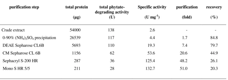
Purification of the phytase
A summary of the purification scheme is given in Table 1.
The phytase activity was eluted as a single sharp activity peak
from each ion-exchange column after application of the
gradient. A 51-fold purification of the enzyme was achieved
with a recovery of 20.3%. The enzyme exhibited an activity of
about 133 U mg-1.
Table 1. Purification scheme for the phytate-degrading enzyme from Aspergillus niger
purification step total protein (µg)
total phytate-degrading activity
(U)
Specific activity (U mg-1)
purification (fold)
recovery (%)
Crude extract
0-90% (NH4)2SO4 precipitation
DEAE Sepharose CL6B
CM Sepharose CL 6B
Sephacryl S-200 HR
Mono S HR 5/5
54000
26539
5693
1156
287
211
138
117
110
62
36
28
2.6
4.4
19.3
53.6
125.4
132.7
-
1.7
7.4
20.6
48.2
51.0
-
84.8
79.7
44.9
26.1
Molecular properties
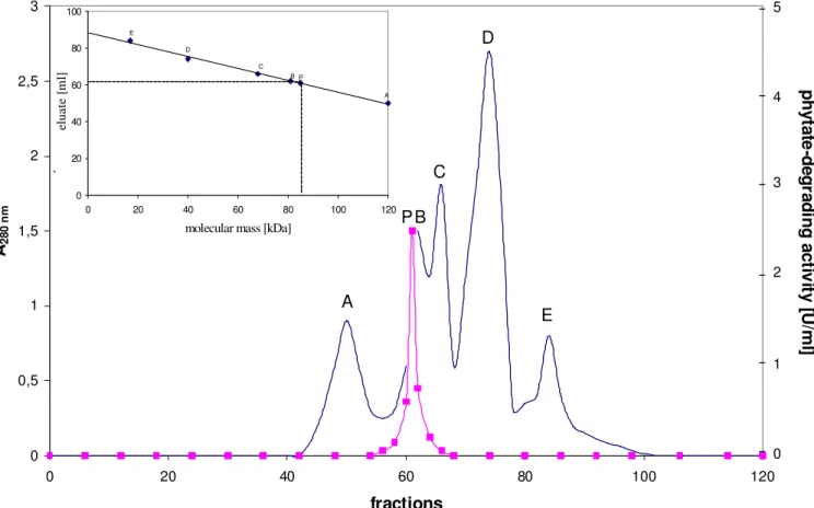
Gel filtration of the native enzyme on a calibrated
Sephacryl S-200 column gave a molecular mass of the phytase
of 85,000 ± 2,500 Da with elution position being measured by
determination of enzyme activity (figure 1). Lower molecular
mass species or higher molecular mass aggregates were not
observed. The phytase preparation gave a single broad protein
band upon SDS gel electrophoresis after Coomassie staining of
the gels (data not shown). This result indicates that the phytase
could be regarded as homogeneous. According to the estimated
molecular masses after SDS-PAGE, the protein band
corresponds to a molecular mass of 85,000 Da. Consequently,
this enzyme is a monomeric protein.
Figure 1. Estimation of the molecular mass of the phytate-degrading enzyme from Aspergillus niger by gel filtration.
The column was calibrated with A: glucose-6-phosphate dehydrogenase (Mr = 120,000), B: creatine kinase (Mr = 81,000), C:
bovine serum albumin (Mr = 68,000), D: ß-lactoglobulin (Mr = 40,000), and E: myoglobin (Mr = 17,000). P: phytate-degrading
enzyme from Aspergillus niger (Mr estimated to be approximately 85,000). Phytate-degrading activity ( ), optical density at 280
nm ( — )
Enzymatic properties pH optimum and pH stability
The standard phytase assay was performed using a variety
of buffers from pH 1.0 to 9.0. Two distinct pH optima were
identified; the highest activity was recorded at pH 5.0 and the
second activity peak occurred at pH 2.8. The activity at pH 5.0
was 1.5 fold higher than at pH 2.8. The enzyme was virtually
inactive above pH 7.0. The effect on enzyme stability was
studied in the pH range 1.0 to 9.0 at 4°C. Within 14 days the
phytase did not lose any activity in the pH range from 3.0 to
8.0. The purified enzyme is also stable at low pH values,
retaining full activity after incubation at pH 2 for 24 h at 4°C 0
0,5 1 1,5 2 2,5 3
0 20 40 60 80 100 120
fractions
A
28
0
n
m
5
4
3
2
1
0
p
h
y
ta
te
-d
e
g
ra
d
in
g
a
c
tiv
it
y
[U
/m
l]
A
B
C
D
E
P
0 20 40 60 80 100
0 20 40 60 80 100 120
molecular mass [kDa]
el
u
a
te
[
m
l]
A B
C D
E
A. niger phytase
and 95% after incubation at pH 2.0 at 37°C.
Temperature optimum and thermal stability
The temperature profile of the purified phytase was
determined from 10 to 80°C using the standard phytase assay.
The temperature optimum was found to be 55°C. The apparent
activation energy was estimated at pH 5.0 from the slope of log
vmax versus 1/T. The data showed excellent linearity from 15 to
50°C. The Arrhenius activation energy for the hydrolysis of
phytate was calculated to be 39.1 kJ mol-1. In order to check thermal stability, the purified enzyme was incubated at different
temperatures, cooled to 4°C and assayed using the standard
phytase assay. The enzyme lost no activity in 10 min at
temperatures up to 55°C. When exposed for 10 min at 70°C, it
retained 45% and at 80°C for 5 min 8% of the initial activity.
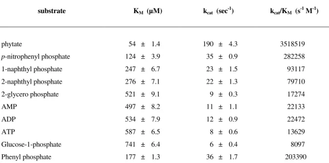
Substrate selectivity and kinetic parameters
In order to determine the substrate selectivity of the
purified phytase, several phosphorylated compounds (ATP,
ADP, AMP, pNPP, phenylphosphate, glucose-phosphate,
1-naphthyl phosphate, 2-naphthyl phosphate, 2-glycero
phosphate), in addition to phytate, were used for KM and vmax
estimation by detecting the release of the phosphate ion during
hydrolysis using formation of a soluble phospho-molybdate
complex in an acidic water-acetone mixture (table 2). Phytate
was identified as a good substrate. All other compounds tested
were only marginally hydrolysed by the purified enzyme. Of all
compounds tested, phytate gave the highest kcat/KM-value.
Therefore, under physiological conditions the likely substrate
for the Aspergillus niger 11T53A9 phytase is phytate. The kinetic parameters for the hydrolysis of phytate were
determined to be KM = 54 µmol l -1
and kcat = 190 sec -1
at pH 5.0
and 37°C. Like other fungal phytases, the purified enzyme
showed a substrate inhibition (15). The activity of the purified
enzyme was inhibited at substrate concentrations > 2.5 mM at
pH 5.0. The maximum amount of phosphate released from
phytate by the purified enzyme suggests myo-inositol monophosphate as the final product of enzymatic phytate
degradation.
Table 2. Kinetic constants for the hydrolysis of phosphorylated compounds by thephytate-degrading enzyme from Aspergillus niger at pH 5.0
substrate KM (µM) kcat (sec -1
) kcat/KM (s -1
M-1)
phytate
p-nitrophenyl phosphate 1-naphthyl phosphate
2-naphthyl phosphate
2-glycero phosphate
AMP
ADP
ATP
Glucose-1-phosphate
Phenyl phosphate
54 ± 1.4
124 ± 3.9
247 ± 6.7
276 ± 7.1
521 ± 9.1
497 ± 8.2
534 ± 7.9
587 ± 6.5
741 ± 6.4
177 ± 1.3
190 ± 4.3
35 ± 0.9
23 ± 1.5
22 ± 1.3
9 ± 0.3
11 ± 1.1
12 ± 0.9
8 ± 0.6
6 ± 0.4
36 ± 1.7
3518519
282258
93117
79710
17274
22133
22472
13629
8097
203390
Effector studies
None of the metal ions used in the study had an activating
effect when used at a concentration between 10-4 and 10-3 M. Mg2+, Ca2+, Mn2+, Co2+, Ag+, Hg2+, Cu2+ had little or no effect on enzyme activity, while Zn2+, Fe2+, and Fe3+ showed strong inhibitory effects. The reduced phytase activity in the presence
of Fe2+ and Fe3+ is attributed to a lower phytate concentration in the enzyme assay because of the appearance of a Fe-phytate
precipitate. When compounds which tend to chelate metal ions,
such as o-phenanthroline, EDTA, oxalate, citrate or tartrate, were tested for their effect on enzyme activity, it was noted that
none of them was inhibitory at a concentration from 10-4 to 10-3 M. Fluoride, a known inhibitor of different phytases from
different origin (15) and the hydrolysis product phosphate as
well as its structural analogous molybdate, wolframate and
vanadate were found to be strong inhibitors of the purified
enzyme. Fluoride inhibited the hydrolysis of phytate with a Ki
value of 158 µmol l-1.
Phytate dephosphorylation pathway
Intermediates of enzymatic myo-inositol hexakisphosphate dephosphorylation
Identification of the hydrolysis products of myo-inositol hexakisphosphate generated by the Aspergillus niger 11T53A9 phytase was performed by HPIC analysis (figure 2). The
chromatographic profile of the zero-time control indicated only
the myo-inositol hexakisphosphate peak. After 30 minutes of incubation, the quantity of myo-inositol hexakisphosphate had decreased and D/L-Ins(1,2,4,5,6)P5 appeared as the major
degradation product, accompanied by significant amounts of
D/L-Ins(1,2,5,6)P4 and low amounts of D/L-Ins(1,2,6)P3 /
Ins(1,2,3)P3. After 60 minutes of incubation, D/L-Ins(1,2,5,6)P4
was the major myo-inositol phosphate present in the incubation mixture. In addition, significant amounts of D/L-Ins(1,2,6)P3 /
Ins(1,2,3)P3 and low amounts of D/L-Ins(1,2)P2 / Ins(2,5)P2 /
D/L-Ins(4,5)P2 appeared. After 90 minutes of incubation, myo
-inositol hexakis- and myo-inositol pentakisphosphate were completely degraded to D/L-Ins(1,2,5,6)P4, D/L-Ins(1,2,6)P3 /
Ins(1,2,3)P3, and D/L-Ins(1,2)P2 / Ins(2,5)P2 / D/L-Ins(4,5)P2.
After 120 minutes of incubation only the myo-inositol tris- and bisphosphate peaks remained. Because all theoretically existing
myo-inositol pentakis- and tetrakisphosphate isomers are well resolved on the high performance ion chromatography system
used, identity of the myo-inositol pentakis- and tetrakisphosphate isomers formed by the Aspergillus niger 11T53A9 phytase as D/L-Ins(1,2,4,5,6)P5 and
D/L-Ins(1,2,5,6)P4 is well established. A clear identification of the
generated myo-inositol tris-, bis- and monophosphate isomers by high performance ion chromatography was not possible,
because not all theoretically existing isomers were available.
Identification of the absolute configuration of the predominantly generated myo-inositol pentakisphosphate isomer
To determine the absolute configuration of the myo-inositol pentakisphosphate isomer generated predominantly by the
phytase from Aspergillus niger 11T53A9, kinetic studies with the purified myo-inositol pentakisphosphate isomers generated either by the phytases from Pantoea agglomerans and the phytase from Aspergillus niger 11T53A9 were performed. The enzymes were added to sequentially diluted solutions of the
purified myo-inositol pentakisphosphate isomers and the kinetic parameters (KM, kcat) were calculated from the
Lineweaver-Burk plots of the data (table 3). KM and kcat for enzymatic
hydrolysis of the major myo-inositol pentakisphosphate isomer generated by the Aspergillus niger 11T53A9 and Pantoea agglomerans phytases were almost identical. Since it is known that the Pantoea agglomerans phytase generates the D-Ins(1,2,4,5,6)P5 isomer (10), D-Ins(1,2,4,5,6)P5 and
D-Ins(1,2,5,6)P4 were the first two predominant breakdown
products of myo-inositol hexakisphosphate dephosphorylation by the phytase from Aspergillus niger 11T53A9.
Identification of the myo-inositol monophosphates formed The end product of the enzymatic dephosphorylation of
A. niger phytase
mass spectrometry as Ins(2)P (data not shown).
Figure 2. High performance ion chromatography analysis of hydrolysis products of phytate by an apparently pure phytate-degrading enzyme from Aspergillus niger
Reference sample: The source of the reference myo-inositol phosphates is as indicated in Skoglund et al. (23); Peaks: (1) Ins(1,2,3,4,5,6)P6; (2) Ins(1,3,4,5,6)P5; (3) D/L-Ins(1,2,4,5,6)P5; (4) D/L-Ins(1,2,3,4,5)P5; (5) Ins(1,2,3,4,6)P5; (6)
D/L-Ins(1,4,5,6)P4; (7) Ins(2,4,5,6)P4; (8) D/L-Ins(1,2,5,6)P4; (9) D/L-Ins(1,3,4,5)P4; (10) D/L-Ins(1,2,4,5)P4; (11) Ins(1,3,4,6)P4; (12)
D/L-Ins(1,2,3,4)P4; (13) D/L-Ins(1,2,4,6)P4; (14) Ins(1,2,3,5)P4; (15) Ins(4,5,6)P3; (16) D/L-Ins(1,5,6)P3; (17) D/L-Ins(1,4,5)P3;
(18) D/L-Ins(1,2,6)P3, Ins(1,2,3)P3; (19) D/L-Ins(1,3,4)P3; (20) D/L-Ins(1,2,4)P3, (21) D/L-Ins(2,4)P2; (22) D/L-Ins(1,2)P2,
Ins(2,5)P2, D/L-Ins(4,5)P2; (23) D/L-Ins(1,4)P2, D/L-Ins(1,6)P2.
0 5 10 15 20 25 30 35
reference sample
0 min
30 min
60 min
90 min
120 min
13 6
8 12
15 16
17 18 19
21 22
23
9
2
5
7 13
14 10
11 20
Table 3. Kinetic constants for enzymatic myo-inositol pentakisphosphate dephosphorylation InsP5 generated by phytase kinetic constant Aspergillus niger
phytase
Pantoea agglomerans phytase
Aspergillus niger Pantoea agglomerans
KM [µ mol l-1]
kcat [s-1]
KM [µ mol l-1]
kcat [s -1
]
156 ± 12a 140 ± 12a 154 ± 5a 141 ± 11a
159 ± 12a 136 ± 7a 155 ± 10a 139 ± 9a Temperature: 37°C; buffer: 100 mM sodium acetate, pH 5.0; enzyme concentration: 25 mU ml-1. For calculation of k
cat the following molecular masses were used: Aspergillus niger, 85 kDa, Pantoea agglomerans, 42 kDa (9). The data are mean values of five independent experiments. ameans within the same line with the same superscripts are not significantly different (P < 0.05)
Elucidation of the major phytate degradation pathway In consideration of Ins(2)P as the final degradation product,
dephosphorylation of D-Ins(1,2,5,6)P4 may yield
D-Ins(1,2,5)P3, D-Ins(1,2,6)P3 and D-Ins(2,5,6)P3 as a myo
-inositol trisphosphate intermediate and D-Ins(1,2)P2,
D-Ins(2,5)P2 and D-Ins(2,6)P2 as a myo-inositol bisphosphate
isomer. According to High-Performance Ion Chromatography,
D-Ins(2,5,6)P3 and D-Ins(2,6)P2 have to be excluded as
intermediates, since these myo-inositol phosphates elute well resolved from the InsP3 and InsP2 peaks, respectively, generated
during myo-inositol hexakisphosphate dephosphorylation by the Aspergillus niger 11T53A9 phytase. It is not possible to discriminate between D-Ins(1,2)P2 and D-Ins(2,5)P2, since these
two isomers co-elute on the High-Performance Ion
chromatography system used. In addition, the discrimination
between D-Ins(1,2,5)P3 and D-Ins(1,2,6)P3 is impossible, since
pure D-Ins(1,2,5)P3 is not available. To determine the absolute
configuration of the myo-inositol trisphosphate isomer
generated predominantly by the phytase from Aspergillus niger 11T53A9, kinetic studies with the purified myo-inositol trisphosphate isomers generated either by the phytases from
Sacharomyces cerevisiae and the phytase from Aspergillus niger 11T53A9 were performed. The enzymes were added to sequentially diluted solutions of the purified myo-inositol pentakisphosphate isomers and the kinetic parameters (KM, kcat)
were calculated from the Lineweaver-Burk plots of the data
(table 4). KM and kcat for enzymatic hydrolysis of the major
myo-inositol pentakisphosphate isomer generated by the Aspergillus niger 11T53A9 and Saccharomyces cerevisiae phytases were almost identical. Since it is known that the
Saccharomyces cerevisiae phytase generates the D-Ins(1,2,6)P3
isomer (12), D-Ins(1,2,6)P3 and D-Ins(1,2)P2 were the most
likely myo-inositol tris- and bisphosphate isomers generated by the action of the phytase from Aspergillus niger 11T53A9 upon phytate.
Table 4. Kinetic constants for enzymatic myo-inositol trisphosphate dephosphorylation
InsP3 generated by phytase kinetic constant Aspergillus niger
phytase
S. cerevisiae phytase
Aspergillus niger S. cerevisae
KM [µ mol l-1]
kcat [s-1]
KM [µ mol l -1
] kcat [s-1]
215 ± 11a 101 ± 9a 284 ± 15a 96 ± 7a
223 ± 14a 106 ± 11a 293 ± 12a 99 ± 9a Temperature: 37°C; buffer: 100 mM sodium acetate, pH 5.0; enzyme concentration: 25 mU ml-1. For calculation of k
A. niger phytase
DISCUSSION
The enzymatic properties of the purified phytase suggest
that this enzyme is very similar to the Aspergillus niger phytases reported in the literature (3, 6, 18, 21, 24-27, 32, 34).
The subunit molecular mass of the purified phytase was
estimated to be 85 kDa by SDS PAGE. This molecular mass is
in close agreement with that of the purified phytase (phy A)
from A. niger var. ficuum NRRL 3135 (27) and A. niger ATCC 9142 (3). Determination of the molecular mass of the
biologically active enzyme was also carried out by gel filtration.
A native molecular mass of 85 kDa was reported (figure 1),
indicating that the catalytically active form of the enzyme is that
of a monomer. This is in accordance with all A. niger phytases reported so far. The enzyme was also found to be glycosylated
(data not shown). Glycosylation is a characteristic of many
fungal extracellular enzymes, including the phytases from A. niger (3, 6, 24, 27).
The pH versus activity profiles of the purified phytase
displayed substantial similarity in having two distinct pH
optima, an identifying characteristic of the phytaseA enzyme
from A. niger (3, 25, 27). As with the phytase from A. niger var. ficcum NRRL 3135, the highest activity was recorded at pH 5.0 and a second activity peak occurs below pH 3.0 (27).
Furthermore, the activity ratio of the two enzymes at pH 5.0 and
pH <3.0 were very similar. A further characteristic both
enzymes have in common is the relatively high stability under
acidic condition. Both enzymes retained about 95% of their
activities after incubation for 24 h at pH 2.0 and 37°C (11).
Measurements of phytase activity as a function of
temperature revealed only minor differences between the
purified phytase and the A. niger phytases reported so far (3, 6, 18, 21, 24-27, 32, 34). The phytases exhibited maximum
activity at about 50-55°C. As the phytase from A. niger var. ficuum NRRL 3135 the purified enzyme retained about 40-45% of its initial activity after incubation for 10 min at 70°C (29). It
is widely accepted that A. niger phytase is not a thermostable enzyme nor does it have the capacity to refold properly after
denaturation (33). This is also in agreement with the
observation of Gibson (7) who stated that the available
industrial phytases which originate from A. niger have a low intrinsic resistance to heat inactivation.
Substrate specificity studies (table 2) showed that the
purified enzyme accepts phytate as a good substrate. All other
compounds tested were only marginally hydrolysed by the
purified enzyme. These results are broadly similar to values
reported for phytases purified from other A. niger strains (3, 6, 21, 24). The kinetic constants were also in close agreement to
those reported for A. niger var. ficuum NRRL 3135 (25). As the phytase activity from A. niger var ficuum NRRL 3135, the activity of the purified enzyme was unaffected by calcium ions
but inhibited by iron (25). In addition fluoride was shown to be
a potent inhibitor of the A. niger phytases.
A detailed characterisation of the hydrolysis pathway of
myo-inositol hexakisphosphate by the phytase from Aspergillus niger 11T53A9 purified to apparent homogeneity revealed that this enzyme dephosphorylates myo-inositol hexakisphosphate via D-Ins(1,2,4,5,6)P5, D-Ins(1,2,5,6)P4, D-Ins(1,2,6)P3,
D-Ins(1,2)P2 to finally Ins(2)P. Therefore, this phytase has to be
considered a 3-phytase (EC. 3.1.3.26). This is in agreement
with the data reported by Ullah and Phillippy (28). In addition,
Chen and Li (4) identified D-Ins(1,2,4,5,6)P5, D-Ins(1,2,5,6)P4,
D-Ins(1,2,6)P3, D-Ins(1,2)P2 as phytate dephosphorylation
products generated by a commercially available phytase derived
from A. niger. In addition they found a further degradation pathway proceeding from D-Ins(1,2,5,6)P4 via D-Ins(1,5,6)P3 to
D-Ins(5,6)P2. The latter pathway could not be confirmed with
the apparently pure phytase preparation used in this study. The
following three reasons point to the correctness of the phytate
degradation pathway given here. Firstly, the phytase preparation
used by Chen and Li (4) was shown to be contaminated with
additional acid phosphatases. It can not be excluded that these
acid phosphatases act upon partially phosphorylated myo -inositol phosphates generated by the A. niger phytase, even if phytate itself is not a substrate for that enzyme. Secondly, the
formation of D-Ins(1,5,6)P3 upon action on phytate would
require hydrolysis of the phosphate residue at position C-2 of
resistant to dephosphorylation by phytases (15). Finally, the
phytate degradation pathway is consistent with the preference
of histidine acid phytases such as the enzyme from A. niger 11T53A9 to continue dephosphorylation after removal of the
first phosphate residue from phytate adjacent to a free
hydroxyl group. The only sequence of dephosphorylation
which would be in agreement with the results obtained by
high performance ion chromatography is 3, 4, 5 and
C-6.
ACKNOWLEDGEMENT
Analysis of the individual myo-inositol phosphate derivatives by N.-G. Carlsson, University of Technology
(Gothenburg, Sweden) is gratefully acknowledged.
REFERENCES
1. Bradford, M. (1976). A rapid and sensitive method for the quantitation of microgram quantities of protein utilizing the principle of protein-dye binding. Anal. Biochem. 72, 248-254.
2. Carrington, A.L.; Calcutt, N.A.; Ettlinger, C.B.; Gustafsson, T.; Tomlinson, D.R. (1993). Effects of treatment with myo-inositol or its 1,2,6-trisphosphate (PP56) on nerve conduction in streptozotocin-diabetes. Eur. J. Pharmacol. 237, 257-263.
3. Casey, A.; Walsh, G. (2003). Purification and characterization of extracellular phytase from Aspergillus niger ATCC 9142. Biores. Technol. 86, 183-188.
4. Chen, Q.-C.; Li, B.W. (2003). Separation of phytic acid and other related inositol phosphates by high-performance ion chromatography and its applications. J. Chromatogr. A 1018, 41–52.
5. Claxon, A.; Morris, C.; Blake, D.; Siren, M.; Halliwell, B.; Gustafsson, T.; Löfkvist, B.; Bergelin, I. (1990). The anti-inflammatory effects of D-myo-inositol-1.2,6-trisphosphate (PP56) on animal models of inflammation. Agents Actions 29, 68-70.
6. Dvoráková, J.; Volfová, O.; Kopecky, J. (1997). Characterisation of phytase produced by Aspergillus niger. Folia Microbiol. 42, 349–352. 7. Gibson, K. (1995). The pelleting stability of animal feed enzymes. In:
Second European Symposium on Feed Enzymes, Noordwijkerhout, The Netherlands, pp. 157-162.
8. Grases, F.; March, J.G.; Prieto, R.M.; Simonet, B.M.; Costa-Bauza, A.; García-Raja, A.; Conte, A. (2000). Urinary phytate in calcium oxalate stones formers and healthy people. Scand. J. Urol. Nephrol. 34, 162-164.
9. Greiner, R. (2004). Purification and Properties of a Phytate-degrading Enzyme from Pantoea agglomerans. The Prot. J. 23, 567-576.
10. Greiner, R. (2004). Degradation of myo-inositol hexakisphosphate by a phytate-degrading enzyme from Pantoea agglomerans. The Prot. J. 23, 577-585.
11. Greiner, R.; Farouk, A. (2007). Purification and characterization of a bacterial phytase whose properties make it exceptionally useful as a feed supplement. The Prot. J. 26, 467-474.
12. Greiner, R.; Larsson Alminger, M.; Carlsson, N.-G. (2001). Stereospecificity of myo-inositol hexakisphosphate dephosphorylation by a phytase of baker´s yeast. J. Agric. Food Chem. 49, 2228-2233.
13. Heinonen, J.K.; Lahti, R.J. (1981). A new and convenient colorimetric determination of inorganic orthophosphate and its application to the assay of inorganic pyrophosphatase. Anal. Biochem. 113, 313-317. 14. Jariwalla, R.J.; Sabin, R.; Lawson, S.; Herman, Z.S. (1990). Lowering
of serum cholesterol and triglycerides and modulation of divalent cations by dietary phytate. J. Appl. Nutr. 42, 18-28.
15. Konietzny, U.; Greiner, R. (2002). Molecular and catalytic properties of phytate-degrading enzymes (phytases). Int. J. Food Sci. Technol. 37, 791-812.
16. Laemmli, U.K. (1970). Cleavage of structural proteins during the assembly of the head of bacteriophage T4. Nature 227, 680-685.
17. Maffucci, T.; Piccolo, E.; Cumashi, A.; Iezzi, M.; Riley, A.M.; Saiardi, A.; Godage, H.Y.; Rossi, C.; Broggini, M.; Iacobelli, S.; Potter, B.V.L.; Innocenti, P.; Falasca, M. (2005). Inhibition of the phosphatidylinositol 3-Kinase/Akt pathway by inositol pentakisphosphate results in antiangiogenic and antitumor effects. Cancer Res.65, 8339-8349. 18. Martin, J.A.; Murphy, R.A.; Power, R.F.G. (2006). Purification and
physico-chemical characterisation of genetically modified phytases expressed in Aspergillus awamori. Biores. Technol. 93,: 1703-1708. 19. Phillippy, B.Q.; Bland, J.M. (1988). Gradient ion chromatography of
inositol phosphates. Anal. Biochem. 175, 162-166.
20. Sandberg, A.-S.; Ahderinne, R. (1986). HPLC method for determination of inositol tri-, tetra-, penta-, and hexaphosphate in food and intestinal contents. J. Food Sci. 51, 547-550.
21. Sariyska, M.V.; Gargova, S.A.; Koleva, L.A.; Angelov, A.I. (2005). Aspergillus niger phytase: Purification and characterization. Biotechnol. Biotechnol. Eq. 19, 98-105.
22. Shears, S.B. (1998). The versatility of inositol phosphates as cellular signals. Biochim. Biophys. Acta 1436, 49-67.
23. Skoglund, E.; Carlsson, N.-G.; Sandberg, A.-S. (1998). High-Performance Chromatographic separation of inositol phosphate isomers on strong anion exchange columns. J. Agric. Food Chem. 46, 1877-1882.
24. Skowro ski, T. (1978). Some properties of partially purified from Aspergillus niger. Acta Microbiol. Polonica 27, 41-48.
A. niger phytase
26. Ullah, A.H.J. (1988). Aspergillus ficuum phytase: Partial primary structure, substrate selectivity, and kinetic characterization. Prep. Biochem. 18, 459-471.
27. Ullah, A.H.J.; Gibson, D.M. (1987). Extracellular phytase (E.C. 3.1.3.8) from Aspergillus ficuum NRRL 3135: Purification and characterization. Prep. Biochem. 17, 63-91.
28. Ullah, A.H.J.; Phillippy, B.Q. (1988). Immobilization of Aspergillus ficuum phytase: Product characterization of the bioreactor. Prep. Biochem. 18, 483-489.
29. Ullah, A.H.J.; Sethumadhavan, K. (2003). PhyA gene product of Aspergillus ficuum and Peniophora lycii produces dissimilar phytases. Biochem. Biophys. Res. Commun. 303, 463-468.
30. Vohra, A.; Satyanarayana, T. (2003). Phytases: Microbial sources, production, purification, and potential biotechnological applications. Crit. Rev. Biotechnol. 23, 29-60.
31. Vucenik, I.; Shamsuddin, A.M. (2003). Cancer inhibition by inositol hexaphosphate (IP6) and inositol: From laboratory to clinic. J. Nutr. 133, 3778S-3784S.
32. Wyss, M.; Brugger, R.; Kronenberger, A.; Remy, R.; Fimbel, R.; Oesterhelt, G.; Lehmann, M.; van Loon, A.P.G.M. (1999). Biochemical characterization of fungal phytases (myo-inositol hexakisphosphate phosphohydrolase): Catalytic properties. Appl. Environm. Microbiol. 65, 367-373.
33. Wyss, M.; Pasamontes, L.; Remy, R.; Kohler, J.; Kusznir, E.; Gadient, M.; Muller, F.; van Loon, A.P.G.M. (1998). Comparison of the thermostability properties of three acid phosphatases from molds: A. fumigatus phytase, A. niger phytase and A. niger pH 2.5 acid phosphatase. Appl. Environ. Microbiol. 64, 4446–4451.