Isolation of a thermostable acid phytase from
Aspergillus niger
UFV-1
with strong proteolysis resistance
Paulo S. Monteiro
1,2, Valéria M. Guimarães
2, Ricardo R. de Melo
2,
Sebastião T. de Rezende
21
Instituto de Ciências Agrárias, Universidade Federal de Viçosa, Rio Paranaíba, MG, Brasil. 2
Departamento de Bioquímica e Biologia Molecular, Universidade Federal de Viçosa, Viçosa, MG, Brasil.
Submitted: August 17, 2012; Approved: June 06, 2014.
Abstract
AnAspergillus nigerUFV-1 phytase was characterized and made available for industrial application. The enzyme was purified via ultrafiltration followed by acid precipitation, ion exchange and gel fil-tration chromatography. This protein exhibited a molecular mass of 161 kDa in gel filfil-tration and 81 kDa in sodium dodecyl sulfate polyacrylamide gel electrophoresis (SDS-PAGE), indicating that it may be a dimer. It presented an optimum temperature of 60 °C and optimum pH of 2.0. TheKMfor so-dium phytate hydrolysis was 30.9 mM, while the kcatandkcat/KMwere 1.46 x10⁵s-1and 4.7 x 10⁶ s-1.M-1, respectively. The purified phytase exhibited broad specificity on a range of phosphorylated compounds, presenting activity on sodium phytate, p-NPP, 2- naphthylphosphate, 1- naphthyl-phosphate, ATP, phenyl-naphthyl-phosphate, glucose-6-naphthyl-phosphate, calcium phytate and other substrates. En-zymatic activity was slightly inhibited by Mg2+, Cd2+, K+and Ca2+, and it was drastically inhibited by F-. The enzyme displayed high thermostability, retaining more than 90% activity at 60 °C during 120 h and displayed a t1/2of 94.5 h and 6.2 h at 70 °C and 80 °C, respectively. The enzyme demon-strated strong resistance toward pepsin and trypsin, and it retained more than 90% residual activity for both enzymes after 1 h treatment. Additionally, the enzyme efficiently hydrolyzed phytate in live-stock feed, liberating 15.3mmol phosphate/mL after 2.5 h of treatment.
Key words:phosphatase, phytic acid, dephosphorylation.
Introduction
Phytic acid (myo-inositol hexakisphosphate) ac-counts for 60-90% of the total phosphorus content in plants, where it serves as a major storage form of phosphorus and divalent cations that can be hydrolyzed by several enzymes during seed germination (Vatset al., 2009). Phytic acid acts as an antinutritional factor in animals because of its ability to complex with ions such as calcium, magnesium, iron and zinc as well as proteins, thus decreasing its bioavailability (Bohnet al., 2008; Fugthong et al., 2010). Additionally, phytic phosphorus cannot be absorbed by monogastric ani-mals including humans because of phytate degrading en-zyme insufficiency or absence. Thus, unutilized phytate phosphorus from plant feed is excreted, which becomes an
environmental pollutant in areas of intensive livestock units (Yaoet al., 2011). One of the alternatives for solving this problem may be supplementation of animal feed with phytase (Maguireet al., 2005).
Phytases (myo-inositol hexakisphosphate phospho-hydrolases (EC 3.1.3.8, 3.1.3.26 and 3.1.3.72)) are phos-phatase enzymes that catalyze the hydrolysis of phosphate moieties from phytate, resulting in stepwise formation of myo-inositol pentakis-, tetrakis-, tris-, bis- and monophos-phate isomers as well as liberation of inorganic phosmonophos-phate and potentially chelated minerals (Bohnet al., 2008; Azeke et al., 2011). Therefore, the benefits of phytase are at least three-fold: increase bio-availability of phosphorus and oth-ers minerals in livestock feed, preserve non-renewable phosphorus sources by reducing the need for
supplement-DOI: http://dx.doi.org/10.1590/S1517-838220120037
Send correspondence to: S.T. Rezende. Department of Biochemistry and Molecular Biology, Universidade Federal de Viçosa, 36570-900 Viçosa, MG, Brazil. E-mail: [email protected].
ing diets with phosphorus and reduce environmental pollu-tion (Yaoet al., 2011).
This paper describes the purification and character-ization of a novel phytase produced from a Aspergillus nigerUFV-1 isolate and its application in a commercial livestock feed.
Materials and Methods
Materials
Substrates including sodium phytate, calcium phy-tate, p-nitrophenyl phosphate (pNPP), adenosine triphos-phate (ATP), adenosine diphostriphos-phate (ADP), adenosine monophosphate (AMP), glucose-6-phosphate, glucose-1-phosphate, 1-naphtyl glucose-1-phosphate, 2-naphtyl glucose-1-phosphate, phenyl phosphate and glycerol 2-phosphate as well as the chemical reagents Tween-20, calcium chloride, mono-potassium phosphate, sodium carbonate, sodium dodecyl sulfate (SDS), b-mercaptoethanol, dithiothreitol (DTT), urea, guanidine chloride, ethylenediaminetetraacetic acid (EDTA) and potato dextrose agar (PDA) were purchased from Sigma Chemical Co. (St. Louis, MO, USA). Peptone was obtained from Himedia Laboratories Co. (Mumbai, Maharashtra, India), and sodium acetate, sucrose, hydro-chloric acid and trichloroacetic acid was acquired from Vetec Fine Chemical (Duque de Caxias, RJ, Brazil). Gly-cine, 2-Morpholinoethanesulfonic acid (MES) and tris(hy-droxymethyl)aminomethane (TRIS) were purchased from Merck (Darmstadt, Germany). Standard molecular weight markers were obtained from Fermentas (USA), and protein standards, trypsin and pepsin were also acquired from Sigma. Potato and commercial pig feed was obtained from the local market. All other reagents used in this study were of analytical grade.
Strain isolation, culture conditions and enzyme production
Fungus was isolated from sugarcane bagasse samples that had been collected at a sugar-alcohol plant located at Urucânia, Minas Gerais, Brazil. Subsequently, the isolates were identified by morphology and physiology (Klich, 2002; Samsonet al., 2010) asAspergillus niger. The identi-fication was performed at the Phytopathology Department of the Federal University of Viçosa. The fungus was main-tained in the culture collection of the Applied Enzymology Laboratory, Federal University of Viçosa (UFV), Brazil. In this study, it is designated asAspergillus nigerUFV-1.
Induction of phytase was achieved in liquid medium according to Gunashree and Venkateswaran (2008) with some modifications. Its content per liter potato broth was: sucrose 10 g, peptone 5 g, Tween-20 2.5 mL and CaCl2 0.10 g. Potato broth was obtained by boiling 200 g fresh po-tato in 1 L distilled water. The final medium pH was ad-justed to 5.5 by addition of a 0.1 M HCl solution. Aliquots of 50 mL medium were dispensed in 250 mL Erlenmeyer
flasks and autoclaved at 121 °C for 20 min. Seven day-old spores grown on solid potato dextrose agar media were har-vested by washing the plate surface with a distillated water solution containing 0.2% (v/v) Tween-20. The medium was inoculated with a spore suspension (106spores mL-1). The flasks were incubated for 8 days at 28 °C on an orbital shaker at 180 RPM. The culture broth was then harvested and submitted to initial filtration through a nylon filter, and the filtrate was centrifuged (10,000 xg, 10 min at 4 °C). The supernatant was stored at 4 °C until purification.
Phytase assay
Phytase activity was determined by measuring the in-organic phosphate released from sodium phytate. For anal-ysis, 600mL 1.5 mM sodium phytate in 100 mM sodium acetate buffer, pH 2.5 and 150mL enzymatic extract were used. The reaction was conducted in a water bath at 50 °C, and it was stopped after 30 min by adding 250mL 10% (v/v) trichloroacetic acid solution. This was followed by the ad-dition of 1000mL colorimetric reagent to the tubes (Har-land and Har(Har-land, 1980). Absorbance was measured at 700 nm, and the measured values were correlated with a standard curve that had been constructed using monopo-tassium phosphate. All of the enzyme activity analyses were performed in triplicate; one unit of phytase activity was defined as the amount of enzyme that released 1mmol
phosphate per minute under the assay conditions.
Enzyme purification
Bradford method using bovine serum albumin as the stan-dard at 0-20mg/mL.
Electrophoretic analysis and molecular mass determination
One dimensional SDS gel electrophoresis was per-formed using a 12.5% (w/v) acrylamide gel in a vertical electrophoresis system, and the proteins bands were visual-ized by silver staining.
For zymogram analysis, one dimensional SDS gel electrophoresis was conducted using a 12.5% (w/v) acryl-amide gel, where SDS andb-mercaptoethanol were omit-ted from the gel and buffer, respectively, and the sample was not heated prior to analysis. After electrophoresis, the gel was equilibrated with 0.2 M sodium acetate buffer, pH 2.5 for 30 min and then incubated in the same buffer con-taining 0.04% (w/v) pNPP at 50 °C for 30 min. After incu-bation, the gel was rinsed with distilled water, and then a 0.5 M Na2CO3solution was added for visualization of yel-low zones, indicating substrate hydrolysis.
Native enzyme molecular mass was determined by gel filtration chromatography using a Sephacryl S-200 (Amersham Biosciences) column (100 x 2.5 cm) and 25 mM sodium acetate buffer, pH 5.0, containing 0.5% (w/v) sucrose as a running buffer. Void volume was deter-mined using blue dextran (2 mg/mL), and the protein stan-dards utilized were albumin (Bovine serum, MW 66,000), alcohol dehydrogenase (yeast, MW 150,000), b-amylase (Sweet potato, MW 200,000), carbonic anhydrase (bovine erythrocytes, MW 29,000) and cytochrome C (Horse heart, MW 12,400).
Effect of temperature and pH on enzyme activity
The temperature profile of the purified enzyme was determined within the range of 20 to 80 °C using the stan-dard phytase assay. The pH profile was determined by mea-suring phytase activity using the following buffers: 0.2 M glycine-HCl (pH 1.0-3.5), 0.2 M sodium acetate (pH 4.0-5.5), 0.2 M MES (pH 6.0-6.5) and 0.2 M Tris-HCl (pH 7.0-8.0).
Enzyme thermal stability and proteolysis resistance
Purified phytase was incubated at 60, 70 and 80 °C for periods of up to 144, 120 and 12 hours, respectively, cooled to 4 °C and assayed in accordance with the phytase assay above.
Phytase resistance to pepsin and trypsin was assessed in accordance with Zhanget al.(2010) with modifications. The purified enzyme (0.2mg/mL) was incubated in 0.1 M glycine-HCl buffer (pH 2.5) containing pepsin (0.2mg/mL, pepsin/enzyme ratio of 1:1 (v/v)) or 0.1 M Tris-HCl buffer (pH 8.0) containing trypsin (0.2mg/mL, trypsin/enzyme ra-tio of 1:1 (v/v)) for 10, 20, 40 and 60 min at 37 °C. As a con-trol, the purified enzyme was incubated at the same
conditions as above but without pepsin or trypsin addition. Residual phytase activity was assessed in accordance with the phytase assay above. The positive control of pepsin and trypsin was performed with azocasein as the substrate, in accordance with Morgaviet al.(2001), with modifications. For analysis, 50 mL 1% azocasein solution and 50 mL 0.2mg/mL pepsin in 100 mM glycine-HCl buffer, pH 2.5 or 0.2mg/mL trypsin in 100 mM Tris-HCl buffer, pH 8.0 were used. The reaction was performed at 37 °C and was stopped after 60 min by adding 100mL 10% (v/v) trichloroacetic acid solution, and the mixture was centrifuged at 12,000 xg for 10 min. An aliquot of the supernatant was mixed with an equal volume of 500 mM NaOH solution, and the concen-tration of digested azocasein was determined spectrophoto-metrically at 440 nm.
Effect of metal ions and others reagents on phytase activity from Aspergillus niger UFV-1
The effects of Mg2+, Cd2+, Mn2+, Cu2+, Zn2+, Ca2+, Hg2+, Na+and F-ions at 5, 10 and 20 mM, EDTA,
b
-mer-captoethanol and DTT at 1 and 5 mM, SDS at 0.1 and 0.5% (v/v) and chaotropic agents (urea and guanidine chloride) at 0.5, 1.0 and 2.0 M on enzyme activity were investigated by incorporating these compounds into the reaction mixtures. Residual phytase activity was then obtained in accordance with the phytase assay above.
Determination of substrate specificity and kinetic parameters
Purified phytase substrate specificity was evaluated by replacing phytic acid in the reaction mixture with other phosphate compounds. The substrates used were sodium phytate, calcium phytate, pNPP, ATP, ADP, AMP, glu-cose-6-phosphate, glucose-1-phosphate, 1-naphtyl phos-phate, 2-naphtyl phosphos-phate, phenyl phosphate and glycerol 2-phosphate at final concentrations of 1.2 mM. Phytase ac-tivity was obtained in accordance with the phytase assay above, except for pNPP, in which the reaction mixture was composed of 400mL 3.0 mM pNPP in 0.2 M glycine-HCl (pH 2.5), 400mL distilled water and 150mL purified phy-tase, which was incubated at 50 °C for 15 min. The reaction was stopped by the addition of 1 mL 0.5 mM Na2CO3, and the p-nitrophenyl (pNP) concentration was measured at 410 nm.
Kinetic properties were investigated using pNPP as a substrate at 0-95 mM. The kinetic enzyme parameters were investigated using a Lineweaver-Burk plot.
Evaluation of phytate hydrolysis in livestock feed
vitamin premix. Autoclaved (121 ºC/20 min) feed samples (1 g) were suspended in 5 mL 0.2 M glycine-HCl buffer (pH 2.5) in a 50 mL conical flask and supplemented with crude phytase (4 U/mL). An autoclaved feed suspension without phytase was used as a control. The flasks were then incubated at 50 °C and 200 rpm in a rotary shaker. Samples were withdrawn at intervals up to 8 h (test and control flasks) and immediately cooled to 4 °C and centrifuged (10000 xg, 10 min, at 4 °C), and the supernatant was used for quantification of colorimetrically released phosphate at 700 nm in accordance with the phytase assay above.
Results and Discussion
Enzyme purification
Enzyme purification was achieved by a combination of ultrafiltration followed by acid precipitation and ion ex-change and gel filtration chromatography. After the ultra-filtration and precipitation steps, the fractions adsorbed on DEAE-Sepharose demonstrated phytase activity (Figu-re 1A). Subsequently, only 1 peak with phytase activity was obtained when this fraction was submitted to gel filtration chromatography (Figure 1B). A purification factor of 64.34 was recorded with 11% yield (Table 1).
Purification to homogeneity was confirmed by SDS PAGE analysis, and a single band was observed on the non-denaturing electrophoresis gels. Identification of this band as phytase on the non-denaturing gels was confirmed by zymogram analysis, where the band stained for protein and enzymatic activity displayed identical retention factor (Rf) values (Figure 2). Previous studies on fungal phytase purifications present similar purification factors and yields. TheA. niger11T53A9 phytase was purified 51 times with a 20.3% yield (Greiner et al., 2009), and Thermomyces lanuginosusTL-7 phytase purification demonstrated a pu-rification factor of 40.75-fold and 3.44% yield (Gulatiet al., 2007).
Molecular mass
Molecular mass was determined by gel filtration chromatography using a standard curve (Figure 3) and SDS-PAGE. The native phytase molecular mass was deter-mined to be 161 kDa by gel filtration, while SDS-PAGE analysis demonstrated a single band of 81 kDa (Figure 2A). A single band of phytase activity was observed on a
non-denaturating gel (Figure 2B), which suggests that the en-zyme could be a dimer, and the molecular mass is in the same range as that observed in previous studies on fungal phytase. The molecular mass of phytase fromA. ficuumas determined by gel filtration analysis was 130 kDa, while SDS-PAGE resulted in approximately 68 kDa (Ullah and Cummins, 1987). In another study, the molecular mass of phytase fromA. nigervan Teighem was demonstrated to be a pentamer of 353 kDa with 66 kDa monomers (Vats and Banerjee, 2005). The molecular mass ofA. niger NCIM 563 phytases (I, II, III and IV) as determined by SDS-PAGE and gel filtration were 66, 264; 150, 148; 87, 85 and Figure 1- Elution profiles of phytase fromAspergillus nigerUFV-1. (A) Ion exchange chromatography of fractions adsorbed on DEAE-Sepharose. The column was initially eluted with sodium acetate buffer (50 mM, pH 5.0) containing 0.5% (w/v) sucrose and subsequently with a linear gradi-ent of 0-0.5 M NaCl in the same buffer. The flow rate was 1.0 mL/min. (B) Gel filtration of fractions displayed phytase activity from DEAE-Sepha-rose on a Sephacryl S-300 HR column (2.6 x 60 cm), which was eluted with sodium acetate buffer (25 mM, pH 5,0) containing 0.5% (w/v) su-crose. The flow rate was 0.8 mL/min. Protein (-◆-), NaCl gradient (-o-),
Phytase activity (-x-).
Table 1- Summary of the steps for phytase purification fromAspergillus nigerUFV-1.
Purification step Total protein (mg) Total activity (U) Specific activity (U/mg) Purification (fold) Yield (%)
Crude extract 47.70 138.6 2.9 1 100
Ultra-filtration 41.30 129.6 3.14 1.08 93.5
Acid treatment 15.36 103.7 6.75 2.33 74.8
DEAE-Sepharose CL-6B 0.988 24.50 24.80 8.55 17.7
120, 120 kDa, respectively, indicating that phytase I con-sists of four subunits and others are monomers (Soniet al., 2010; Bhavsaret al., 2013). Bhavsaret al.(2011) identified a phytase fromA. nigerNCIM 563 at 87 kDa. Spieret al. (2011) studied a 108 kDa phytase fromA. nigerFS3, while anotherA. niger11T53A9 phytase was identified at 85 kDa (Greineret al., 2009).
Effects of temperature and pH on enzyme activity
Purified phytase fromA. nigerUFV-1 demonstrated optimum activity at 60 °C (Figure 4 A), similar to other fun-gal phytases (Fugthonget al., 2010; Bhavsaret al., 2013), and its optimum pH was 2.0 with a decrease in enzyme ac-tivity as the pH approached neutral (Figure 4 B).
Figure 2- SDS-PAGE (A) and zymogram (B) analysis ofAspergillus nigerUFV-1 phytase. Lane M: molecular mass marker; Lane 1: Purified phytase; Lane 2: Non-denaturing gel electrophoresis; Lane 3: Phytase ac-tivity on a non-denaturing gel.
Figure 3- Native molecular mass determination of purified phytase by Sephacryl S-200 gel filtration chromatography.
Enzyme thermal stability and proteolysis resistance
Purified phytase ofA. nigerUFV-1 retained more than 90% activity at 60 °C during 120 h. At 70 °C, approx-imately 80% activity was retained after 72 h with a t1/2of 94.5 h. The enzyme maintained more than 70% activity at 80 °C after 4 h with t1/2of 6.2 h (Figure 5). Thermostability and resistance to proteolysis in the animal digestive tracts are important and useful criteria for industrial phytase ap-plication (Yaoet al., 2011). The purified phytase fromA. ficuum NTG-23 demonstrated complete activity loss when maintained at 80 °C for 10 min (Zhanget al., 2010), while the purified phytase fromA. nigerFS3 maintained
25.1% residual activity after 15 min at 80 °C (Spieret al., 2011).
Purified phytase fromA. nigerUFV-1 demonstrated high stability in the presence of proteolytic enzymes at a 1:1 ratio (pepsin or trypsin/phytase, w/w), while retaining more than 90% residual activity for both proteolytic enzymes (Figure 6). The positive control of pepsin and trypsin dem-onstrated that the enzymes exhibited proteolytic activity under the experimental conditions (data not shown). The recombinant phytase from Eupenicillium parvum BCC 17694 was expressed inPichia pastorisand presented low trypsin resistance at a protease/phytase ratio of 1:500 or
Figure 5- Thermal stability of the purifiedAspergillus nigerUFV-1 phytase at 60 °C (●), 70 °C (O) and 80 °C (▼). The activity of an unheated phytase sample was defined as 100%. Phytase activity was expressed as the mean±SD (n = 3).
Figure 6- Proteolysis resistance of the purifiedAspergillus nigerUFV-1 phytase at 37 °C, phytase control, pH 2.5 (●); phytase + pepsin, pH 2.5 (O);
higher (Fugthonget al., 2010). The A. fumigatus WY-2 phytase was also evaluated for proteolysis resistance, and it was very sensitive to trypsin at a protease/phytase ratio of 1:200, thus maintaining approximately 30% of its initial ac-tivity after 2 h of treatment (Wanget al., 2007).
Effect of metal ions and other reagents on phytase activity fromAspergillus nigerUFV-1
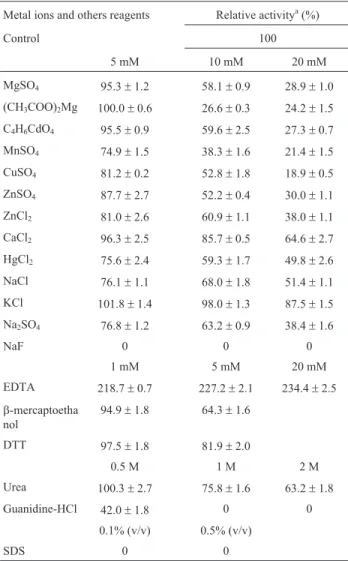
The effects of metal ions and other reagents are shown in Table 2. At 5 mM, Mg2+, Cd2+ and Ca2+ ions slightly inhibited phytase activity, while Mn2+, Cu2+, Zn2+, Hg2+and Na+had a moderate inhibitory effect. However, the F- ion highly inhibited enzyme activity. Fluoride is a strong competitive inhibitor of several acid bacterial, fun-gal and plant phytases (Konietzny and Greiner, 2002). Other than the F-ion, the other ions evaluated inhibited
en-zyme activity at 10 and 20 mM. Metal ions modulate phytase activity, while it is suggested that the inhibitory ef-fect of various metal ions results from the formation of metal ion-phytate complexes that display low solubility. Moreover, most phytases that have been characterized to date are greatly inhibited by Cu2+and Zn2+(Konietzny and Greiner, 2002). The Sporotrichum thermophile phytase demonstrates 30.0, 12.4, 16.6, 58.2 and 10.5% inhibition when evaluated in the presence of 5 mM Ca2+, Mn2+, Cu2+, Zn2+and Na+ions, respectively (Singh and Satyanarayana, 2009). In the presence of 1 mM Zn2+, Mg2+, Ca2+, Mn2+, K+ and Na+ions,Cladosporiumsp. FP-1 phytase was inhibited by 46.0, 45.0, 44.0, 54.0, 39.0 and 34.0%, respectively (Quanet al., 2004). The phytase fromA. nigerNCIM 563 was moderately stimulated in the presence of 5 mM Ca2+, Fe2+,Fe3+, Ba2+and Pb2+and was inhibited in the presence of 1 mM Hg2+, Ni2+, Zn2+, Cu2+, Ag2+, Fe2+, Fe3+, Pb2+and Ca2+(Bhavsaret al., 2011; Bhavsaret al., 2013).
The purified phytase ofA. nigerUFV-1 was moder-ately inhibited by 5 mM of the reducing agents
b-mercaptoethanol (35.7% decrease) and DTT (18.1% de-crease), which is different from other phytases that were not inhibited by these agents (Quan et al., 2004; Singh and Satyanarayana, 2009). The phytases (I and II) fromA. niger NCIM 563 were not significantly affected by DTT and
b-mercaptoethanol at 1 mM and 0.1% concentrations, re-spectively (Soniet al., 2010). However, the phytase fromE. parvum(BCC17694) demonstrated a similar inhibitory ef-fect when evaluated in the presence ofb-mercaptoethanol and DTT at 5 mM (Fugthonget al., 2010). This outcome suggested that the sulfhydryl group may be involved in the catalytic activity of the enzyme. Most phytases have a num-ber of cysteine residues, which may be implicated in disul-fide bonds, as described in A. ficuum (Kostrewa et al., 1997) andEscherichia coli(Limet al., 2000). The chelat-ing agent EDTA did not inhibit enzyme activity, suggestchelat-ing that this enzyme does not require metal ions for its activity. EDTA enhanced phytase activity at 1-20 mM by approxi-mately 130%. This behavior is similar to that of most phytases except the alkaline phytases ofBacillusspp., for example, which are calcium dependent (Ohet al., 2004). The enzyme activity of A. ficuum NTG-23 phytase was stimulated by EDTA concentrations of 1.25-10 mM, in-creasing enzyme activity by approximately 10% (Zhanget al., 2010). A similar result was demonstrated for the phy-tase fromA. nigervan Teighem; enzymatic activity was en-hanced by approximately 50% with 0.1-2.0 mM EDTA (Vats and Banerjee, 2005). However, this result differs from other studies where the presence of EDTA at 1-5 mM inhibited phytase enzyme activity (Quanet al., 2004; Gula-tiet al., 2007; Fugthonget al., 2010). Chaotropic agents such as urea and guanidinium chloride inhibited phytase activity, particularly guanidinium chloride at 1-2 M, which severely inhibited phytase activity. It is suggested that non-covalent forces such as H-bonds and van der Waals in-Table 2- Effect of metal ions and other reagents on phytase activity from
Aspergillus nigerUFV-1
Metal ions and others reagents Relative activitya(%)
Control 100
5 mM 10 mM 20 mM
MgSO4 95.3±1.2 58.1±0.9 28.9±1.0 (CH3COO)2Mg 100.0±0.6 26.6±0.3 24.2±1.5 C4H6CdO4 95.5±0.9 59.6±2.5 27.3±0.7 MnSO4 74.9±1.5 38.3±1.6 21.4±1.5 CuSO4 81.2±0.2 52.8±1.8 18.9±0.5 ZnSO4 87.7±2.7 52.2±0.4 30.0±1.1 ZnCl2 81.0±2.6 60.9±1.1 38.0±1.1 CaCl2 96.3±2.5 85.7±0.5 64.6±2.7 HgCl2 75.6±2.4 59.3±1.7 49.8±2.6 NaCl 76.1±1.1 68.0±1.8 51.4±1.1 KCl 101.8±1.4 98.0±1.3 87.5±1.5 Na2SO4 76.8±1.2 63.2±0.9 38.4±1.6
NaF 0 0 0
1 mM 5 mM 20 mM
EDTA 218.7±0.7 227.2±2.1 234.4±2.5
b-mercaptoetha nol
94.9±1.8 64.3±1.6
DTT 97.5±1.8 81.9±2.0
0.5 M 1 M 2 M
Urea 100.3±2.7 75.8±1.6 63.2±1.8
Guanidine-HCl 42.0±1.8 0 0
0.1% (v/v) 0.5% (v/v)
SDS 0 0
*Metal ions and other reagent concentrations were calculated as 5 mM, 10 mM and 20 mM of Mg2+, Cd2+(C4H6CdO4= cadmium acetate), Mn2+, Cu2+, Zn2+, Ca2+, Hg2+, Na+, K+and F-; 1 mM, 5 mM and 20 mM EDTA,
b-mercaptoethanol and DTT; 0.5 M, 1 M and 2 M Urea, Guanidine-HCl and SDS.aPhytase activity was expressed as the mean
teractions play a role in maintaining the active conforma-tion of the enzyme (Vats and Banerjee, 2005; Gulatiet al., 2007). The anionic detergent SDS, even at low concentra-tions, fully inhibited enzymatic activity. The phytase from A. nigervan Teighem was also inhibited by SDS as a con-centration of 0.1% (v/v), resulting in a 92% loss in enzy-matic activity (Vats and Banerjee, 2005). Anionic detergents can bind to proteins and induce structural chan-ges that inhibit enzymatic activity (Singh and Satyanaraya-na, 2009).
Substrate specificity and kinetic parameters
The purified phytase exhibited broad substrate speci-ficity on a range of phosphorylated compounds (Table 3), presenting activity on pNPP, 2-naphthyl phosphate,
1-naphthyl phosphate, and ATP of more than 3-fold greater enzymatic activity than sodium phytate. In vitro experi-ments with livestock feed suggest that phytate degrading enzymes with broad substrate specificity are better suited for animal nutrition purposes than phytate-degrading en-zymes with narrow substrate specificity (Wysset al., 1999; Escobin-Mopera et al., 2012). Fungal phytases demon-strated different substrate specificity. The phytase of A. ficuum NTG-23 displayed a broad substrate specificity, with enzyme activity on ATP, fructose-6-phosphate, and pNPP more than 2-fold higher than sodium phytate (Zhang et al., 2010). A study of the catalytic properties of fungal phytase demonstrated thatA. nigeracid phosphatase andA. fumigatusphytase displayed rather broad substrate speci-ficity, while phytases from A. niger,A. terreus CBS, A. terreus9A1 andE. coliwere more specific to phytic acid (Wyss et al., 1999). The phytase from Rhizopus oligosporushad a rather broad substrate specificity but was more specific to p-NPP than sodium phytate (Azekeet al., 2011). Similar results were shown for theR. oligosporus ATCC 22959 phytase, which displayed rather broad sub-strate specificity; however, it was more specific to phytate than other phosphate compounds (Casey and Walsh, 2004).
Upon incubation of the purified phytase with various pNPP concentrations (up to 95.0 mM), its kinetic properties were determined from a Lineweaver-Burk plot. TheKMand Vmax values were 30.9 mM and 7.48 mmol/min, respec-tively. The estimatedkcatof the enzyme was 1.46 x 10⁵s-1, and the catalytic efficient (kcat/KM) was 4.7 x 10⁶s-1.M-1. Generally, enzymatic hydrolysis presents classical Michae-lis-Menten kinetics, whereas relatively lowKMvalues have been reported for phytate-degrading enzymes fromA. niger (10-40 mM), A. terreus (11-23 mM), A. fumigatus (< 10mM),Schwanniomyces castelli(38mM),Klebsiella aerogenes (62 mM), cattail pollen (17 mM), maize root
Figure 7-In vitrophosphate liberation from commercial livestock feed at 50 °C, enzymatic hydrolysis (●) and control (O). Phytase activity was ex-pressed as the mean±SD (n = 3).
Table 3- Substrate specificity of the purified phytase fromAspergillus nigerUFV-1.
Substrate Relative activitya(%)
Sodium phytate 100.0±1.3
p-NPP 384.1±2.3
2-naphthyl phosphate 377.2±3.4 1-naphthyl phosphate 359.1±3.3
ATP 343.6±3.6
Phenyl phosphate 272.8±2.5
Glucose 6-phosphate 189.2±2.4
ADP 101.7±1.7
Calcium phytate 97.0±1.2
Glycerol 2-phosphate 67.3±0.3
Glucose 1-phosphate 58.2±2.8
AMP 11.0±0.3
aPhytase activity was expressed as the mean
(24-43mM), tomato root (38mM), oat (30mM), wheat bran (PHY1: 48mM, PHY2: 77mM), barley (P1: 72mM), soy-bean (48-61mM), and lupine (L11: 80mM) (Konietzny and Greiner, 2002; Bohnet al., 2008).
Evaluation of phytate hydrolysis in livestock feed
Crude phytase extract fromA. nigerUFV-1 was able to effectively hydrolyze phytate and other phosphate com-pounds in the animal feed tested. After 2.5 h, 15.3mmol phosphate/mL had already been liberated, and this rate was maintained up to 8 h (Figure 7).
Because of the amount of phosphorus released in a short time, this result is very significant. Dephosphoryla-tion of phytic acid in livestock feed using phytase fromA. nigervan Teighem at 8.4 U/mL demonstrated a maximum liberation of 0.048mmol Pi/mL at 55 °C after 48 h treatment (Vatset al., 2009).
Conclusions
Because of the excellent features displayed, this phy-tase appears to be a promising enzyme for use in animal feed. Its high thermostability suggests that the enzyme is suitable for industrial use because high temperatures are usually encountered in industrial animal food processing. Its high ability to hydrolyze phytate in acidic conditions as well as its high resistance to proteolytic enzymes also sug-gest that it may effectively release phytic phosphorus in the animal digestive tract.
Acknowledgments
This study was supported by grants from the Fundação de Amparo à Pesquisa do Estado de Minas Gerais -FAPEMIG and Conselho Nacional de Desenvolvimento Científico e Tecnológico - CNPq, Brazil.
References
Azeke MA, Greiner R, Jany K-D (2011) Purification and charac-terization of two intracellular phytases from the tempeh fun-gusRhizopus oligosporus. J Food Biochem 35:213-227. Bhavsar K, Buddhiwant P, Soni SKet al. (2013) Phytase
iso-zymes fromAspergillus nigerNCIM 563 under solid state fermentation: Biochemical characterization and their corre-lation with submerged phytases. Process Biochem 48:1618-1625.
Bhavsar K, Kumar VR, Khire JM (2011) High level phytase pro-duction byAspergillus nigerNCIM 563 in solid state cul-ture: response surface optimization, up-scaling, and its par-tial characterization. J Ind Microbiol Biotechnol 38:1407-1417.
Bohn L, Meyer A, Rasmussen S (2008) Phytate: impact on envi-ronment and human nutrition. A challenge for molecular breeding. J Zhejiang Univ Sci B 9:165-191.
Casey A, Walsh G (2004) Identification and characterization of a phytase of potential commercial interest. J Biotechnol 110:313-322.
Escobin-Mopera L, Ohtani M, Sekiguchi Set al.(2012) Purifica-tion and characterizaPurifica-tion of phytase from Klebsiella pneumoniae9-3B. J Biosci Bioeng 113:562-567.
Fugthong A, Boonyapakron K, Sornlek Wet al.(2010) Biochemi-cal characterization and in vitro digestibility assay of
Eupenicillium parvum (BCC17694) phytase expressed in
Pichia pastoris. Protein Expr Purif 70:60-67.
Greiner R, Silva LG, Couri S (2009) Purification and character-ization of an extracellular phytase fromAspergillus niger
11T53A9. Braz J Microbiol 40:795-807.
Gulati HK, Chadha BS, Saini HS (2007) Production, purification and characterization of thermostable phytase from thermo-philic fungus Thermomyces lanuginosus TL-7. Acta Mi-crobiol Immunol Hung 54:121-138.
Gunashree B, Venkateswaran G (2008) Effect of different cultural conditions for phytase production byAspergillus nigerCFR 335 in submerged and solid-state fermentations. J Ind Microbiol Biotechnol 35:1587-1596.
Harland BF, Harland J (1980) Fermentative reduction of phytate in rye, white, and whole wheat breads. Cereal Chem 57:226-229.
Klich MA (2002) Identification of commonAspergillusspecies. Centraalbureau voor Schimmelcultures, Utrecht, The Neth-erlands.
Konietzny U, Greiner R (2002) Molecular and catalytic properties of phytate-degrading enzymes (phytases). Int J Food Sci Technol 37:791-812.
Kostrewa D, Gruninger-Leitch F, D’Arcy Aet al.(1997) Crystal structure of phytase fromAspergillus ficuumat 2.5 Ä resolu-tion. Nat Struct Mol Biol 4:185-190.
Lim D, Golovan S, Fosberg CWet al.(2000) Crystal structures of
Escherichia coliphytase and its complex with phytate. Nat Struct Mol Biol 7:108-113.
Maguire RO, Dou Z, Sims JTet al.(2005) Dietary strategies for reduced phosphorus excretion and improved water quality. J Environ Qual 34:2093-2103.
Morgavi DP, Beauchemin KA, Nsereko VLet al.(2001) Resis-tance of feed enzymes to proteolytic inactivation by rumen microorganisms and gastrointestinal proteases. J Anim Sci 79:1621-1630.
Oh B, Choi W, Park Set al.(2004) Biochemical properties and structure specificities of alkaline and histidine acid phytases. Appl Microbiol Biotechnol 63:362-372.
Quan C, Tian W, Fan Set al.(2004) Purification and properties of a low-molecular-weight phytase from Cladosporium sp. FP-1. J Biosci Bioen 97:260-266.
Samson RA, Houbraken J, Thrane Uet al.(2010) Food and indoor fungi. Centraalbureau voor Schimmelcultures, Utrecht, The Netherlands.
Singh B, Satyanarayana T (2009) Characterization of a HAP-phytase from a thermophilic mould Sporotrichum thermophile. Bioresour Technol 100:2046-2051.
Soni SK, Magdum A, Khire JM (2010) Purification and character-ization of two distinct acidic phytases with broad pH stabil-ity from Aspergillus niger NCIM 563.World J Microbiol Biotechnol 26:2009-2018.
Spier MR, Fendrich RC, Almeida PCet al.(2011) Phytase pro-duced on citric byproducts: purification and characteriza-tion. World J Microbiol Biotechnol 27:267-274.
phosphatase (E.C. 3.1.3.2) fromAspergillus ficuum. Prep Biochem 17:397-422.
Vats P, Banerjee UC (2005) Biochemical characterization of extracellular phytase (myo-inositol hexakisphosphate phos-phohydrolase) from hyper-producing strain ofAspergillus nigervan Teighem. J Ind Microbiol Biotechnol 32:141-147.
Vats P, Bhushan B, Banerjee UC (2009) Studies on the dephos-phorylation of phytic acid in livestock feed using phytase from Aspergillus niger van Teighem. Bioresour Technol 100:287-291.
Wang Y, Gao X, Su Qet al.(2007) Cloning, expression, and en-zyme characterization of an acid heat-stable phytase from
Aspergillus fumigatusWY-2. Curr Microbiol 55:65-70.
Wyss M, Brugger R, Kronenberger Aet al.(1999) Biochemical Characterization of fungal phytases (myo-inositol hexakis-phosphate phosphohydrolases): Catalytic properties. Appl Environ Microbiol 65:367-373.
Yao M-Z, Zhang Y-H, Lu W-Let al.(2011) Phytases: crystal structures, protein engineering and potential biotechnologi-cal applications.J Appl Microbiol 112:1-14.
Zhang GQ, Dong XF, Wang ZHet al.(2010) Purification, charac-terization, and cloning of a novel phytase with low pH opti-mum and strong proteolysis resistance from Aspergillus ficuumNTG-23. Bioresour Technol 101:4125-4131.
Associate Editor: Eleni Gomes