65
J Bras Patol Med Lab, v. 53, n. 1, p. 65-67, February 2017 CASE REPORT
Applications of electron microscopy in health:
the example of epidermolysis bullosa
Aplicações da microscopia eletrônica em saúde: o exemplo da epidermólise bolhosa
Maiara A. Floriani1; Ana Elisa K. Bau1-3; Raquel P. Silva1; Carla Graziadio1-3; Luiza Emy Dorfman1; Tatiana D. Zen1; Rafael Fabiano M. Rosa1-3; Paulo Ricardo G. Zen1-3
1. Universidade Federal de Ciências da Saúde de Porto Alegre (UFCSPA), Rio Grande do Sul, Brazil. 2. Hospital da Criança Santo Antônio (HCSA), Rio Grande do Sul, Brazil. 3. Irmandade Santa Casa de Misericórdia de Porto Alegre (ISCMPA), Rio Grande do Sul, Brazil.
First submission on 14/11/16; last submission on 16/11/16; accepted for publication on 19/12/16; published on 20/02/17
ABSTRACT
We report the case of a patient with dystrophic epidermolysis bullosa (DEB) diagnosed by transmission electron microscopy (TEM), emphasizing the applications and importance of this technique in the health area. The patient was a male, the only child of young and non-consanguineous parents without similar cases in the family. The patient underwent a cutaneous biopsy in which TEM revealed sub-basal membrane involvement, conirming the diagnosis of DEB. Despite technological advances, TEM continues to play an important role in diagnosis and clinical research and is considered the best option for conirmation of diagnosis and subtypes of diseases such as epidermolysis bullosa (EB).
Key words: epidermolysis bullosa; epidermolysis bullosa dystrophica; epidermis; basement membrane; electron microscopy.
INTRODUCTION
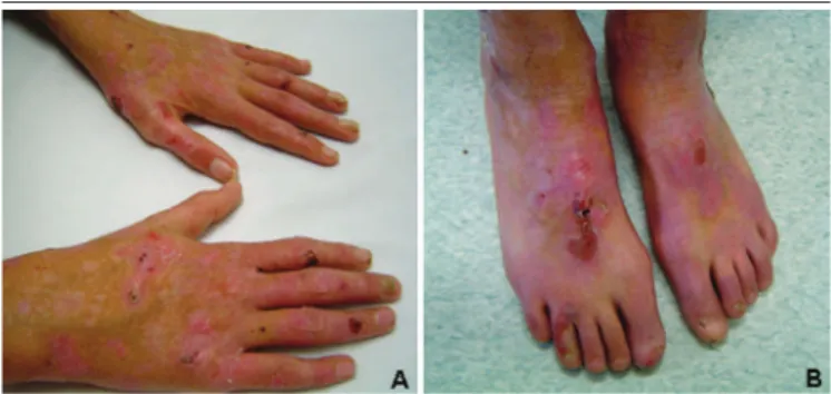
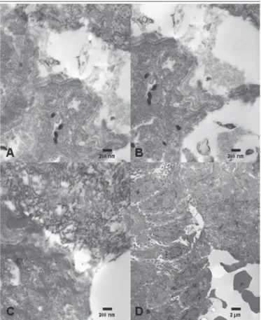
Our aim was to report the case of a patient with dystrophic epidermolysis bullosa (DEB), whose diagnosis was reached through transmission electron microscopy (TEM), highlighting the applications and importance of this technique in health. The patient was an 18-year-old Caucasian man, the only child of young and non-consanguineous parents without similar cases in the family. According to his mother, the bullous lesions appeared after 15 days of life, affecting the skin of hands, shoulders, elbows, back, knees, ankles and feet, besides oral and nasal mucosa. Some of the lesions had blood inside and the developed scars were atrophic (erythematous in the region of the knees and feet). The lesions frequently get infected, causing itching when dry (Figure 1). When the patient was 8 years old he underwent a skin biopsy, whose histopathological analysis showed indings indicative of a subepithelial blister, with only one intrapapillary projection in the central area. The adjacent dermis showed mild edema and minimal lymphocytic iniltrate. TEM performed from the biopsy of a bullous lesion caused by skin friction showed sub-basement membrane involvement, conirming the diagnosis of epidermolysis bullosa (EB) of the dystrophic type
(Figure 2). An increased number of lesions appeared with the
patient growth, especially after puberty. At age 13, epidermal cysts (milia) and dystrophic nails were also present. Dysphagia for solid foods and bleeding of the oral mucosa were also reported. Endoscopy and colonoscopy were normal. He presented abdominal pain at age 17 years, being diagnosed with secondary duodenal stenosis due to an aorto-mesenteric impingement. The patient underwent a duodenal-jejunal anastomosis surgery that progressed well, without the occurrence of stenosis or constipation.
FIGURE 1 − Images of the patient’s hands and feet showing blistering and scarring skin
lesions, in addition to dystrophic and missing nails (especially in the feet), secondary to EB
EB: epidermolysis bullosa.
66
EB is an etiologically heterogeneous group of rare inherited disorders with different patterns of inheritance, characterized by blistering of the skin and mucous membranes after minimal
trauma(1, 2). EB can be classiied based on the histopathological
inding of blistering level into four main types: simplex (SEB), junctional (JEB), dystrophic (DEB) and Kindler syndrome. Thus, the separation of the tissue may occur in the intraepidermal (SEB), lamina lucid (JEB) or sublamina densa (DEB) level. Kindler syndrome is a mixed type, with several cleavage planes. The different degrees of vulnerability of the skin are caused by mutations in structural proteins involved in the adhesion of the basement membrane zone (BMZ) of the epithelium(2-4). EB groups
can be further subdivided according to pattern of inheritance, morphology of the injury and mutation type. The overall incidence of EB is 19:1,000,000 live births, occurring with similar frequency
in both sexes(2, 5). In Brazil, there are no epidemiological data
about the disease. All EB types and subtypes are associated with mechanical fragility of the skin, emergence of milia, dystrophic or absent nails, and scars that may appear at birth or after months
or years of life(5), as seen in our patient.
After the initial diagnosis based on clinical manifestations, the EB diagnosis can be conirmed through three main techniques: immunoluorescence antigen mapping (IFM), TEM and mutational analysis from blood and skin samples (ibroblasts and keratinocytes)(2, 3, 5). TEM, the technique used in our case,
is able to identify the level of skin cleavage. It is an extremely lexible technique that may reveal several biological pieces of information by providing lat and greatly magniied images, which allow tissue visualization and organization of organelles at the nanoscale(6). Methodologically, the sample is obtained by
biopsy of a newly formed skin blister. It is immediately immersed in ixative, avoiding cell and tissue damaging and drying. The skin fragment must be of suficient size to be inely cut (~ 0.5-1 mm of thickness) and perfectly ixed, allowing visualization of the level of blister division and the structure of the dermal-epidermal junction, relevant for the EB diagnosis. Examination by TEM can be also supplemented by IFM(7). However, some EB subtypes
may have overlapped immunohistochemical indings, leading to limitation of the IFM. The use of TEM also allows the semi-direct quantiication of speciic structures, as keratin ilaments, hemidesmosomes and anchoring ibrils (AF), which are important for disease classiication(8).
DEB, the form diagnosed in our patient, is considered the second most common type of EB(9). It has a prevalence of about
2-6 cases per million births and is associated with both autosomal dominant (DDEB) and recessive (RDEB) patterns of inheritance, according to its subtypes(2). In these cases, mutations of the
COL7A1 gene, responsible for collagen VII codiication, promotes
sublamina densa cleavage. Currently, more than 300 pathogenic
mutations in COL7A1 have been described in patients with DEB(2, 3, 5, 9, 10). Most patients with DEB are already symptomatic in
the irst year of life. The blisters are spread, constant and leave scars. However, there are variations in the clinical presentations, ranging from nail dystrophy and light blisters to the development of widespread blisters and mutilation, formation of esophageal blistering, growth deicit, and premature death. Problems such as swallowing dificulty and constipation are the result of extensive blisters that mainly affect mouth, esophagus, eye and anal canal
membranes(2, 3, 5, 9, 10).
Therefore, TEM not only can conirm the diagnosis of EB but also determine its type. In DEB, TEM allows direct visualization and semiquantitative assessment of structural deicits in the BMZ, being able to reveal the lack of collagen VII, the rudimentary structure of the skin, and the lack of AFs(8). It can also identify
microsplits and subtle changes in the dermal-epidermal junction that cannot be detected through IFM. Prenatal diagnosis of DEB can also be performed through TEM, from fetal skin samples
FIGURE 2 − Images obtained through TEM, in scales of 200 nm (A-C) and 2 µm (D),
showing cleavage at the level of sublamina densa, which is consistent with the diagnosis of DEB
TEM: transmission electron microscopy; DEB: dystrophic epidermolysis bullosa.
67
REFERENCES
1. McGrath JA. Recently identiied forms of epidermolysis bullosa. Ann Dermatol. 2015; 27: 658-66.
2. Shinkuma S. Dystrophic epidermolysis bullosa: a review. Clin Cosmet Investig Dermatol. 2015; 8: 275-84.
3. Intong LRA, Murrell DF. Inherited epidermolysis bullosa: new diagnostic criteria and classiication. Clin Dermatol. 2012; 30: 70-7.
4. Fine JD, Bruckner-Tuderman L, Eady RA, et al. Inherited epidermolysis bullosa: updated recommendations on diagnosis and classiication. J Am Acad Dermatol. 2014; 70: 1103-26.
5. Fine JD. Inherited epidermolysis bullosa. Orphanet J Rare Dis. 2010; 5: 12. 6. Müller SA, Aebi U, Engel A. What transmission electron microscopes can visualize now and in the future. J Struct Biol. 2008; 163: 235-45. 7. Eady RAJ, Dopping-Hepenstal PJC. Transmission electron microscopy for the diagnosis of epidermolysis bullosa. Dermatol Clin. 2010; 28: 211-22.
obtained by fetoscopy(2, 5). Currently, despite the obtained advances,
non-molecular diagnostic tests such as TEM are considered the best option for diagnosis of EB. However, the number of laboratories with experience in this type of technique and diagnosis is quite small in our midst. Molecular analysis is most appropriate in cases selected for a screening of silent mutations in families with severely affected individuals(2, 5).
Other genetic conditions can also be diagnosed through TEM. For example, changes in the glomerular basement membrane (GBM), represented by irregular alternation of lamina densa of
GBM in renal biopsy samples, may be detected by TEM and are indicative of the diagnosis of Alport syndrome(11). In primary ciliary
dyskinesia (PCD), the identiication of ciliary abnormalities in the nasal epithelium through TEM has been considered the gold standard for the disease diagnosis in recent years(12, 13).
Thus, despite the technological advances, TEM continues to have an important role in the diagnosis and clinical research of diseases such as EB. Accurate diagnosis allows proper management, prognosis determination and genetic counseling of the individual
and his family.
RESUMO
Relatamos o caso de um paciente com epidermólise bolhosa distrófica (EBD) diagnosticado por microscopia eletrônica de transmissão (MET), destacando aplicações e importância desta técnica na área da saúde. Paciente do sexo masculino, filho único de pais jovens não consanguíneos, sem histórico de caso familial. O paciente foi submetido à biópsia cutânea, na qual a MET revelou comprometimento da membrana sub-basal, confirmando o diagnóstico de EBD. Apesar dos avanços tecnológicos, a MET continua tendo papel importante no diagnóstico e na pesquisa clínica, sendo considerada a melhor opção para a confirmação do diagnóstico e dos subtipos de doenças como a epidermólise bolhosa (EB).
Unitermos: epidermólise bolhosa; epidermólise bolhosa distrófica; epiderme; membrana basal; microscopia eletrônica.
8. Bruckner-Tuderman L. Dystrophic epidermolysis bullosa: pathogenesis and clinical features. Dermatol Clin. 2010; 28: 107-14.
9. Gürtler TGR, Diniz LM, Souza Filho JB. Epidermólise bolhosa distróica recessiva mitis – relato de caso clínico. An Bras Dermatol. 2005; 80: 503-8.
10. Dang N, Murrell DF. Mutation analysis and characterization of COL7A1 mutations in dystrophic epidermolysis bullosa. Exp Dermatol. 2008; 17: 533-68.
11. Yao XD, Chen X, Huang GY, et al. Challenge in pathologic diagnosis
of Alport syndrome: evidence from correction of previous misdiagnosis. Orphanet J Rare Dis. 2012; 7: 100.
12. Snijders D, Bertozzi I, Barbato A. Nasal NO, High-speed video microscopy, electron microscopy, and genetics: a primary ciliary dyskinesia puzzle to complete. Pediatr Res. 2014; 76(3): 321.
13. Honoré I, Burgel PR. Primary ciliary dyskinesia in adults. Rev Mal Respir. 2015; 33: 165-89.
CORRESPONDING AUTHOR
Paulo Ricardo Gazzola Zen
Genética Clínica; UFCSPA; Rua Sarmento Leite, 245/403; Centro; CEP: 90050-170; Porto Alegre-RS, Brasil; Phone: +55 (51) 8771; Fax: +55 (51) 3303-8710; e-mail: paulozen@ufcspa.edu.br.

