* Study conducted in the Pulmonology Department of the Faculdade de Medicina da Universidade de São Paulo – FMUSP, University of São Paulo School of Medicine – São Paulo (SP) Brazil.
1. Resident in Pulmonology at the Faculdade de Medicina da Universidade de São Paulo – FMUSP, University of São Paulo School of Medicine – São Paulo (SP) Brazil. 2. PhD student in Pulmonology at the Faculdade de Medicina da Universidade de São Paulo – FMUSP, University of São Paulo School of Medicine – São Paulo (SP) Brazil.
3. Head of the Pulmonary Function Laboratory at the Instituto do Coração – InCor, Heart Institute – of the Hospital das Clínicas da Faculdade de Medicina da Universidade de São Paulo – HCFMUSP, University of São Paulo School of Medicine Hospital das Clínicas – São Paulo (SP) Brazil.
4. Head of the Pulmonology-Oncology Group of the Department of Pulmonology at the Faculdade de Medicina da Universidade de São Paulo – FMUSP, University of São Paulo School of Medicine – São Paulo (SP) Brazil.
Correspondence to: Bruno Guedes Baldi. Av. Dr. Enéas de Carvalho Aguiar, 255, Sala 7079, CEP 05403-900, São Paulo, SP, Brasil. Tel 55 11 3069-7202. E-mail: [email protected]
Submitted: 24 May 2006. Accepted, after review: 29 September 2006.
Abstract
Benign tracheal tumors are rare, recurrent papillomatosis being the most common. They often simulate obstructive pulmonary diseases, such as asthma and chronic obstructive pulmonary disease, and patients with benign tracheal tumors often undergo long-term treatment for such diseases, without any improvement, Therefore, these tumors should be included in the differential diagnosis in patients presenting tracheobronchial tree obstruction. This report describes the case of a patient with a tracheal polyp. The patient presented symptoms for three years, and the spirometry findings suggested intrathoracic obstruction. The patient presented complete clinical and spirometric recovery after bronchoscopic resection of the tumor.
Keywords: Polyps; Airway obstruction; Spirometry; Bronchoscopy.
Introduction
Primary tracheal tumors account for only approximately 2% of all tumors of the upper respiratory tract, recurrent benign papillomatosis being the most common. Despite being rare, their clinical presentation frequently simulates obstructive pulmonary diseases, such as asthma and chronic obstructive pulmonary disease (COPD), often creating diffi-culty in the diagnosis and errors in the treatment. Therefore, they should be included in the differential diagnosis of profiles of tracheobronchial tree obstruction.(1)
In this report, we present the case of a patient with dyspnea upon moderate exertion and repeated respiratory infections secondary to the presence of tracheal polyp, which evolved to complete clinical and spirometric recovery after bronchoscopic excision of the tumor.
Case report
The patient was then submitted to laryngotra-cheobronchoscopy, which revealed a pedunculated vegetative tracheal lesion located 9 cm from the vocal folds and 4 cm from the carina, obstructing approximately 80% of the lumen, with discrete reduction of the stenosis during inspiration. The lesion was removed piecemeal, but completely, with forceps.
The anatomopathological evaluation of the sample, which had a diameter of approximately 2 cm and weighed 5 g, revealed fibroepithelial polyp, with squamous metaplasia of the epithelial covering. There was mononuclear inflammatory infiltrate in the chorion, vascular congestion and reactive fibroblasts (Figure 2b).
After the lesion had been excised, the patient evolved to complete resolution of the clinical status and to functional normalization (Figure 3).
Discussion
Approximately 80% of primary tracheal tumors are malignant, the remainder consisting of benign lesions, among which are recurrent papilloma-tosis (most frequent), lipoma, fibroma, leiomyoma,
hemangioma, and polyps.(1)
disease. He was not a smoker or an alcoholic, and he did not use illicit drugs. Peripheral oxygen satu-ration at rest on room air was 95%, as determined using pulse oximetry.
The physical examination and the chest X-ray did not present alterations.
He was submitted to spirometry, which revealed the presence of obstructive respiratory disorder, characterized by a decrease in forced expiratory volume in one second (FEV1) and in the ratio of
FEV1 to forced vital capacity (FEV1/FVC), although
FVC was normal. The morphological characteris-tics of the flow-volume loop indicated a pattern of intrathoracic and variable obstruction of the airways: decreased peak expiratory flow and flat-tening of the expiratory loop of the flow-volume curve; and the inspiratory loop was preserved. The ratio between the forced expiratory and inspiratory flows at 50% of the FVC (FEF50%/FIF50%) was lower than one (Figure 1).
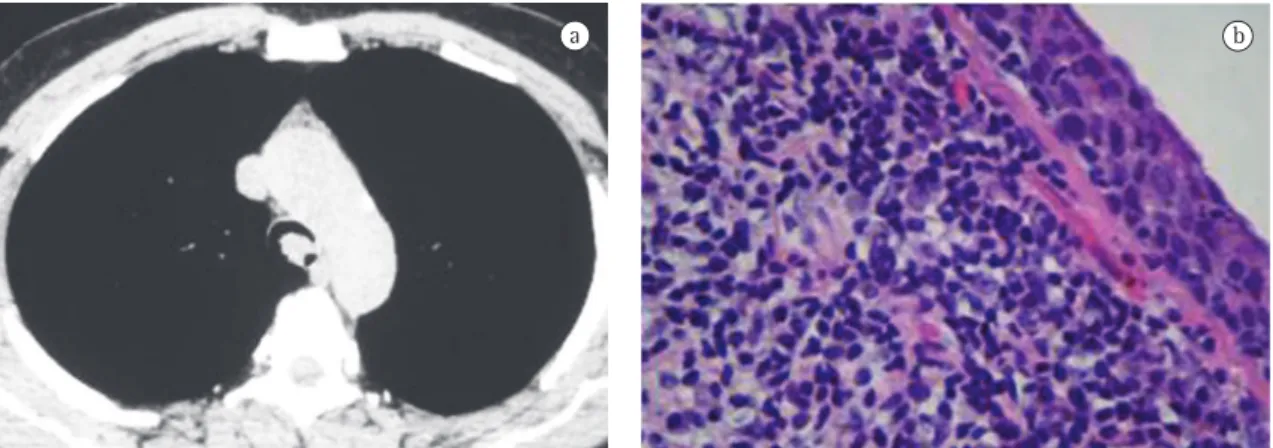
Computed tomography of the chest revealed a vegetative lesion in the posterior region of the trachea, obstructing 70-80% of the tracheal lumen and located approximately 3 cm from the carina (Figure 2a ). The tomography scan revealed no alterations of the pulmonary parenchyma.
Predicted Value % of Predicted)
FVC (L) FEV1 (L) FEV1/FVC FEF25-75%(L/sec) PEF (L/sec)
4.13 5.60 136 3.35 2.17 65 0.81 0.39 48 3.48 1.76 50 8.28 4.87 59 Flow-volume curve
12 FN
8
8 –4
–8 –12
0 4 6
the tracheobronchial tree, all of which lead to lesion of the local mucosa.(1-3) Initially there is edema or
erosion, with progression to ulceration, and then the attempt at regeneration through vascular congestion, deposition of fibrous connective tissue, and local migration of inflammatory cells, espe-cially neutrophils and lymphocytes.(1,4,5) The polyps
are covered with stratified squamous epithelium or Although endotracheal polyps are rarely found,
they should be included in the differential diagnosis of partial or complete obstruction of the trache-obronchial tree.(1,2) They present as protruding
structures, predominantly inflammatory, single or multiple, resulting from the inhalation of a foreign body, smoke, toxic gases or other irritants, as well as from infection or the insertion of prostheses into
a b
Figure 2 - a) Computed tomography of the chest showing vegetative lesion in the posterior region of the trachea, obstructing 70-80% of its lumen; and b) Anatomopathological evaluation demonstrating squamous metaplasia of the epithelial covering, chorion with mononuclear inflammatory infiltrate, vascular congestion, and reactive fibroblasts, compatible with fibroepithelial polyp.
Predicted Value % of Predicted
FVC (L) FEV1 (L) FEV1/FVC FEF25-75%(L/sec) PEF (L/sec)
4.13 5.33 130 3.35 4.42 133 0.81 0.83 102 3.48 4.76 138 8.28 9.34 113 Flow-volume curve
12 8
8 6 4 4
0
–12 –8 –4
FN
tion and the airway compliance in the region of obstruction:
1) Pattern of variable extrathoracic obstruction, characterized by the flattening of the inspiratory loop and preservation of the expiratory loop of the curve, with the FEF50%/FIF50% > 1.(8-10)
2) Pattern of variable intrathoracic obstruction, characterized by the flattening of the expira-tory loop and preservation of inspiraexpira-tory loop of the curve, with the FEF50%/FIF50% < 1.(8-10)
3) Pattern of fixed obstruction of central airways, in which there is flattening of the inspiratory and expiratory loops of the curve, related to the approximately equal decrease in the forced flows at 50% of the FVC (FEF50%/FIF50%≈ 1).(8-10)
In the case presented here, the expiratory flow-volume loop reveals decrease of the flows in the initial phase of the expiration, still at a volume near total lung capacity, following practically constant expiratory flow during most of the expiration. Close to residual volume, where the expiratory flows are naturally lower as a result of the weaker traction of lung elastic recoil, the flow-volume loop shows normal behavior again, indicating that the limitation to the flow imposed by the tracheal lesion turned to be less critical than the natural limitation to the airflow at that pulmonary volume. These character-istics are fundamental to differentiate the nature of the obstruction in the presented case in relation to the pattern observed in diffuse and distal obstruc-tive diseases of the airways (asthma or COPD), in which the limitation to the flow is more pronounced at low pulmonary volumes.
The gold standard method for diagnosing obstruction of the central airways is bronchoscopy (rigid or flexible), which allows the lesion to be biopsied for histopathologic evaluation and treat-ment planning.(7,11)
The treatment of tracheal polyps varies according to the size of the lesion, the presence of symptoms, and the viability of performing bron-choscopic procedures.(1) Small lesions provoking
few symptoms can be treated with corticosteroids and antibiotics.(1-6) In most cases, lesions of greater
volume that provoke more symptoms can be extir-pated through bronchoscopic procedures, such as curettage, laser, electrocauterization, or cryosurgery, according to the equipment available and the local normal tracheobronchial mucosa and,
macroscopi-cally, are reddish in color, similar to normal mucosa, being either sessile or pedunculated.(1,6)
Regarding the clinical status, patients can be asymptomatic or present dyspnea, which might be progressive in nature, occurring only upon exertion, or be intermittent (as a result of the closing of the tracheal lumen secondary to the mass movement, similar to a valvular mechanism). In addition to alterations related to secondary respiratory infec-tion, such as fever, suppurative cough, and asthenia, wheezing and hemoptysis can occur.(1,4,6,7) Generally,
dyspnea upon exertion occurs when the tracheal lumen is smaller than 8 mm, and at rest when it is smaller than 5 mm.(7) Individuals with endotracheal
polyps are often misdiagnosed as suffering from obstructive pulmonary diseases such as asthma or COPD and are treated for long periods, with less than satisfactory clinical results.(7) Upon physical
examination, there might be wheezing, stridor, tach-ypnea, or accessory respiratory muscle use, together with signs of infection, such as fever.(4,5) Weight loss
can also occur.(4)
Although chest X-ray is the initial radiolog-ical examination to be obtained in the evaluation of patients with symptoms of airway obstruc-tion, although it is rarely diagnostic.(7) Computed
tomography of the chest reveals nodules or masses (single or multiple) obstructing the tracheal lumen, allowing the size and mobility of the lesion, as well as its relationship with adjacent structures and distal impairment, to be evaluated.(3,7)
It is fundamental to perform spirometry in the evaluation of patients with suspicion of central airway obstruction, principally for the observation of the morphology of the flow-volume curve, which might suggest the location of the obstruction, whether extrathoracic (impairment of the pharynx, larynx, or extrathoracic trachea) or intrathoracic (involvement of the intrathoracic trachea or of the main bronchi). In addition, spirometry can indicate the dynamic behavior of the obstruction (variable or fixed) according to the difference or lack of a difference between the inspiratory and expiratory loops.(8-10) Spirometry also provides quantitative
data, which are important for the assessment of severity.
loca-surgery: a late complication of use of a double-lumen endobronchial tube. Anesthesiology. 1996;84(5):1234-6. 3. Yoon YC, Lee KS, Kim TS, Seo JB, Han J. Benign
bronchopulmonary tumors: radiologic and pathologic findings. J Comput Assist Tomogr. 2002;26(5):784-96. 4. Kahn B, Amer NS. Multiple bronchial polyps. Chest.
1970;57(3):279-83.
5. Tedeschi LG, Libertini R, Conte B. Endobronchial polyp. Chest. 1973;63(1):110-2.
6. Berman DE, Wright ES, Edstrom HW. Endobronchial inflammatory polyp associated with a foreign body. Successful treatment with corticosteroids. Chest. 1984;86(3):483-4. 7. Ernst A, Feller-Kopman D, Becker HD, Mehta AC.
Central airway obstruction. Am J Respir Crit Care Med. 2004;169(12):1278-97.
8. Aboussouan LS, Stoller JK. Diagnosis and management of upper airway obstruction. Clin Chest Med. 1994;15(1):35-53.
9. Sociedade Brasileira de Pneumologia e Tisiologia. Diretrizes para Testes de Função Pulmonar. J Pneumol. 2002;28(Supl 3):1-221.
10. Pellegrino R, Viegi G, Brusasco V, Crapo RO, Burgos F, Casaburi R, et al. Interpretative strategies for lung function tests. Eur Respir J. 2005;26(5):948-68.
11. Seijo LM, Sterman DH. Interventional pulmonology. N Engl J Med. 2001;344(10):740-9.
12. Moorjani N, Beeson JE, Evans JM, Maiwand MO. Cryosurgery for the treatment of benign tracheo-bronchial lesions. Interact Cardiovasc Thorac Surg. 2004;3(4):547-50.
experience. Surgery (thoracotomy or sternotomy) is rarely necessary.(1,7,12)
The present case can serve to illustrate the fact that, although rare, benign tumors of the airways, such as tracheal polyps, should be remembered in the cases of suspicion of tracheobronchial tree obstruction, especially in patients who are thought to be as suffering from obstructive pulmonary diseases and are treated for a long period without presenting a satisfactory response. This diagnosis is suggested principally by the morphology of the inspiratory and expiratory loops of the flow-volume curve, as in this report, in which we observed flat-tened expiratory loop and preserved inspiratory loop, suggesting variable intrathoracic obstruction, which was subsequently confirmed in the broncho-scopic evaluation.
References
1. Gamblin TC, Farmer LA, Dean RJ, Bradley RA, Dalton ML. Tracheal polyp. Ann Thorac Surg. 2002;73(4):1286-7. 2. Ikeda M, Ishida H, Tsujimoto S, Kato H. Endobronchial