Abst ract
Submitted: June 3, 2016 0RGL¿FDWLRQ$XJXVW Accepted: September 20, 2016
7ROOOLNHUHFHSWRUDJRQLVWV
Porphyrom onas gingivalis LPS and
CpG different ially regulat e I L- 10
com pet ency and frequencies of m ouse
B10 cells
,/H[SUHVVLQJUHJXODWRU\%FHOOV%SOD\DNH\UROHLQLPPXQHV\VWHP EDODQFHE\OLPLWLQJH[FHVVLYHLQÀDPPDWRU\UHVSRQVHV(IIHFWVRIWROOOLNHUHFHSWRU
signaling and co- st im ulat or y m olecules on B1 0 act iv it y dur ing innat e and adapt ive im m une responses are not fully underst ood. Obj ect ive: This st udy is
t o det erm ine t he effect s of P. gingivalis LPS and CpG on B10 cell expansion and
I L- 10 com pet ency in vit ro. Mat erial and Met hods: Spleen B cells w ere isolat ed
IURP&%/-PLFHZLWKRUZLWKRXWIRUPDOLQ¿[HGP. gingivalis im m unizat ion.
B cells w er e cult ur ed for 48 hour s under t he follow ing condit ions: CD40L,
CD40L+ LPS, CD40L+ CpG, and CD40L+ LPS+ CpG in t he presence or absence of
¿[HGP. gingivalis. Percent ages of CD1dhiCD5+%FHOOVZHUHPHDVXUHGE\ÀRZ
cyt om et ry. I L- 10 m RNA expression and secret ed I L- 10 w ere m easured by real-t im e quanreal-t ireal-t areal-t ive PCR and by ELI SA respecreal-t ively. Resulreal-t s: P. gingivalis LPS plus
&'/VLJQL¿FDQWO\LQFUHDVHG&'GKL&'%FHOOSHUFHQWDJHVDQGVHFUHWHG,/
10 levels in bot h im m unized and non- im m unized m ice B cells in t he presence
or absence of P. gingivalis, com pared w it h cont rol group. Secret ed I L- 10 levels
ZHUHVLJQL¿FDQWO\LQFUHDVHGLQ&'//36WUHDWHGJURXSFRPSDUHGZLWK&'/
t reat m ent group in t he absence of P. gingivalis&S*SOXV&'/VLJQL¿FDQWO\
decreased CD1dhiCD5+ B cell percent ages, but great ly elevat ed secret ed I
L-10 levels in im m unized and non- im m unized m ice B cells in t he absence of P. gingivalis, com pared w it h CD40L t reat m ent group. Conclusions: P. gingivalis
LPS and CpG different ially enhance I L- 10 secret ion and expansion of m ouse B10
cells during innat e and adapt ive im m une responses.
Ke yw or ds: I L- 10. Porphyrom onas gingivalis LPS.
Zhiqiang LIU1,2
Yang HU1
Pei YU1,3
Mei LIN2
Grace HUANG1
Toshihisa KAWAI1
Martin TAUBMAN1
Zuomin WANG2
Xiaozhe HAN1
http://dx.doi.org/10.1590/1678-77572016-0277
1The Forsyt h I nst it ut e, Depart m ent of I m m unology and I nfect ious Diseases, Cam bridge,
Massachuset t s, Unit ed St at es.
2Capit al Medical Universit y, Beij ing ChaoYang Hospit al, Depart m ent of St om at ology,
Beij ing, China.
3Sichuan Universit y, West China School of St om at ology, St at e Key Laborat ory of Oral
Diseases, Chengdu, Sichuan, China.
I nt roduct ion
I L- 1 0 ex pr essing r egulat or y B cells ( B1 0 ) is a
VSHFL¿F ,/ FRPSHWHQW UHJXODWRU\ % FHOO VXEVHW WKDW KDV EHHQ UHFHQWO\ LGHQWL¿HG LQ ERWK PLFH DQG
h u m an s2 7. B1 0 cell d ow n - r eg u lat es au t oim m u n e
GLVHDVHLQÀDPPDWLRQDQGLPPXQHUHVSRQVHVWKURXJK
I L- 10 expression, playing crucial regulat ory roles in
innat e and adapt ive im m unit y27. Though m ouse B10
FHOOVVKDUHVRPHRYHUODSSLQJSKHQRW\SLFPDUNHUVZLWK RWKHUPXOWLSOHSKHQRW\SLFDOO\GH¿QHG%FHOOVXEVHWV
t hey have been found t o be pr edom inant ly enr iched
in spleen CD1dhighCD5+ B cells27.
7ROOOLNHUHFHSWRUV7/5VZKLFKEHORQJWRSDWWHUQ
recognit ion recept ors, are specialized t ransm em brane p r ot ein s t h at m ed iat e in n at e im m u n it y t h r ou g h
d et ect in g com m on st r u ct u r es of m an y m icr ob ial
species such as bact erial lipopolysaccharides ( LPS)
or v ir al n u cleic acid s1 7 , 2 5. Up on r ecog n it ion of a
pat h ogen , TLRs in it iat e a sign alin g cascade t h at
OHDGVWRH[SUHVVLRQDQGUHOHDVHRISURLQÀDPPDWRU\
F\WRNLQHV FKHPRNLQHV DQG 7\SH, LQWHUIHURQV8, 21.
Porphyrom onas gingivalis (P. gingivalis) LPS has been show n t o be able t o act ivat e bot h TLR2 and TLR4
due t o it s unique st r uct ur e and funct ion2,6, and CpG
LVNQRZQDV7/5DJRQLVWWRVWLPXODWHWKHLPPXQH
r esponses9,16.
I n t e r a ct i o n b e t w e e n CD 4 0 Li g a n d ( CD 4 0 L)
and CD40 plays an im por t ant r ole in t he init iat ion
and pr ogr ession of cellular and hum oral adapt iv e
im m unit y15. The act ivat ion of CD40 on B cells by CD40L
is cr ucial for T cell- dependent B cell pr oliferat ion,
different iat ion, and ant ibody isot ype sw it ching11,13,14.
Recen t st u d i es d em o n st r a t ed t h a t cu l t u r i n g
spleen B cells w it h LPS or CD40L for 48 h induced
VLJQL¿FDQWO\ KLJKHU IUHTXHQFLHV RI F\WRSODVPLF ,/
10 product ion in B cells t han cont rol in vit ro20. LPS
st im ulat ion of spleen B cells for 24 h induced m or e
I L- 10 t han unst im ulat ed cells28. Spleen B cells w it h a
CD1dhighCD21+CD23- MZ phenot ype can produce I L- 10
LQUHVSRQVHWR&S*VWLPXODWLRQLQPLFHZLWKOXSXVOLNH
aut oim m une disease30. How ever, despit e all t hese
¿QGLQJVWKHHIIHFWVRI7/5DJRQLVWVDORQJZLWKFR
st im ulat ory m olecules, such as CD40L, on B10 act ivit y
dur ing innat e and adapt ive im m une r esponses ar e
not clear ly under st ood. Fur t her m or e, t her e is lim it ed infor m at ion on t he r ole of B10 cells dur ing im m une
r esp on ses t o or al d iseases, su ch as p er iod on t al
disease, w hen encount er ing oral pat hogens and t heir
derivat ives. I n t he present st udy, spleen B cells from P. gingivalis non- im m unized and im m unized m ice w er e co- st im ulat ed w it h TLR4, TLR9, and CD40 signals t o
invest igat e t heir effect s on B10 cell expansion and
I L- 10 com pet ency in vit ro.
Mat er ial and Met hods
P. gingivalis
FXOWXUHDQG¿[DWLRQ
P. gingivalis ( st rain ATCC 33277) w er e gr ow n on
anaer obic blood agar plat es ( NHK agar, Nor t heast Laborat or y, Wat er ville, ME, U.S.A.) in an anaer obic
cham ber w it h 85% N2, 5% H2, and 10% CO2. Single
colony of P. gingivalis was isolat ed fr om t he plat e
and gr ow n in ATCC Medium 2722. Aft er incubat ion at 37° C for 4 d, bact er ia num ber in cult ur e m edium was
det erm ined by reading opt ical densit y values using
spect rophot om et er and com paring t hem w it h a curve
der ived fr om a st andar d plat e count . Bact er ia w er e
FROOHFWHGDQG¿[HGZLWKSDUDIRUPDOGHK\GH3)$
for 30 m in at r oom t em perat ur e, t hen washed t hr ee
t im es wit h st erile phosphat e- buffered saline ( PBS) and
resuspended in PBS at t he concent rat ion of 5× 10⁸/ m L.
Anim als
&%/-PLFH-DFNVRQ/DERUDWRU\%DU+DUERU 0( 86$ DJLQJ ZHHNV ZHUH HTXDOO\ DQG
random ly divided int o four gr oups. Gr oup 1 and 2 w er e set as non- im m unized m ice gr oups in w hich
PLFHZHUHVDFUL¿FHGGLUHFWO\IRUVSOHHQ%FHOOLVRODWLRQ
Gr oup 3 and 4 w er e set as im m unized m ice gr oups
and m ice were im m unized by 1× 10⁸¿[HGP. gingivalis
int raperit oneal inj ect ion at day 0, t hen follow ed by
1× 10⁷¿[HGP. gingivalis inj ect ion at day 7 t o enhance
WKH LPPXQL]DWLRQ 0LFH ZHUH VDFUL¿FHG IRU % FHOO
isolat ion at day 10. All m ice used in t he st udy w er e
m aint ained under pat hogen- free condit ions in lam inar
ÀRZFDELQHWV([SHULPHQWDOSURWRFROVZHUHDSSURYHG
by t he I nst it ut ional Anim al Car e and Use Com m it t ee of t he For syt h I nst it ut e.
B cell isolat ion
Mice w ere eut hanized in CO2 cham ber and spleens
w ere harvest ed. Single splenic cells w ere yielded by
JULQGHGRQDVWHHOPHVKDQGWKHQ¿OWHUHGZLWK NjP&HOO6WUDLQHUV$IWHUUHGEORRGFHOOVUHPRYDOE\
Am m onium - Chlor ide- Pot assium ( ACK) ly sis buffer
( Life Technologies, Carlsbad, CA, USA) , splenic cells
Cell St rainers. Then non- B cells w ere m agnet ically
ODEHOHGXVLQJ3DQ%FHOOLVRODWLRQNLW0LOWHQ\L%LRWHF &DPEULGJH 0$ 86$ %ULHÀ\ VLQJOH VSOHQLF FHOO
suspensions w ere incubat ed w it h biot in- conj ugat ed
m on oclon al an t ibodies again st n on - B cell su r face
PDUNHUV &' &'F &'E &' *U DQG
Ter 119) at 4° C for 10 m in follow ed by incubat ion w it h m agn et ic m icr obeads con j u gat ed an t i- biot in
ant ibodies at 4° C for 15 m in. Magnet ically labeled
cells w er e t h en deplet ed by passin g t h r ou gh LD colum ns ( Milt enyi Biot ec, Cam bridge, MA, USA) under
WKHPDJQHWLF¿HOGRIWKH4XDGUR0$&66HSDUDWRU
( Milt eny i Biot ec, Cam br idge, MA, USA) . Unlabeled
cells t hat passed t hr ough LD colum n w er e collect ed ( cont ained > 98.5% CD19+ cells) .
B cell cult ure
B cell num ber was count ed by hem acyt om et er.
Ea c h 1 × 1 06 % FHOOV ZHUH FXOWXUHG LQ Nj/
I MDM+ Glut aMAXTM ( Life Technologies, Car lsbad, CA,
USA) com plet e m edium ( cont ains 10% FCS, 100 U/ m L
penicillin, 100 m g/ m L st rept om ycin, 2 m M L- glut am ine,
NjJP/$PSKRWHULFLQ%DQGNj00(LQZHOO
plat es under t he following condit ions: cont rol, CD40L,
CD40L+ LPS, CD40L+ CpG, or CD40L+ LPS+ CpG in t he
DEVHQFHRULQWKHSUHVHQFHRI¿[HGP. gingivalis. Final
concent rat ions of t hese st im ulant s w ere as follow s:
&'/H%LRVFLHQFH6DQ'LHJR&$86$NjJP/
P. gingivalis LPS ( I nvivogen, San Diego, CA, USA, 10
NjJP/PRXVH&S*'1$+\FXOW3O\PRXWK0HHWLQJ
3$86$Nj0DQG¿[HGP. gingivalis ( 5× 10⁶/ 1× 10⁶
cells) . P. gingivalis LPS was used as TLR4 agonist and
m ouse CpG- DNA( 5’-TCCATGACGTTCCTGATGCT - 3’)
was used as TLR9 agonist . B cells cult ured w it hout st im ulat ion w ere used as cont rol. Cells w ere cult ured
LQDKXPLGL¿HGLQFXEDWRUDW&ZLWK&22 for 48
K7KLUW\RINj/PHGLXPLQHDFKZHOOZDVXVHG
t o det er m ine CD1dhighCD5+ B cell per cent ages and
rem aining cells w ere used t o det erm ine I L- 10 m RNA
expr ession levels. Cult ur e super nat ant was used for
secr et ed I L- 10 levels m easur em ent .
CD1d
highCD5
+B cells percentages determ ination
B cells w er e washed w it h PBS and Fc r ecept or s
ZHUH EORFNHG E\ LQFXEDWLQJ ZLWK 7UX6WDLQ IF;TM
( BioLegend, San Diego, CA, USA) on ice for 10 m in,
t hen followed by incubat ion wit h PE ant i- m ouse CD1d ( BioLegend, San Diego, CA, USA) and Alexa Fluor 647
DQWLPRXVH&'ÀXRUHVFHQFHFRQMXJDWHGDQWLERGLHV
( BioLegend, San Diego, CA, USA) on ice for 30 m using
predet erm ined opt im al concent rat ions. Then, all cells
ZHUH FRXQWHG E\ ÀRZ F\WRPHWHUV %' %LRVFLHQFHV
San Jose, CA, USA) an d d at a w er e an aly zed b y
Flow Jo v10 soft ware. For each sam ple, t he sam e gat e
was applied t o t he ot her sam ples t o det er m ine t heir
CD1dhighCD5+ B cell per cent ages. Since m ouse B10
cells has been found t o be pr edom inant ly enr iched
in spleen CD1dhighCD5+ B cells, CD1dhighCD5+ B cell
percent age is considered as t he proport ional indicat or of B10 cell per cent age.
I L- 10 m RNA expr ession m easur em ent
7RWDO P51$ RI % FHOOV ZDV LVRODWHG E\ 3XUH/LQN
RNA Mini Kit ( Life Technologies, Carlsbad, CA, USA) follow ing t he m anufact ur er ’s inst r uct ions. I solat ed
m RNA was t hen r ever se t ranscr ibed t o cDNA using
t he Super Scr ipt ™ I I Rever se Transcr ipt ase sy st em
( I nv it r ogen, San Diego, CA, USA) in t he pr esence of r an d om p r im er s f ollow in g t h e m an u f act u r er ’s
inst ruct ions. Then, real- t im e quant it at ive PCR (
RT-T3&5 ZDV FDUULHG RXW LQ D Nj/ UHDFWLRQ V\VWHP XVLQJ/LJKW&\FOHU6<%5*UHHQ,0DVWHUNLW5RFKH
Diagnost ics, I ndianapolis, I N, USA) and Light Cycler
480 I nst rum ent ( Roche Diagnost ics, I ndianapolis, I N,
86$Nj/F'1$WHPSODWHZDVXVHGIRUHDFKVDPSOH
and m easur ed in duplicat e. 250 nM pr e- designed I L- 10 ( I nvit r ogen, San Diego, CA, USA) or GAPDH
pr im er s ( Sigm a, St . Lou is, MO, USA) w er e u sed
and t heir sequences w er e as follow s: I L- 10, for war d
5’- GACCAGCTGGACAACATACTGCTAA- 3’ and reverse 5 ’ - GATAAGGCTTGGCAACCCAAGTAA- 3 ’ ; GAPD H,
f or w ar d 5 ’- CCCCAGCAAGGACACTGAGCAA- 3 ’ an d
reverse 5’- GTGGGTGCAGCGAACTTTATTGATG- 3’.
RT-qPCR condit ions w ere: 95° C for 10 m in, follow ed by 45 cycles of 95° C for 10 s, 55° C for 15 s, and 72° C
IRUV0HOWLQJFXUYHVZHUHDFTXLUHGWRFKHFNWKH WDUJHW F'1$ DPSOL¿FDWLRQ VSHFL¿FLW\ ,/ P51$
ex pr ession lev el w er e pr esen t ed as fold ch an ges
r elat ive t o GAPDH r efer ence.
Secr et ed I L- 10 level m easur em ent
Secret ed I L- 10 levels in t he cult ured supernat ant w er e m easur ed by Mouse I L- 10 ELI SA MAX St andar d
Kit ( BioLegend, San Diego, CA, USA) follow ing t he
m anufact urer ’s m anual. All sam ples were im m ediat ely
1 : 1 d i l u t ed p r i o r t o m easu r e an d m easu r ed i n duplicat e. Absor bance values w er e r ead by Syner gy
at 450 nm and I L- 10 concent rat ions w er e calculat ed
accor ding t o st andar d cur ve and dilut ion rat io.
St at ist ical analysis
All quant it at ive dat a were expressed as m eans± SD. St at ist ical analy sis was per for m ed using St udent ’s
t-t est f or com par ison s of t w o gr ou ps. St at ist ical
VLJQL¿FDQFHZDVVHWDWS
Result s
Non - im m u n ized m ice CD1 d
h ig hCD5
+B cell
e x p a n s i o n w i t h CD 4 0 L, LPS, a n d Cp G
t r eat m en t w i t h / w i t h o u t
P. g i n g i v al i s
co
-st im ulat ion
B cells sep ar at ed f r om n on - im m u n ized m ice
splenocyt es w er e cult ur ed for 48 h under m ult iple condit ions including CD40L, CD40L+ LPS, CD40L+ CpG,
an d CD4 0 L+ LPS+ Cp G ( Fig u r e 1 a) ; P. gin giv alis,
P. g in g iv alis+ CD4 0 L, P. g in g iv alis+ CD4 0 L+ LPS,
P.g+ CD40L+ CpG, and P. gingivalis+ CD40L+ LPS+ CpG ( Figur e 1b) . The per cent age of CD1dhighCD5+ B cell
ZDVPHDVXUHGDQGTXDQWL¿HGE\ÀRZF\WRPHWU\IRU
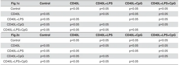
each gr oup. Com par ed w it h non- t r eat m ent cont r ol
g r ou p, CD4 0 L sig n if ican t ly in d u ced CD1 dh ig hCD5+
B ce l l e x p a n si o n a n d CD 4 0 L+ LPS h a d si m i l a r
si g n i f i ca n t i n d u ct i o n ; h o w e v e r, a d d i t i o n a l Cp G
VLJQL¿FDQWO\VXSSUHVVHG&'/LQGXFHG&'GhighCD5+
B cell ex pansion w it h or w it hout LPS ( Figur e 1c) . CD1 dh ighCD5+ % FHOO SRSXODWLRQ KDG QR VLJQL¿FDQW
FKDQJH XQGHU ¿[HGP. gin giv alis t r eat m en t on ly, com paring wit h non- t reat m ent cont rol group; and t he
induct ion by CD40L and CD40L+ LPS or suppression by addit ional CpG w ere not affect ed by addit ional P.
gingivalisWUHDWPHQW)LJXUHG7DNHQWRJHWKHUZLWK
or w it hout P. gingivalis t r eat m ent on non- im m unized m ice splen ocy t e B cells, CD4 0 L an d CD4 0 L+ LPS
VLJQL¿FDQWO\ LQGXFHG &'GhighCD5+ B cell expansion
DQG&S*UHGXFHGWKLVH[SDQVLRQVLJQL¿FDQWO\7DEOH
1) .
I m m unized m ice CD1d
highCD5
+B cell expansion
w it h CD40L, LPS, and CpG t r eat m ent w it h/
w it hout
P. gingivalis
co- st im ulat ion
B cells separat ed from im m unized m ice splenocyt es
w er e cult ur ed for 48 h under t he sam e condit ions
SUHYLRXVO\PHQWLRQHGDQGPHDVXUHGE\ÀRZF\WRPHWU\
( Figures 2a, 2b) . Com pared wit h non- t reat m ent cont rol
JURXS&'/VLJQL¿FDQWO\LQGXFHG&'GhighCD5+ B cell
expansion and addit ional LPS slight ly r educed t his
induct ion; also, addit ional CpG lar gely suppr essed
CD40L- induced CD1dhighCD5+ B cell expansion w it h
or w it hout LPS ( Figur e 2c) . For im m unized m ice,
CD1dhighCD5+%FHOOSRSXODWLRQDOVRKDGQRVLJQL¿FDQW
FKDQJH ZLWK ¿[HGP. gin giv alis t r eat m en t alon e, com paring wit h non- t reat m ent cont rol group; and t he
induct ion by CD40L or suppression by addit ional CpG
were not affect ed by addit ional P. gingivalis t reat m ent .
How ever, addit ional LPS inhibit ion effect on CD40L induct ion was vanished w it h P. gingivalis t r eat m ent
( Figure 2d) com paring wit h no P. gingivalis t reat m ent
( Figur e 2 c) in im m unized m ice B cells ( Table 2 ) .
These r esult s indicat ed t hat B cells fr om P. gingivalis
im m unized m ice had par t ial sim ilar r esponses for
CD40L, CD40L+ LPS, and CD40L+ CpG t reat m ent wit h
or wit hout P. gingivalis co- st im ulat ion, com pared wit h
B cells fr om non- im m unized m ice. Ot her t han t hat ,
/36VLJQL¿FDQWO\UHGXFHGWKHH[SDQVLRQRILPPXQL]HG
CD1dhighCD5+ B cell w it hout P. gingivalis ( Figure 2c) ,
but had no effect on non- im m unized CD1dhighCD5+ B
cell ( Figure 1c) ; also, com pared wit h P. gingivalis only t r eat m ent gr oup, CD40L+P. gingivalis t r eat m ent had
QR VLJQL¿FDQW LQGXFWLRQ LQ LPPXQL]HG &'GhighCD5+
%FHOO)LJXUHGEXWLQGXFHGVLJQL¿FDQWH[SDQVLRQ
in non- im m unized CD1dhighCD5+ B cell ( Figure 1d) .
I L 1 0 m RNA l ev el s i n B cel l s f r o m n o n
-im m unized and -im m unized m ice w it h CD40L,
LPS, an d Cp G t r eat m en t w it h / w it h ou t
P.
gingivalis
co- st im ulat ion
I L- 1 0 m RN A l e v e l s w e r e m e a s u r e d a n d
analyzed by RT- qPCR in cult ured B cells separat ed
f r om n on - im m u n ized m ice ( Fig u r es 3 a an d 3 b ) an d i m m u n i zed m i ce ( Fi g u r es 3 c an d 3 d ) w i t h
m ult iple t r eat m ent s including CD40L, CD40L+ LPS,
CD4 0 L+ Cp G, an d CD4 0 L+ LPS+ Cp G ( Fig u r es 3 a an d 3 c) ; P. g i n g i v al i s, P. g i n g i v al i s+ CD 4 0 L, P.
gingiv alis+ CD40L+ LPS, P. gingiv alis+ CD40L+ CpG,
and P. gingivalis+ CD40L+ LPS+ CpG ( Figures 3b and
3d) . Com par ing w it h non- t r eat m ent cont r ol gr oup,
&'/VLJQL¿FDQWO\LQFUHDVHG,/P51$H[SUHVVLRQ
and addit ional LPS enhanced t his incr ease in B cells
fr om non- im m unized m ice ( Figur e 3a) , but not fr om
im m unized m ice ( Figur e 3 c) . How ev er, addit ional Cp G l a r g e l y i n cr e a se d I L- 1 0 m RNA e x p r e ssi o n
com par ed w it h CD40L t r eat m ent w it h or w it hout LPS
in B cells from bot h t ypes of m ice ( Figures 3a and
Figure 1-%FHOOH[SDQVLRQLQQRQLPPXQL]HGPRXVHVSOHQRF\WH%FHOOVDIWHU&'//36DQG&S*WUHDWPHQWZLWKZLWKRXWP. gingivalis
FRVWLPXODWLRQ6SOHQRF\WH%FHOOVZHUHVHSDUDWHGIURPQRQLPPXQL]HG&%/-PLFHDQGFXOWXUHGKRXUVZLWK&'/Pg/mL), CD40L (1 Pg/mL)+P. gingivalis LPS (10 Pg/mL), CD40L(1 Pg/mL)+CpG (10 PM), and CD40L (1 Pg/mL)+P. gingivalis LPS (10 Pg/mL)+CpG (10 P0LQWKHDEVHQFHRULQWKHSUHVHQFHRI¿[HGP. gingivalis (5×10⁶per 1×10⁶ cells). CD1highCD5+%FHOOVZHUHGHWHFWHGXVLQJÀRZ
F\WRPHWU\LQFRQWURODQGWUHDWPHQWJURXSVZLWKRXWP. gingivalisDDQGZLWKP. gingivalisE;D[LV&'3(VWDLQLQJ<D[LV&'G$3& staining). The percentage of CD1highCD5+%FHOOVZDVTXDQWL¿HGDQGDQDO\]HGE\)ORZ-RVRIWZDUHLQFRQWURODQGWUHDWPHQWJURXSVZLWKRXW
I L- 1 0 m RNA ex p r ession , an d t h is en h an cem en t
ZDVVLJQL¿FDQWO\VXSSUHVVHGZLWKDGGLWLRQDO&'/
CD40L+ LPS, CD40L+ CpG, and CD40L+ LPS+ CpG in B cells from bot h t ypes of m ice ( Figures 3b and 3d) .
7DNHQWRJHWKHU&G//36DQG&'/&S*LQGXFHG VLJQL¿FDQWLQFUHDVHRI,/P51$H[SUHVVLRQZLWKRXW
P. gingiv alis t r eat m ent ; how ever, t hese addit ional com binat ions suppressed t he induct ion of I L- 10 m RNA
expression caused by P. gingivalis.
Secr et ed I L- 10 levels in B cells fr om
non-im m unized and non-im m unized m ice w it h CD40L,
LPS, an d Cp G t r eat m en t w it h / w it h ou t
P.
gingivalis
co- st im ulat ion
Se c r e t e d I L- 1 0 l e v e l s w e r e m e a s u r e d a n d
analyzed by ELI SA fr om super nat ant of cult ur ed B
cells separat ed fr om non- im m unized m ice ( Figur e 4a
and 4b) and im m unized m ice ( Figur es 4c and 4d) .
Com par ing w it h non- t r eat m ent cont r ol gr oup, CD40L
VLJQL¿FDQWO\LQFUHDVHG,/VHFUHWLRQLQ%FHOOVIURP
non- im m unized m ice only and addit ional LPS enhanced t his increase in B cells from bot h t ype of m ice ( Figures
4a and 4c) . Also, addit ional CpG largely increased
I L- 1 0 secr et ion com par ed w it h CD4 0 L t r eat m en t
w it h or w it hout LPS in B cells fr om bot h t y pes of m ice ( Figur es 4a and 4c) . P. gingivalis st im ulat ion
DORQH VLJQL¿FDQWO\ LQFUHDVHG ,/ VHFUHWLRQ DQG
t h is in d u ct ion w as sig n if ican t ly su p p r essed w it h addit ional CD40L+ CpG, but addit ional CD40L and
Cd40L+ LPS had no im pact s on t his induct ion ( Figures
4b, 4d) . These result s suggest ed t hat Cd40L+ LPS and
&'/&S* VLJQL¿FDQWO\ LQFUHDVHG ,/ VHFUHWLRQ
wit hout P. gingivalis t reat m ent ; however, P. gingivalis
t r eat m en t sig n if ican t ly in d u ced I L- 1 0 secr et ion ,
addit ional Cd40L+ LPS had no effect , and CD40L+ CpG
VLJQL¿FDQWO\LQKLELWHGWKLVLQGXFWLRQ
Fig.1c Control CD40L CD40L+LPS CD40L+CpG CD40L+LPS+CpG
Control S S S S
CD40L S S! S S
CD40L+LPS S S! S S
CD40L+CpG S S S S!
CD40L+LPS+CpG S S S S!
Fig.2c Control CD40L CD40L+LPS CD40L+CpG CD40L+LPS+CpG
Control S S S S
CD40L S S S S
CD40L+LPS S S S S
CD40L+CpG S S S S!
CD40L+LPS+CpG S S S S!
Table 1- Analysis of CD1dhighCD5+%FHOOVSHUFHQWDJHVWDWLVWLFVLQJURXSVZLWKRXWP. gingivalis treatment
Fig.1d Control P.g P.g+CD40L P.g+CD40L+LPS P.g+CD40L+CpG P.g+CD40L+LPS+CpG
Control S S S S S
P.g S S S S S
P.g+CD40L S S S! S S
P.g+CD40L+LPS S S S! S S
P.g+CD40L+CpG S S S S S!
P.g+CD40L+LPS+CpG S S S S S!
Fig.2d Control P.g P.g+CD40L P.g+CD40L+LPS P.g+CD40L+CpG P.g+CD40L+LPS+CpG
Control S S S S S
P.g S S! S! S S
P.g+CD40L S S! S! S S
P.g+CD40L+LPS S S! S! S S
P.g+CD40L+CpG S S S S S!
P.g+CD40L+LPS+CpG S S S S S!
Figure 2-%FHOOH[SDQVLRQLQLPPXQL]HGPRXVHVSOHQRF\WH%FHOOVDIWHU&'//36DQG&S*WUHDWPHQWZLWKZLWKRXWP. gingivalis
FRVWLPXODWLRQ&%/-PLFHZHUHLPPXQL]HGE\LQWUDSHULWRQHDOLQMHFWLRQRI¿[HGP. gingivalis on day 0 (1×10⁶) and day 7 (1×10⁵).
6SOHQRF\WH%FHOOVZHUHVHSDUDWHGIURPLPPXQL]HGPLFHRQGD\DQGFXOWXUHGKRXUVZLWK&'/Pg/mL), CD40L (1 Pg/mL)+P. gingivalis LPS (10 Pg/mL), CD40L(1 Pg/mL)+CpG (10 PM), and CD40L (1 Pg/mL)+P. gingivalis LPS (10 Pg/mL)+CpG (10 PM) in the
DEVHQFHRULQWKHSUHVHQFHRI¿[HGP. gingivalis (5×10⁶per 1×10⁶ cells). CD1highCD5+%FHOOVZHUHGHWHFWHGXVLQJÀRZF\WRPHWU\LQ
FRQWURODQGWUHDWPHQWJURXSVZLWKRXWP. gingivalisDDQGZLWKP. gingivalisE;D[LV&'3(VWDLQLQJ<D[LV&'G$3&VWDLQLQJ7KH percentage of CD1highCD5+%FHOOVZDVTXDQWL¿HGDQGDQDO\]HGE\)ORZ-RVRIWZDUHLQFRQWURODQGWUHDWPHQWJURXSVZLWKRXWP. gingivalis
Figure 3- ,/ P51$ H[SUHVVLRQ LQ % FHOOV IURP QRQLPPXQL]HG DQG LPPXQL]HG PLFH ZLWK &'/ /36 DQG &S* WUHDWPHQW ZLWK
ZLWKRXWP. gingivalisFRVWLPXODWLRQ6SOHQRF\WH%FHOOVZHUHVHSDUDWHGDQGFXOWXUHGKRXUVZLWK&'/Pg/mL), CD40L (1 Pg/mL)+P. gingivalis LPS (10 Pg/mL), CD40L (1 Pg/mL)+CpG (10 PM), and CD40L (1 Pg/mL)+P. gingivalis LPS (10 Pg/mL)+CpG (10 PM) in the
DEVHQFHRULQWKHSUHVHQFHRI¿[HGP. gingivalis (5×10⁶per 1×10⁶FHOOV,/H[SUHVVLRQVZHUHGHWHUPLQHGE\57T3&5LQFRQWURODQG
WUHDWPHQWJURXSVZLWKRXWP. gingivalisDDQGZLWKP. gingivalis (b) from non-immunized C57/BL6J mice, and same groups from immunized
Figure 4-6HFUHWHG,/OHYHOVLQ%FHOOVIURPQRQLPPXQL]HGDQGLPPXQL]HGPLFHZLWK&'//36DQG&S*WUHDWPHQWZLWKZLWKRXWP. gingivalisFRVWLPXODWLRQ6SOHQRF\WH%FHOOVZHUHVHSDUDWHGDQGFXOWXUHGKRXUVZLWK&'/Pg/mL), CD40L (1 Pg/mL)+P. gingivalis LPS (10 Pg/mL), CD40L (1 Pg/mL)+CpG (10 PM), and CD40L (1 Pg/mL)+P. gingivalis LPS (10 Pg/mL)+CpG (10 PM) in the absence or in
WKHSUHVHQFHRI¿[HGP. gingivalis (5×10⁶per 1×10⁶FHOOV0HGLXPVXSHUQDWDQWVZHUHFROOHFWHGDQGVHFUHWHG,/SURWHLQOHYHOVZHUH
PHDVXUHGE\(/,6$LQFRQWURODQGWUHDWPHQWJURXSVZLWKRXWP. gingivalisDDQGZLWKP. gingivalis (b) from non-immunized C57/BL6J mice,
Discussion
I L- 10 pr oducing B10 cells play an essent ial r ole
in im m une syst em balance by suppressing excessive
LQÀDPPDWRU\UHVSRQVHV18,20,30+RZHYHUOLWWOHLVNQRZQ
about t he effect s of co- st im ulat ion by m ult iple TLR
agonist s and CD40 act ivat or on B10 cells under different
im m unological condit ions. I n t he present st udy, w e invest igat ed t he changes of B10 cell populat ion and
I L- 10 secret ion by com bined t reat m ent of P. gingivalis
LPS ( TLR4 agonist ) , CpG ( TLR9 agonist ) , and CD40L
in t he cont ex t of innat e im m unit y ( cells fr om non-im m unized m ice) and adapt ive non-im m unit y ( cells from
im m unized m ice) . The result s showed t hat P. gingivalis
LPS enhanced I L- 10 secret ion by B10 cells in m ice in vit ro during innat e and adapt ive im m une responses
wit h increased CD1dhighCD5+ B cells; However, CpG was
m ore effect ive t han P. gingivalis LPS t o enhance I L- 10
com pet ency during t hese responses w it h decreased CD1dhighCD5+ B cells.
P. g i n g i v a l i s L PS i s a p u r i f i e d p r o d u c t o f
l i p o p o l y sacch ar i d e f r o m Gr am - n eg at i v e b act er i a
Por ph y r om on as gin giv alis, w h ich is con sider ed as t he m ain pat hogen of periodont al disease3,12,26. LPS
is t he m aj or com ponent of Gram negat ive bact er ia
t hat act ivat es t he innat e im m une syst em4,29. How ever,
P. gin giv alis LPS h as a u n iqu e an d h et er ogen ou s chem ical st ruct ure, which is different from t radit ionally
recognized ent eric bact erium - derived LPS such as E.
coli LPS7,22,23. P. gingivalis LPS and E. coli LPS have been
VKRZQWRWULJJHUGLIIHUHQWLQWUDFHOOXODULQÀDPPDWRU\
signaling pat hways7 DQG F\WRNLQH SURGXFWLRQV1,22. I t
was suggest ed t hat t he st r uct ural het er ogeneit y of
P. gingivalis lipid A cont ribut es t o t he unusual innat e
host response t o t his LPS and it s abilit y t o int eract wit h bot h TLR2 and TLR42,6. This m ay explain t he difference
of t he induct ion effect s on B10 populat ion and I L- 10
secret ion bet ween co- st im ulat ing P. gingivalis LPS plus
CD40L and E. coli LPS plus CD40L in non- im m unized m ice19. I n non- im m unized m ice w it hout P. gingivalis
t reat m ent , addit ional P. gingivalis LPS did not furt her increase CD1dhighCD5+ B cells percent ages ( Figure 1c) ,
EXW VLJQL¿FDQWO\ LQFUHDVHG ,/ P51$ H[SUHVVLRQ
( Figure 3a) and I L- 10 secret ion ( Figure 4a) com pared
w it h CD4 0 L on ly g r ou p. Mor eov er, in im m u n ized
m ice w it hout P. gingiv alis t r eat m ent, P. gingiv alis
LPS suppressed t he expansion of CD1dhighCD5+ cells
induced by CD40L ( Figure 2c) w it h an increase of I
L-10 secret ion ( Figure 4c) . These result s suggest t hat P.
gingivalis LPS st im ulat ion has different effect s on innat e
and adapt ive im m une responses, and it m ay enhance
t he I L- 10 secret ion from fewer CD1dhighCD5+ B cells wit h
higher com pet ence or from increased B10 cells ot her
t han CD1dhighCD5+ cell subset . These differences and
possible m echanism s need t o be invest igat ed in fut ure st udies. Porphyrom onas gingivalis synt hesizes t wo LPS,
O- LPS and A- LPS. The st ruct ures of t he O- PS and A- PS
repeat ing unit s, t he core oligosaccharide ( OS) , and t he
OLQNDJHRIWKHUHSHDWLQJXQLWWRWKHFRUHLQ2/36DQG
A- LPS have been ext ensively st udied24. Analysis of t he
det ailed st ruct ure of P. gingivalis LPS is essent ial for
furt her m echanist ic invest igat ion of t he ant igenicit y of
t his im port ant periodont al pat hogen.
P. gin giv alis in du ces per iodon t it is t h r ou gh t h e
disrupt ion of t he host t issue hom eost asis and adapt ive
im m une response, which allows uncont rolled growt h of
t he com m ensal m icrobial com m unit y in oral cavit y5,10.
I n our st udy, P. gingiv alis alone show ed no effect
on expansion or reduct ion of CD1dhighCD5+ cells in B
cells from bot h im m unized and non- im m unized m ice.
However, P. gingivalisWUHDWPHQWVLJQL¿FDQWO\LQFUHDVHG
I L- 10 secret ion in bot h im m unized and non- im m unized
m ice B cells, suggest ing t his induct ion was caused by
cells ot her t han CD1dhighCD5+ B cells or by increasing
t he com pet ence of B10 cells in CD1dhighCD5+ subset .
Fu r t h er m or e, P. gin giv alis t r eat m en t sig n if ican t ly
dim inished t he CpG- induced I L- 10 product ion ( Figures
4b and 4d) com pared wit h groups wit hout P. gingivalis
t r eat m ent ( Figur es 4a and 4c) in bot h innat e and adapt ive im m une responses, suggest ing t hat t he I L- 10
secret ion induced by TLR9 signaling m ay be inhibit ed
by com ponent s of P. gingivalis. The m echanism of how
P. gingivalis induces I L- 10 secret ion and inhibit s TLR9 signaling induced I L- 10 secret ion in splenocyt es B cell
needs t o be furt her invest igat ed.
Conclusions
Wit h CD4 0 L, P. gin giv alis LPS en h an ced I L- 1 0
com pet ency of B10 cells and B10 cell expansion in t he absence, but not in t he presence of P. gingivalis;
how ever, CpG induced t he st ronger I L- 10 com pet ency
of B10 cells but inhibit ed B10 expansion under t he
sam e condit ions.
$FNQRZOHGJHPHQWV
7KLV VWXG\ ZDV VXSSRUWHG E\ 1,+ 1,'&5 JUDQW
References
$QGUXNKRY2(UWOVFKZHLJHU60RULW]$%DQWOHRQ+35DXVFK)DQ;
Different effect s of P. gingivalis LPS and E. coli LPS on t he expression
RI LQWHUOHXNLQ LQ KXPDQ JLQJLYDO ¿EUREODVWV $FWD 2GRQWRO 6FDQG
2014; 72( 5) : 337- 45.
2- Bainbridge BW, Coat s SR, Darveau RP. Porphyrom onas gingivalis
lipopolysaccharide displays funct ionally diverse int eract ions w it h t he innat e host defense syst em . Ann Periodont ol. 2002; 7( 1) : 29- 37. 3- Ballini A, Cant ore S, Farronat o D, Cirulli N, I nchingolo F, Papa F, et
DO3HULRGRQWDOGLVHDVHDQGERQHSDWKRJHQHVLVWKHFURVVWDONEHWZHHQ F\WRNLQHVDQGPorphyrom onas gingivalis. J Biol Regul Hom eost Agent s. 2015; 29( 2) : 273- 81.
%HXWOHU%3ROWRUDN$7KHVROHJDWHZD\WRHQGRWR[LQUHVSRQVHKRZ /36ZDVLGHQWL¿HGDV7OUDQGLWVUROHLQLQQDWHLPPXQLW\'UXJ0HWDE
Dispos. 2001; 29( 4 Pt 2) : 474- 8.
5- Blasco- Baque V, Garidou L, Pom ié C, Escoula Q, Loubieres P, Le Gall- David S, et al. Periodont it is induced by Porphyrom onas gingivalis
dr iv es per iodon t al m icr obiot a dy sbiosis an d in su lin r esist an ce v ia an im pair ed adapt iv e im m une r esponse. Gut . 2016. doi 10. 1136/ gut j nl- 2015- 309897. Epub ahead of print .
6- Darveau RP, Pham TT, Lem ley K, Reife RA, Bainbridge BW, Coat s SR, et al. Porphyrom onas gingivalis lipopolysaccharide cont ains m ult iple
OLSLG$VSHFLHVWKDWIXQFWLRQDOO\LQWHUDFWZLWKERWKWROOOLNHUHFHSWRUV
and 4. I nfect I m m un. 2004; 72( 9) : 5041- 51.
7- Diya Z, Lili C, Shenglai L, Zhiyuan G, Jie Y. Lipopolysaccharide ( LPS) of Por phy r om onas gingiv alis induces I L- 1bet a, TNF- alpha and I L- 6 product ion by THP- 1 cells in a way different from t hat of Escherichia coli LPS. I nnat e I m m un. 2008; 14( 2) : 99- 107.
)UD]mR-%(UUDQWH35&RQGLQR1HWR$7ROOOLNHUHFHSWRUVSDWKZD\
dist urbances are associat ed w it h increased suscept ibilit y t o infect ions in hum ans. Arch I m m unol Ther Exp ( Warsz) . 2013; 61( 6) : 427- 43.
*DR0+D7=KDQJ;:DQJ;/LX/.DOEÀHLVFK-HWDO7KHWROO OLNHUHFHSWRUOLJDQG&S*ROLJRGHR[\QXFOHRWLGHDWWHQXDWHVFDUGLDF
d y sf u n ct ion in p oly m icr ob ial sep sis, in v olv in g act iv at ion of b ot h
SKRVSKRLQRVLWLGHNLQDVH$NWDQGH[WUDFHOOXODUVLJQDOUHODWHGNLQDVH
signaling. J I nfect Dis. 2013; 207( 9) : 1471- 9.
*|O]/0HPPHUW65DWK'HVFKQHU%-lJHU$$SSHO7%DXPJDUWHQ
G, et al. LPS from P. gingivalis and hypoxia increases oxidat ive st ress
LQ SHULRGRQWDO OLJDPHQW ¿EUREODVWV DQG FRQWULEXWHV WR SHULRGRQWLWLV 0HGLDWRUV,QÀDPP
*RUEDFKHYD 9 $\DVRX¿ . )DQ 5 %DOGZLQ :0 UG 9DOXMVNLNK
A. B cell act ivat ing fact or ( BAFF) and a proliferat ion inducing ligand ( APRI L) m ediat e CD40- independent help by m em ory CD4 T cells. Am J Transplant . 2015; 15( 2) : 346- 57.
12- Gully N, Bright R, Marino V, Marchant C, Cant ley M, Haynes D, et al. Por phy r om onas gingiv alis SHSWLG\ODUJLQLQH GHLPLQDVH D NH\
cont ribut or in t he pat hogenesis of experim ent al periodont al disease and experim ent al art hrit is. PloS One. 2014; 9( 6) : e100838.
+DVVDQ*60HUKL<0RXUDG:&'OLJDQGDQHRLQÀDPPDWRU\
m olecule in vascular diseases. I m m unobiology. 2012; 217( 5) : 521- 32. 1 4 - Jen ab ian MA, Pat el M, Kem a I , Vy b oh K, Kan ag ar at h am C, Radzioch D, et al. Soluble CD40- ligand ( sCD40L, sCD154) plays an im m unosuppressive role via regulat ory T cell expansion in HI V infect ion. Clin Exp I m m unol. 2014; 178( 1) : 102- 11.
15- Kawabe T, Mat sushim a M, Hashim ot o N, I m aizum i K, Hasegawa Y. CD40/ CD40 ligand int eract ions in im m une responses and pulm onary im m unit y. Nagoya J Med Sci. 2011; 73( 3- 4) : 69- 78.
.DZDVDNL7.DZDL77ROOOLNHUHFHSWRUVLJQDOLQJSDWKZD\V)URQW
I m m unol. 2014; 5: 461.
/HVWHU61/L.7ROOOLNHUHFHSWRUVLQDQWLYLUDOLQQDWHLPPXQLW\-I m m unol. 2014; 426( 6) : 1246- 64.
18- Li J, Shen C, Liu Y, Li Y, Sun L, Jiao L, et al. I m paired funct ion of CD5+ CD19+ CD1dhi B10 cells on I gE secret ion in an at opic derm at it
is-OLNHPRXVHPRGHO3OR62QHH
1 9 - Lin M, Lin J, Wan g Y, Bon h eu r N, Kaw ai T, Wan g Z , et al. Lipopoly sacchar ide at t enuat es CD40 ligand- induced r egulat or y B10 cell expansion and I L- 10 pr oduct ion in m ouse splenocyt es. Open J I m m unol. 2015; 5( 1) : 1- 8.
/\NNHQ -0 &DQGDQGR .0 7HGGHU 7) 5HJXODWRU\ % FHOO
developm ent and funct ion. I nt I m m unol. 2015; 27( 10) : 471- 7. 21- Miao J, Zheng L, Zhang J, Ma Z, Zhu W, Zou S. The effect of t aurine
RQ WKH WROOOLNH UHFHSWRUVQXFOHDU IDFWRU NDSSD % 7/5V1)NDSSD%
signaling pat hway in St rept ococcus uberis- induced m ast it is in rat s. I nt I m m unopharm acol. 2011; 11( 11) : 1740- 6.
22- Nebel D, Arvidsson J, Lillqvist J, Holm A, Nilsson BO. Different ial effect s of LPS from Escherichia coli and Porphyrom onas gingivalis on I L- 6 pr oduct ion in hum an per iodont al ligam ent cells. Act a Odont ol Scand. 2013; 71( 3- 4) : 892- 8.
23- Ogawa T, Suda Y, Kashihara W, Hayashi T, Shim oyam a T, Kusum ot o
6HWDO,PPXQRELRORJLFDODFWLYLWLHVRIFKHPLFDOO\GH¿QHGOLSLG$IURP
Helicobact er pylori LPS in com parison w it h Porphyrom onas gingivalis
lipid A and Escherichia coli- t ype synt het ic lipid A ( com pound 506) . Vaccine. 1997; 15( 15) : 1598- 605.
3DUDPRQRY1$GXVH2SRNX-+DVKLP$5DQJDUDMDQ0&XUWLV0$ ,GHQWL¿FDWLRQRIWKHOLQNDJHEHWZHHQ$SRO\VDFFKDULGHDQGWKHFRUHLQ
t he A- lipopolysaccharide of Porphyrom onas gingivalis W50. J Bact eriol. 2015; 197( 10) : 1735- 46.
25- Pellegr ini A, Guiñazu N, Gior danengo L, Cano RC, Gea S. The
UROHRIWROOOLNHUHFHSWRUVDQGDGDSWLYHLPPXQLW\LQWKHGHYHORSPHQW
of p r ot ect iv e or p at h olog ical im m u n e r esp on se t r ig g er ed b y t h e
Trypanosom a cruzi prot ozoan. Fut ure Microbiol. 2011; 6( 12) : 1521- 33. 2 6 - Si n g h r a o SK, H a r d i n g A, Po o l e S, Ke sa v a l u L, Cr e a n S.
Porphyrom onas gingivalisSHULRGRQWDOLQIHFWLRQDQGLWVSXWDWLYHOLQNV
ZLWK$O]KHLPHUVGLVHDVH0HGLDWRUV,QÀDPP 7HGGHU7)%FHOOVDIXQFWLRQDOO\GH¿QHGUHJXODWRU\%FHOOVXEVHW
J I m m unol. 2015; 194( 4) : 1395- 401.
28- Xu H, Liew LN, Kuo I C, Huang CH, Goh DL, Chua KY. The m odulat ory effect s of lipopolysaccharide- st im ulat ed B cells on different ial T- cell polarizat ion. I m m unology. 2008; 125( 2) : 218- 28.
29- Yadav R, Zam m it DJ, Lefrancois L, Vella AT. Effect s of LPS- m ediat ed
E\VWDQGHU DFWLYDWLRQ LQ WKH LQQDWH LPPXQH V\VWHP - /HXNRF %LRO
2006; 80( 6) : 1251- 61.
![Figure 1-%FHOOH[SDQVLRQLQQRQLPPXQL]HGPRXVHVSOHQRF\WH%FHOOVDIWHU&'//36DQG&S*WUHDWPHQWZLWKZLWKRXWP](https://thumb-eu.123doks.com/thumbv2/123dok_br/14984698.511596/5.892.177.717.96.1050/figure-fhooh-sdqvlrqlqqrqlppxql-hgprxvhvsohqrf-wh-fhoovdiwhu-dqg-wuhdwphqwzlwkzlwkrxwp.webp)
![Figure 2-%FHOOH[SDQVLRQLQLPPXQL]HGPRXVHVSOHQRF\WH%FHOOVDIWHU&'//36DQG&S*WUHDWPHQWZLWKZLWKRXWP](https://thumb-eu.123doks.com/thumbv2/123dok_br/14984698.511596/7.892.181.694.74.1046/figure-fhooh-sdqvlrqlqlppxql-hgprxvhvsohqrf-wh-fhoovdiwhu-dqg-wuhdwphqwzlwkzlwkrxwp.webp)
![Figure 4-6HFUHWHG,/OHYHOVLQ%FHOOVIURPQRQLPPXQL]HGDQGLPPXQL]HGPLFHZLWK&'//36DQG&S*WUHDWPHQWZLWKZLWKRXWP](https://thumb-eu.123doks.com/thumbv2/123dok_br/14984698.511596/9.892.245.650.102.1067/figure-hfuhwhg-ohyhovlq-fhooviurpqrqlppxql-hgdqglppxql-hgplfhzlwk-dqg-wuhdwphqwzlwkzlwkrxwp.webp)
