Abstract
Submitted: September 11, 2015
0RGL¿FDWLRQ0D\
Accepted: June 21, 2016
Is the bonding of self-adhesive
cement sensitive to root region and
curing mode?
in different regions of root dentin. Material and Methods: Twenty single-rooted
premolars were endodontically treated, and the post spaces were prepared.
The roots were randomly divided into two groups (n=10), according to the
activation mode of the resin cement RelyX™ U200 (3M ESPE Saint Paul, MN, USA): conventional (continuous activation mode) and soft-start activation
mode (Ramp). The posts (WhitePost DC/FGM) were cemented according to the
micro-Raman spectroscopy and the BS was evaluated by the push-out test. The D=0.05). Results: Neither the activation mode nor the root regions affected the DC of the resin
cement. Higher BS was achieved in the soft-start group (p=0.036); lower BS
was observed in the apical third compared to the other root regions (p<0.001).
Irrespective of the activation mode and root region, the mixed failure mode
be improved by soft-started polymerization. The DC was not affected by the
curing mode.
Ke yw or ds: Resin cements. Raman spectroscopy. Dental bonding. Thaynara Faelly BOING1
Giovana Mongruel GOMES1
João Carlos GOMES1
Alessandra REIS1
Osnara Maria Mongruel GOMES1
http://dx.doi.org/10.1590/1678-77572015-0430
1Universidade Estadual de Ponta Grossa, Departamento de Odontologia, Ponta Grossa,
PR, Brazil.
Introduction
Endodontically treated teeth usually demonstrate
an extensive loss of dental structure and require the
29.In this context,the cementation
restorations that resemble the natural dental structure because the modulus of elasticity of the adhesive
14.
Unfortunately, several factors can affect the
histological and anatomical characteristics of the
root canal, density and orientation of the dentinal
tubules in the different root canal regions11, as well as accessibility to the different root canal regions12.
Different areas of the same root canal also do not
respond to acid etching, and thus the ability of
adhesion to root dentin may be different at different depths in the same root canal11. Higher bond strength
values at the cervical third are generally expected
due to the ease of conditioning, and polymerization
of the cements in this region9. However, this is still controversial3.
Failures are
still the major clinical failure18.The polymerization
of these materials may exceed their bond strength, resulting in gaps forming at the
dentin-resin cement interface, loss of retention and the
displacement of the posts4. This may be caused by the
generated in the root canal by the composition of resin
cements and different light curing techniques10,23.
During the pre-gel phase,
is low due to the high flowability of the resin material. However, when the gel point is reached, the
material’s ; then the stress generated
is transferred to the remaining tooth structure,
causing adhesive failures and several other adverse consequences such as tooth fracture and
in the material itself6,23.
This situation is even worse due to the high and
unfavorable factor (C-factor) of
the root space28. Some authors have attributed the
gap formation and low bond strength to the high
C-factor of the root space26. One way to control this stress is by reducing the light
intensity of the curing unit during the polymerization
of the material. The use of soft-start activation
provides low light intensity during the initial seconds
of activation, increasing the period during which
the resinous material remains in the pre-gel phase. Delaying photoactivation decreased the studied
post-23 8.
Although this technique has shown promising
results when employed in composite resin specimens17,
Thus, this in vit ro study, aimed to compare the degree
of conversion and the in
different regions of root dentin, using both conventional
and soft-start polymerization techniques. The null
hypothesis tested was that the degree of conversion and bond strength of the resin cement is not affected
by curing mode or by root region.
Material and methods
The research project was approved by the Ethics
Committee of the Dental School of the State University
of Ponta Grossa, under protocol number 109.876. Twenty extracted human mandibular premolars were
stored in distilled water at 4°C and used within 6
months from the extraction time. The inclusion criteria
was that teeth were absent of restoration, caries or
posts or crowns and absent of severe root curvatures.
Further, a root length of 14±1 mm measured from the
cement-enamel junction was required.
Specimen preparation
Teeth were transversally sectioned at the
cement-enamel junction using a low-speed diamond saw
(Maillefer, Dentsply Ind. e Com. Ltda., Petrópolis, RJ,
Brazil) into each canal until it was visible at the apical
foramen. One millimeter was subtracted from this
technique was used for instrumentation with Gates
Glidden drills #2 to #4 (Maillefer, Dentsply Ind. e Com.
Ltda., Petrópolis, RJ, Brazil). Apical enlargement was
performed to size 40, 0.2 taper (Maillefer, Dentsply Ind. e Com. Ltda., Petrópolis, RJ, Brazil). Irrigation
was performed after every change of instrument by
of 17% EDTA (Biodinâmica Química e Farmacêuitca
Ltda., Ibiporã, PR, Brazil) for 5 minutes. Roots were
dried with paper points (Maillefer, Dentsply Ind. e Com.
based sealer (AH Plus, Dentsply DeTrey, Konstanz,
Germany) and gutta-percha points using the warm
vertical condensation technique. The root access was
cement (Vitro Fil LC, Nova DFL, Taquara, RJ, Brazil).
The roots were stored at 37°C and 100% humidity
using the Gates Glidden burs (Maillefer, Dentsply Ind.
e Com. Ltda., Petrópolis, RJ, Brazil), leaving 4 mm of the apical seal and the post space was prepared with
a low-speed bur provided by the post manufacturer
depth of 10 mm from the cement-enamel junction. The root canals were irrigated with 10 mL of distilled
water and dried with paper points (Maillefer, Dentsply
Ind. e Com. Ltda., Petrópolis, RJ, Brazil).
Ex pe r im e n t a l gr ou ps
At this point, the teeth were randomly divided into
2 groups (n=10) according to the activation mode
of the resin cement in the root canal. In half of the
teeth, a conventional activation mode (continuous light intensity, energy density of 40 J/s) was employed,
while in the other half a soft-start polymerization
(with initial low light intensity and an energy density
of approximately 38.8 J/s) was employed. The light intensity of the device was measured before the
beginning of the experiment using a Led Kondortech
radiometer (Kondortech Equip. Odontológicos. Ltda
horizontally sectioned with a water-cooled diamond
rotary cutting instrument (#2200 diamond bur, KG
Sorensen, Barueri, SP, Brazil) so that a total length
with 70% alcohol for 5 s. Ten millimeters of the post
lengths were cemented inside the root canal, while
the remaining cervical 3 mm served as a guide to standardize the distance of the light-curing device
from the cervical root region.
cemented with a dual, self-adhesive resin cement RelyX™ U200 (3M ESPE, Saint Paul, MN, USA), which
was mixed according to the manufacturer’s instructions
and introduced into the root canal space with a Centrix
syringe (DFL, Rio de Janeiro, RJ, Brazil) before seating
excess resin cement was removed.
Then, the resin cement was immediately
polymerized. In the conventional activation mode,
a light intensity of 1200 mW/cm2 remained constant
throughout the exposure time of 40 s. In the soft-start group, the light intensity increased linearly from 0 to
1200 mW/cm2
remained at 1200 mW/cm2 in the next 35 s. The light curing unit LED Raddi Plus (SDI Limited, Victoria,
Australia) was employed in this experiment.
After the post luting procedures, the roots with
cemented posts were covered with the conventional glass-ionomer cement, (Vitro Fil LC, Nova DFL,
Taquara, RJ, Brazil), and all samples subsequently
Sample preparation for Raman spectroscopy
and push-out tests
The roots were placed in separate polyvinylchloride
tubes and embedded in a Duralay acrylic resin
(Reliance Dental, Alsip, IL, USA). The portion of
sectioned perpendicular to the long axis into six
1-mm-create two cervical, two medium, and two apical slices of each tooth. Then, all specimens were observed
order to identify any artifacts caused by the sectioning
procedure. If any defects were observed, the slices were discarded.
Evaluation of the degree of conversion
One slice of each third of all teeth was submitted to
GmbH, Ettlingen, Germany) evaluation.After polishing
and cleaning, each slice was placed under the
microscope of the spectrometer. The micro-Raman
following micro-Raman parameters were used: 20
mW neon laser with a 532 nm wavelength, spatial resolution of 3 μm, spectral resolution of 5 cm-1,
accumulation time of 30 s with 6 co-additions, and
μ
used to represent the degree of conversion per slice.
The Raman spectra at 1,637 cm-1 indicates
unreacted C=C double bonds of the adhesive, while at 1,608 cm-1 it represents the C-C bonds in aromatic
rings in the Bis-GMA molecules, used as internal
cement were also recorded. For this purpose the uncured resin cement was placed on a glass slide and
area at 1,637 and 1,608 cm-1 for both the uncured and cured resin cement allowed for the calculation of
the degree of conversion of the material according to
the following equations:
-1
1,608 cm-1
-1
1,608 cm-1
(3) Degree of conversion (%) = (1 – Rcured/ Runcured) x 100
Evaluation of bond strength
All slices, including the one that was used in the
micro-Raman analysis, were submitted to a push-out
Mitutoyo digital caliper (Kyoto, Japan) with an accuracy
of 0.01 mm. The slices were also photographed on
both sides with an optical microscope (Olympus, model BX 51,
calculate the cervical and apical diameters of the root
canal (post + resin cement)14 for the calculation of the
with the UTHSCSA ImageTool 3.0 software (University
of Texas Health Science Center, San Antonio, TX, USA).
Each specimen (slice) was subjected to a
push-out bond strength test using a universal testing
at a crosshead speed of 0.5 mm/min. The load was
applied in the apical-cervical direction until post
pin on the center of the post surface without stressing
the surrounding post space walls. Different sizes of
punch pins were used to match the diameter of the post at the different root thirds. Three sizes of punch
pins were selected, one representative for each root
canal region: cervical (1.4 mm), medium (1.0 mm)
and apical (0.6 mm).
The maximum failure load was recorded in Newtons
and converted into MPa by dividing the applied load
by the bonded area (lateral area of the root canal).
The bonded area was the lateral surface of a truncated
+ r)[(h2 + (R – r)2]1/2
root canal radius (cervical post + resin cement radius),
r= apical root canal radius (apical post + resin cement
13.
Failure mode analysis
After the push-out evaluation, the failure modes of
all specimens were evaluated under a stereomicroscope
(40X magnification), only to identify the main substrates where the failures occurred (dentin, resin
calibrated operators analyzed each fractured specimen.
If any disagreement occurred between the evaluators, a consensus had to be obtained by discussion.
Then, according to the failure mode the samples
dentin and resin cement; 2. adhesive failure between resin cement and post; 3. cohesive failure within resin
cement; 4. cohesive failure within the post; 5. cohesive
failure within dentin; 6. mixed failure4,5,13,29.
Statistical analysis
Before running the parametric statistical analysis,
we tested whether or not the assumptions of normality
of the data and equality of variances were valid, using
The data obtained on the degree of conversion and
bond strength were subjected to two-way repeated
level of 5%. The repeated factor was the root third.
or soft-start) and root region (cervical, medium and
apical). The data of the failure modes were compared All calculations
were performed using the SPSS® statistical software
( for the Social Sciences, version
21.0 Mac, SPSS Inc., Chicago, IL, USA).
Results
None of the specimens observed presented artifacts caused by the sectioning procedure, therefore all slices
were tested.
Degree of conversion
cross-product interaction (p=0.949) degree
of conversion of the resin cement. The degree of conversion of all groups was statistically similar (Table
1).
Bond strength
The average values of bond strength in MPa (mean and standard deviation) can be seen in Table
(p=0.634), but the main factors activation mode
(p=0.036) and root third (p<0.001) were. Higher bond strength values were obtained for the soft-start
activation mode and in the apical third of the root canal.
Failure mode analysis
The absolute and relative distributions (%) of the
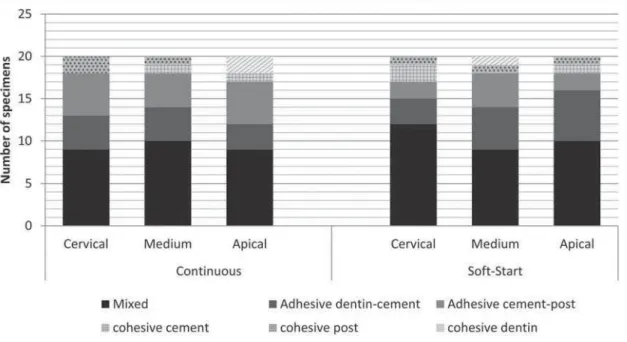
failure modes are shown in Figure 1. Irrespective
of the activation mode and root region, the mixed failure mode was the most prevalent. Representative
images from the optical microscopy of each mode are
illustrated in Figure 2.
Discussion
Despite recent studies that have evaluated the
6,23, the literature is still
limited regarding the use of different curing techniques
and their effects on the aforementioned properties. In the present investigation we opted to use the push
out bond strength test to evaluate the strength of
Activation mode Root region
Cervical Medium Apical
Continuous 72.4±6.9 77.0±7.8 73.6±7.0
Soft-Start 74.1±8.9 78.2±5.5 74.7±7.7
Table 1- Mean and standard deviation of the degree of conversion (%) of the resin cement using the different activation modes (continuous and soft-start) for the different root regions (cervical, medium and apical)
Activation mode Root region
Cervical Medium Apical
Continuous 16.2±4.1B 17.2±4.4B 25.8±7.4A
Soft-Start 20.2±4.2B 22.1±7.4B 26.9±6.3A
0HDQVLGHQWL¿HGZLWKWKHVDPHXSSHUFDVHOHWWHUVDUHQRWVWDWLVWLFDOO\GLIIHUHQW
Table 2-0HDQDQGVWDQGDUGGHYLDWLRQRIERQGVWUHQJWK03DRI¿EHUSRVWVWRURRWGHQWLQXVLQJWKHGLIIHUHQWDFWLYDWLRQPRGHVFRQWLQXRXV
and soft-start) for the different root regions (cervical, medium and apical)
under varying curing techniques, as this test closely
simulates the clinical condition15. According to previous studies, the push-out test provides a better estimation
of the bonding strength than the conventional shear
test because the fracture occurs parallel to the
dentin-15.
Higher bond strength values were observed in the
apical area of root canals irrespectively of the other
factors. Although this is not in agreement with many
7, it is in agreement with
other authors3,22
controversy is the type of resin cement employed
in the different experimental designs. Studies that
reported higher bond strength in the apical third3 employed self-adhesive resin cements while the others
employed conventional, dual-cure luting materials7.
The self-adhesive resin cement used (RelyX U200) is
a new product, released as a substitute for the RelyX U100 resin cement. This resin cement has the same
bonding mechanism of its predecessor, RelyX U100.
Both products have micromechanical retention, but it
seems that their bonding relies mainly on the chemical
adhesion to hydroxyapatite25. This may be the rationale
behind the product’s good performance in the apical third of the root canal in the present and earlier
study3, different from what occurs with conventional
resin cements.
diminishes toward the apical region of the intraradicular
dentin11. In the apical third of the root, there are fewer
dentinal tubules11, the dentin is irregular, and it may
be devoid of dentin tubules9. When present, these
resemble those from peritubular dentin26. Altogether
these factors increase the availability of calcium for
chemical adhesion with the self-adhesive RelyX U200 in the apical region, which yields a higher bond strength
at this third, as observed in this study. Conventional
resin cements, on the other hand, rely mostly on
micromechanical retention. Therefore, better bonding is expected to occur in areas with a high density of
dentinal tubules, such as the cervical region9.
Another factor that favors self-adhesive cements,
as claimed by manufacturers, is that that this type
Figure 2-2SWLFDO0LFURVFRS\5HSUHVHQWDWLYH;PDJQL¿FDWLRQRIHDFKIDLOXUHPRGH$PL[HGIDLOXUH%DGKHVLYHIDLOXUHEHWZHHQ
of resin cement was shown to be more tolerant to
control in such areas of the root canal. While slight variations in the moisture degree of root dentin may
jeopardize dentin bonding with conventional resin
cement13, this had not yet been demonstrated for
self-adhesive resin cements. Additionally, self-self-adhesive resin cements have both the characteristics of resin
cement and glass ionomer cement. They have a rapid
polymerization reaction initiated by light irradiation, and a slow acid-base reaction between the reactive
water20. These materials suffer from effects of water
biocompatibility and the dimensional and color stability
of polymeric-based cements2. A slight water sorption
may have an essential effect in compensating the
improving marginal seal by decreasing gaps12.
During the cementation of endodontic posts to root
canal dentin (in the worst case scenario) the C-factor exceeds 20028.
cement dentin bond strength, causing debonding of
the cement from the dentin. This is a clear limitation
soft-start polymerization has been claimed to reduce
increasing the period that
the resinous material remained in a low modulus of elasticity (pre-gel phase). This is possible through the
which enables an accommodation of molecules and
relief by reducing the speed at which the polymerization occurs17.
This probably explains the higher bond strength
values observed within the groups where soft-start polymerization was employed. Although a similar
degree of conversion was observed in both activation
methods, in the soft-start group debonding might
have occurred less often than in the continuous mode. In the continuous activation mode, the light intensity
remained constant from the beginning to the end of
the polymerization process, decreasing in the pre-gel
phase. This in turn produced a polymeric material with potential for stress relief. In
interface, which may have debonded in some areas,
producing low bond strength for this group8,23.
Another technique used in this study was
micro-Raman spectroscopy, which has been proven to be
well suited for the characterization of the chemical structure and characterization of adhesive resins,
collagen and minerals at a resolution of up to 1 mm.
It is also very useful in determining the degree of
conversion of dental adhesives by providing a direct measurement of the percentage of converted double
bonds16,19.
For light cured and dual cured resin cements, an adequate curing of the resin material by light
is essential. Light intensity is higher at the cervical
third27, yielding a higher degree of conversion than
other regions9,24. This does not seem to be essential for self-adhesive resin cements, as the degree of
conversion of Rely X U200 was neither affected by the
curing technique, nor the root region. This was also
observed in another published study29 that employed similar resin cement. These cited studies employed
conventional, dual-cure resin cements and not
self-adhesive cements, as were used in the present study.
Little has been published on the light-curing potential of conventional dual-cure cements. Earlier
to achieve maximum cement hardening7 and this
was also seen to be true for more recent resin-luting cements21. Perhaps, the dual-cure self-adhesive
resin cements are capable of reaching maximum
mechanical properties under light or chemical cure
modes, explaining the similar degree of conversion observed in the different root regions. Additionally,
both groups employed very similar energy densities,
which may also be the reason for the similar degree of
conversion between the two groups. However, further
Regarding failure modes, there was no statistically
difference between the two activation modes and root regions. The most frequent failure mode was
the mixed type, which agrees with the results of some
authors1,13 who used self-adhesive resin cement. The
present study evaluated only one type of resin cement (dual self-adhesive), which does not necessarily
future studies should investigate more resin cements
in order to investigate the differences between the
The present study has some limitations, for
instance, no thermal cycling or mechanical stress was
of the study results to clinical conditions. Another
limitation is that only one resin cement was employed
to investigate the research question. As resin cements differ in their chemical and mechanical properties,
caution should be used when applying the results of
the present study to other materials available on the
Conclusion
be improved by soft-started polymerization and the
degree conversion was not affected by the curing mode
of the resin cement.
The authors are very grateful to Prof. Dr. Fabio
André dos Santos for his support with the statistical
analyses. This study was partially supported by grant number 304105/2013-9 from the National Council
Brazil).
References
adhesive resin cements. Restor Dent Endod. 2013;38:234-40. 2- Attar N, Tam LE, McComb D. Mechanical and physical properties of contemporary dental luting agents. J Prosthet Dent. 2003;89:127-34.
K, Kielbassa AM. Effects of luting agent and thermocycling on bond strengths to root canal dentine. Int Endod J. 2006;39:809-18.
et al. Randomized clinical trial comparing the effects of post placement on failure rate of postendodontic restorations: preliminary results of a mean period of 32 months. J Endod. 2009;35:1477-82.
5- Cuadros-Sanchez J, Szesz A, Hass V, Patzlaff RT, Reis A, Loguercio
posts to root canals. J Endod. 2014;40:1201-5.
AC, Vaz RR, Moreira AN, et al. The effect of light-curing access and
Dent. 2014;39:E93-100.
7- El-Badrawy WA, el-Mowafy OM. Chemical versus dual curing of resin inlay cements. J Prosthet Dent. 1995;73:515-24.
8- Faria-e-Silva A, Boaro L, Braga R, Piva E, Arias V, Martins L. Effect of
stress of dual cure resin cements. Oper Dent. 2011;36:196-204. 9- Faria e Silva AL, Casselli DS, Ambrosano GM, Martins LR. Effect of the
bond strength to dentin. J Endod. 2007;33:1078-81
10- Faria-e-Silva AL, Peixoto AC, Borges MG, Menezes MS, Moraes RR. Immediate and delayed photoactivation of self-adhesive resin cements
11- Ferrari M, Mannocci F, Vichi A, Cagidiaco MC, Mjör IA. Bonding to root canal: structural characteristics of the substrate. Am J Dent. 2000;13:255-60.
techniques on bonding to root canal walls: an SEM investigation. Dent Mater. 2001;17:422-9.
13- Gomes GM, Rezende EC, Gomes OM, Gomes JC, Loguercio AD,
2014;16:71-8.
14- Goracci C, Ferrari M. Current perspectives on post systems: a literature review. Aust Dent J. 2011;56:77-83.
15- Goracci C, Tavares AU, Fabianelli A, Monticelli F, Raffaelli O,
walls: comparison between microtensile and push-out bond strength measurements. Eur J Oral Sci. 2004;112(4):353-61.
LA, Rodrigues Accorinte ML, et al. Correlation between degree of
etch-and-rinse adhesives. Dent Mater. 2013;29:921-8.
arc vs. halogen standard or soft-start irradiation on polymerization
93.
M. Four-year survival of endodontically treated premolars restored with
19- Navarra CO, Breschi L, Turco G, Diolosà M, Fontanive L, Manzoli L, et al. Degree of conversion of two-step etch-and-rinse adhesives:
in sit u micro-Raman analysis. J Dent. 2012;40:711-7.
20- Nicholson JW. Chemistry of glass-ionomer cements: a review. Biomaterials. 1998;19:485-94.
21- Pegoraro TA, Butignon LE, Filho WB, Pegoraro LF, Carvalho RM. Curing mode and aging affect monomer conversion and tensile strength of dual-cured resin-cements. Dent Mater. 2013;29:e52.
22- Pereira PC, Melo RM, Chaves C, Galhano GA, Bottino MA, Balducci I.
conversion of dual resin cement. J Appl Oral Sci. 2010;18:477-81. 23- Pereira RD, Valdívia A, Bicalho AA, Franco SD, Tantbirojn D, Versluis A, et al. Effect of photoactivation timing on the mechanical properties
Oper Dent. 2015;40:E206-21.
HJ, Gomes JC, et al. An in sit u evaluation of the polymerization
used for luting fiber posts. J Prosthet Dent. 2016. pii: S0022-3913(16)00161-X. Epub ahead of print.
25- Radovic I, Monticelli F, Goracci C, Vulicevic ZR, Ferrari M. Self-adhesive resin cements: a literature review. J Adhes Dent. 2008;10:251-8.
26- Reginato CF, Oliveira AS, Kaizer MR, Jardim PS, Moraes RR.
and bonding to root dentin. J Prosthodont Res. 2013;57:20-3. 27- Reis KR, Spyrides GM, Oliveira JA, Jnoub AA, Dias KR, Bonfantes G. Effect of cement type and water storage time on the push-out bond
28- Tay FR, Loushine RJ, Lambrechts P, Weller RN, Pashley DH. Geometric factors affecting dentin bonding in root canals: a theoretical modeling approach. J Endod. 2005;31:584-9.