Abstract
Submitted: May 18, 2016
Accepted: September 15, 2016
Effect of two desensitizing agents on
dentin permeability
in vit r o
Objective: The aim of this in v it r o study was to investigate the effect
of two desensitizing agents and water on hydraulic conductance in human
dentin. Material and Methods: GLUMA Desensitizer PowerGel (GLU) contains
glutaraldehyde (GA) and 2-hydroxyethyl methacrylate (HEMA), and Teethmate
Desensitizer (TD) is a powder comprising tetracalcium phosphate (TTCP) and
dicalcium phosphate anhydrous (DCPA) that is mixed with water. Deionized water
were cut from the coronal dentin of the third molars and cleaned with 0.5 M
EDTA (pH 7.4). After being mounted in a split-chamber device, the discs were
a baseline value (BL). Following the application of GLU, TD, and CTR (n=10),
hydraulic conductance was remeasured with intermittent storage in water after
15 min, 1 d, 1 w, and 1 m. Reduction in permeability (PR%) was calculated from
hydraulic conductance. Data were statistically analyzed using nonparametric
methods (D<0.05). Representative discs were inspected by SEM. Results: PR% for GLU and TD were 30-50% 15 min and 1 m after their application. Post hoc tests
SEM examinations showed noncollapsed collagen meshes at the tubular entrances
whereas CTR specimens showed typical patterns of etched dentin. Conclusions:
The present study on hydraulic conductance in dentin discs treated with two
chemically different desensitizing agents and water as a control demonstrated
that both products may be characterized as effective.
Ke y w or d s: Dentin sensitivity. Dentin permeability. Dentin desensitizing
agents. Calcium phosphates.
Hiroshi ISHIHATA1
Masafumi KANEHIRA2
Werner J. FINGER3
Hidekazu TAKAHASHI4
Makoto TOMITA5
Keiichi SASAKI6
http://dx.doi.org/10.1590/1678-77572016-0228
1
Endodontology, Department of Oral Biology, Sendai, Japan.
2
Department of Restorative Dentistry, Sendai, Japan.
3
Sendai, Japan.
4
5 6
Dentistry, Department of Oral Function and Morphology, Sendai, Japan.
Corresponding address: Masafumi Kanehira Department of Restorative Dentistry - Division of Operative Dentistry - Tohoku University Graduate School of Dentistry, 4-1, Seiryo-machi, Aoba-ku, Sendai, Miyagi 980-8575, Japan. Phone: +81-22-717-8343 - Fax: +81-22-717-8344
Introduction
Dentin hypersensitivity (DH) is a frequently
reported pain condition. Depending on whether
examination, its prevalence varies widely, ranging
between 3 – 98%4. This variation may be attributed
to study samples, the types of practices in which data
are collected, and regional variations. Therefore, a
careful differential diagnosis is essential because other
conditions may produce similar pain. DH is commonly
considered a diagnosis of exclusion. The internationally
Advisory Board, describes DH as “short, sharp pain
arising from exposed dentin in response to stimuli
typically thermal, evaporative, tactile, osmotic or
chemical and which cannot be ascribed to any other
form of dental defect or disease”3.
globally accepted explanation for DH1. Hypersensitive
dentin is mostly found in buccal tooth areas, in which
enamel is missing because of abrasion, attrition, or
erosion. A precondition for DH is that the dentinal
tubules should be open at both ends. In short, external
stimuli on exposed dentin lead to inward or outward
odontoblasts. Based on this theory, a reasonable and
logical treatment approach is the occlusion/sealing
of peripheral dentin tubules. This concept is widely
used to treat DH with an array of different types of
desensitizing agents20.
Evaluations of dentin desensitizing agents
are mostly performed in laboratory studies that
predominantly use a human dentin disc model to
assess hydraulic conductance, a measure for the ease
with desensitizing agents7,9,10,15,21,23. Dentin discs may
also be used to study morphological details, such as
tubular occlusion on the treated surface as well as the
partial or total obturation of tubules after fracturing the
discs, thereby allowing inspections inside the tubules
by SEM or light microscopy11,18. I n vit ro testing requires
carefully simulated clinical conditions in order to
behavior with a reasonably high probability. This is
particularly important when considering the number
at frequent intervals. I n vit r o
only documentation available for newly introduced
products. However, randomized, placebo-controlled,
blinded clinical trials are the gold standard to prove
the primary efficacy documentation. Subsequent
in vit ro investigations
indicate whether the laboratory set-up used has
adequately simulated clinical conditions12. However,
this procedure is time-consuming and expensive. As
from laboratory tests are often published. These data
a product’s or a technique’s clinical adequacy.
Recently, the calcium phosphate desensitizer
compound Teethmate Desensitizer (TD: Kuraray
in Japan, North America, and Europe, has gained
considerable professional interest because of the
product’s high biocompatibility. The main components
of TD are tetracalcium phosphate (TTCP: Ca4(PO4)2O) and dicalcium phosphate anhydrous (DCPA: CaHPO4), which are eventually transformed in an aqueous
environment to hydroxyapatite (HA: Ca10(PO4)6(OH)2), the principal mineral in enamel and dentin. This
novel product has been evaluated in several in vit r o
investigations8,11,19,26,27 and in a randomized, controlled,
double-blinded six-month clinical trial, in which it was
compared “head-to-head” with GLUMA Desensitizer
PowerGel (GLU: Heraeus Kulzer, Hanau, Germany).
GLU contains glutaraldehyde (GA)/2-hydroxyethyl
methacrylate (HEMA) in aqueous fumed silica
dispersion and is used as a positive control to assess
equivalency or superiority17.
The aim of the present in vit r o study was to assess
hydraulic conductance in two desensitizer agents
(TD and GLU) relative to a control at different times
difference among the investigated agents.
Material and methods
Specimen preparation
Thirty intact human third molars, stored in
distillated water immediately after extraction for a
maximum of one month, were collected and used with
1.2 mm were cut with a diamond wafer saw microtome
(Model SP 1600; Leica Microsystems GmbH, Wetzlar,
Germany) under water-cooling from mid-coronal
dentin perpendicular to the long axis of the teeth. The
apical sides of the discs were cut as close as possible
to the pulp horns, and the coronal aspects were free
from enamel (Figure 1). The discs were rinsed with
water, immersed in 0.5 M EDTA solution (pH 7.4), and
ultrasonicated for two minutes in order to remove the
cutting smear and open the dentinal tubules before a
Specimens were randomly allocated to three
groups (n=10): GLU application, TD treatment, and
water as control agent (CTR). Compositions and
applications are listed in Figure 2.
The schematic drawing in Figure 3 shows the
set-up and function of the measuring device. Dentin discs
cleaned with EDTA were placed between two O-rings
inside a split-chamber device in order to measure
hydraulic conductance. Each specimen was mounted
on a ring style retainer in which the circumference of
resin. The split chamber always accepted the retainer
to reproduce an identical position for the measurement
area of the specimen. Water was pressurized through
the discs from the pulpal side at simulated pulpal
(LG16-0150, Sensirion AG, Staefa ZH, Switzerland).
was measured at intervals of 0.1 seconds. The system
μ
mean value of the last four minutes from the cycle
and registered on a personal computer. After a three
minutes interruption, each of the two subsequent
measurement cycles were performed as above.
of the specimen were then obtained at the alternative
pressure.
After baseline (BL) permeability measurement,
specimens were stored in deionized water, mounted
in exactly the same position, and remeasured 15
treatment with the respective desensitizing agent or
water control using the same procedure described
above. In the case of GLU application, the occlusal
of 2% bovine albumin solution (Albumin from Bovine
Serum, Cohn Fraction V, pH 5.2; SIGMA ALDRICH, St
Louis, MO, USA). On the opposite side of the disc, a
vacuum-connected chamber was placed to aspirate
the albumin solution into the dentin tubules. After a
short rinse of the surface with water, the pretreated
specimen was remounted and GLU was applied
following the manufacturer’s instructions.
Hydraulic conductance (Lp) was calculated from
The unit for Lp is μL min-1 cm-2 cmH 2O
-1. Q is the
μ -1, P is the hydrostatic
pressure across dentin in cmH2O, and A is the surface area in cm2 circumscribed by the O-ring.
Material Manufacturer Batch # Composition Application
Teethmate Desensitizer (TD)
Kuraray Noritake Dental Inc., Niigata,
Japan
Liquid: 000059"
"TTCP, DCPA, others
micro-applicator (15 s), rub (60 s), rinse
GLUMA Desensitizer
Heraeus Kulzer GmbH, Hanau, Germany
010205 glutardialdehyde, HEMA, pyrogenic
spray
Figure 2- Materials used
permeability and at each of the four times after the
desensitizer had been applied. In order to normalize
the data and be independent from the widely scattering
Lp values, the percentage permeability reduction
relative to BL (PR%) was calculated at each stage.
Statistical analyses
The PR% values of the three groups at each time
Steel-Dwass post hoc test. Difference between PR%
at 15 minutes and the other times of each group
Bonferroni correction. Calculations were performed
using JMP version 11 (SAS Institute Inc., Cary, NC,
SEM observation
three characteristic specimens were selected from
each group of discs. After being passed through
ascending grades of alcohol, the discs were immersed
in hexamethyldisilazane for 10 minutes and then
Dentin discs were fractured using a dental chisel
perpendicular through the surface, coated with Au,
treated and fractured surfaces (SU-6600, Hitachi
High-Results
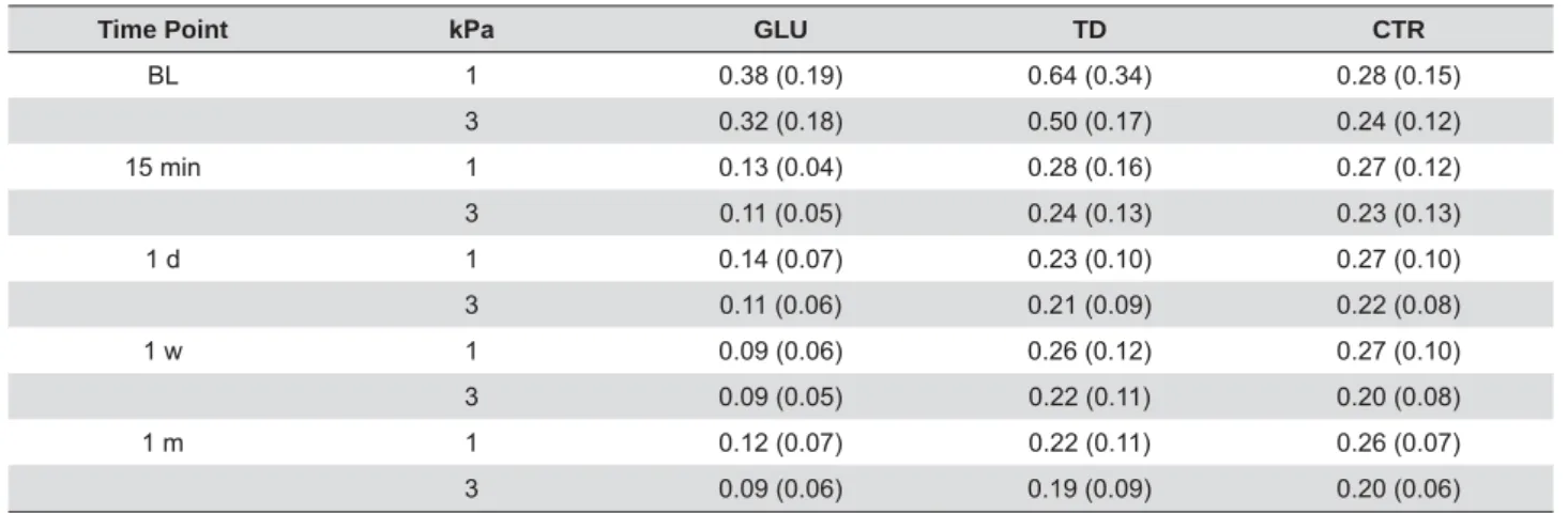
Table 1 shows the means and standard deviations
of hydraulic conductance by material, pressure, and
time. BL variations in the three groups were very
TD group than in the two other groups.
Figure 4 shows PR% in hydraulic conductance by
times regardless of the pulpal pressures used. The
Steel-Dwass test indicated that the PR% of CTR was
at all times, whereas the PR% of TD and GLU were
observed at the four times until 1 month of post
treatment period.
Figure 5 shows the treated and fractured surfaces
of representative disc specimens from each group
one month of storage. SEM photographs 4A and 4B
show aspects of free and fractured surfaces after the
GLU treatment. Under the EDTA cleaning condition,
demineralized, the exposed collagen mesh was not
collapsed. Collagen strings were supposedly
cross-collagen mesh was detected to a depth of several
micrometers. SEMs C and D show the free surface
and a view of the fractured specimen of dentin treated
with TD. All tubules and some intertubular dentin were
closed and covered with a crystalline grainy substance.
considered to be dehydration gaps that had occurred
under the high vacuum during sputtering and/or
observed to a depth of several micrometers (white
arrow), and were presumably precipitates from the
dissolution process of the primary TD phosphates.
The corresponding surfaces of the control specimen
after 1-month storage in water are shown in E and F.
Time Point N3D GLU TD CTR
BL 1 0.38 (0.19) 0.64 (0.34) 0.28 (0.15)
3 0.32 (0.18) 0.50 (0.17) 0.24 (0.12)
15 min 1 0.13 (0.04) 0.28 (0.16) 0.27 (0.12)
3 0.11 (0.05) 0.24 (0.13) 0.23 (0.13)
1 d 1 0.14 (0.07) 0.23 (0.10) 0.27 (0.10)
3 0.11 (0.06) 0.21 (0.09) 0.22 (0.08)
1 0.09 (0.06) 0.26 (0.12) 0.27 (0.10)
3 0.09 (0.05) 0.22 (0.11) 0.20 (0.08)
1 m 1 0.12 (0.07) 0.22 (0.11) 0.26 (0.07)
3 0.09 (0.06) 0.19 (0.09) 0.20 (0.06)
Table 1- Hydraulic conductance (Lp) μL min-1 cm-2 cmH 2O
Figure 3- Schematic illustration of the device used to measure the permeability of dentin discs. Water is perfused under adjusted pressure
4-Free surface and dentinal tubular walls were clean.
No remnants of collagen or mineralized precipitates
were detected.
Discussion
The present in v it r o investigation has provided
evidence to support GLU and TD being effective
agents for immediate and lasting reductions in
dentin disc permeability. Hydraulic conductance after
the application of the two commercial products was
times tested from BL until 1 month. Therefore, the null
difference among the three agents investigated was
rejected.
The active components in the plasma protein
precipitant GLU are GA and HEMA. In a spectroscopic
investigation, the reaction mechanism between GA
and HEMA was described as a two-step reaction. GA
reacts with serum albumin to induce precipitation,
which mediates the second step of the polymerization
of HEMA24. Using confocal laser scanning microscopy
and scanning and transmission electron microscopy,
25 (1997) visualized intrinsically
μ
tubules following the in v iv o application of GLUMA
desensitizer liquid and processing of the extracted
teeth. In an in vit r o
with a protein solution to enable the coagulation
reaction, which reduces hydraulic conductance. In the
bovine albumin solution, which did not affect the BL
permeability of the specimens13.
TTCP and DCPA powder that upon mixing with water
or an aqueous solution is readily transformed into
hydroxyapatite (HA). This transformation is based on
a dissolution-precipitation reaction mechanism6,14,28.
In an aqueous environment, TTCP and DCPA dissolve
and supply Ca2+ and PO 4
3-. Since this solution is
supersaturated concerning apatite, the less soluble
compound HA is precipitated. In the oral cavity, new
continuously formed HA crystals may be precipitated
because of the supersaturation of human saliva with
calcium phosphate salts16.
The results of the present study showed wide
variations in hydraulic conductance values, particularly
at BL. Possible reasons for this relatively large
scattering may be the age of the donors teeth, the
location of the slice cut from coronal dentin, regional
variability, and tubule density and diameter. The
as substrates for in vit r o experiments represents one
testing of dentin.
We selected a very low pressure in order to
simulated human pulpal tissue pressure, as reported
by Ciucchi, et al.5 (1995) and Pashley, et al.22 (1981).
The importance of this variable appears to be greatly
underestimated in the literature, and, even in
applied when studying desensitizing agents using a
conventional dentin disc model27
the study conducted almost 20 years ago, in which
hydraulic conductance decreased with time in the
three middle pressure ranges, but remained constant
2. The authors concluded that
use of high pressure is not recommended because
intratubular obstacles. Only a low, physiological
pressure is suggested to be suitable for measuring
hydraulic conductance in dentin. In contrast to the
highly sensitive device used in the present study,
the accuracy and resolution of many commonly used
why other researchers have disregarded this important
pressures.
In this in v it r o study, we stored specimens in
deionized water to exclude inadvertent mineralization
from confounding the results obtained, which is
is supersaturated with calcium and phosphate ions.
between BL and the end of the 1-month specimen
storage in water. Nevertheless, in future studies,
saliva as the storage liquid.
In contrast to previous studies, we measured
ultrasonicated in neutral EDTA. Thus, free surfaces and
at least tubular entrances were cleaned of smear and
the aldehyde actions of GLU, and, as observed in SEMs,
the collagen surface is noncollapsed. There were no
signs of septa bridging the tubules at deeper layers,
25 (1997). With TD,
the precipitation of HA or precursor phases depends on
the presence of nucleation sites for safe bonding to the
27 (2013)
reported that the application of TD occluded dentinal
tubules and reduced dentin permeability by up to 92%
permeability reductions in the present study. The
SEM of TD-treated dentin surfaces clearly shows
the presence of crystallites closing dentinal tubular
the reaction product was observed inside tubules, and
may have been due to the large amount of nucleation
points present in peritubular dentin. Due to the
supersaturation of saliva with calcium and phosphate
in the oral cavity, the ongoing precipitation of HA may
occur as a clinically important self-sustaining process.
It was of interest to compare the characteristics
of the two products in v iv o and in v it r o. In a
six-month clinical trial, all patients showed a reduction
from 30 to 40% in BL sensitivity for GLU and TD,
respectively, both immediately after their application
and throughout the six-month duration of the
study17. Similarly, the present in vit r o results showed
reductions from 30 to 45% in BL and Lp, both early
storage period. The immediate decreases in hydraulic
conductance after their application and the sustained
reductions observed over time were similar to the
immediate decrease and sustainability in sensitivity
reductions measured clinically on a VAS pain scale.
Therefore, the present laboratory test performed
under the experimental conditions selected is, to
of desensitizing compounds.
In summary, the present study on hydraulic
conductance in dentin discs treated with two
chemically different desensitizing agents and in a
untreated control demonstrated that both products,
according to previous clinical trial data, may be
characterized as effective.
for donation of the materials.
This study was supported by JSPS KAKENHI Grant
no. 26462870.
References
from the dentin. J Dent Res. 1964;43:619-25.
2- Camps J, Giustiniani S, Dejou J, Franquin JC. Low versus high pressure for in vit r o determination of hydraulic conductance of human dentine. Arch Oral Biol. 1997;42:293-8.
3- Canadian Advisory Board on Dentin Hypersensitivity. Consensus-based recommendations for the diagnosis and management of dentin hypersensitivity. J Can Dent Assoc. 2003;69:221-6.
of cervical dentine sensitivity. J Oral Rehabil. 1997;24:15-9.
in human teeth, in vivo. J Endodon. 1995;21:191-4.
Mater Med. 2014;24:1335-63.
evaluation of HEMA-based (desensitizing) products using split-chamber model following in vivo application in the dog. J Oral Rehabil. 2005;32:34-8.
et al. Evaluation of a calcium phosphate desensitizer using an ultrasonic device. Dent Mater J. 2013;32:456-61.
9- Gillam DG, Mordan NJ, Newman HN. The dentine disc surface: a plausible model for dentine physiology and dentine sensitivity evaluation. Adv Dent Res. 1997;11:487-501.
10- Greenhill JD, Pashley DH. The effects of desensitizing agents on the hydraulic conductance of human dentin in vit r o. J Dent Res. 1981;60:686-98.
Am J Dent. 2015;28:90-4.
Guidelines for the design and conduct of clinical trials on dentine hypersensitivity. J Clin Periodontol. 1997;24:808-13.
13- Ishihata H, Finger WJ, Kanehira M, Shimauchi H, Komatsu M. I n vit r o dentin permeability after application of Gluma® desensitizer as aqueous solution or aqueous fumed silica dispersion. J Appl Oral Sci. 2011;19:147-53.
phosphate cements with different amounts of tetracalcium phosphate and dicalcium phosphate anhydrous. J Biomed Mater Res. 1999;46:504-10.
desensitizing agents on dentin permeability and dentin tubule occlusion. J Adhes Dent. 2002;4:211-21.
16- Larsen MJ, Pearce EI. Saturation of human saliva with respect to calcium salts. Arch Oral Biol. 2003;48:317-22.
Acta Odontol Scand. 2014;72:936-41.
18- Mordan NJ, Barber PM, Gillam DG. The dentine disc. A review of its applicability as a model for the in v it r o testing of dentine hypersensitivity. J Oral Rehabil. 1997;24:148-56.
various materials on dentin permeability for the treatment of dentin hypersensitivity. Jpn J Conserv Dent. 2013;56:516-25.
20- Pashley DH. Dentin permeability, dentin sensitivity, and treatment through tubule occlusion. J Endod. 1986;12:465-74.
21- Pashley DH, Galloway SE. The effects of oxalate treatment on the smear layer of ground surfaces of human dentine. Arch Oral Biol. 1985;30:731-7.
22- Pashley DH, Nelson R, Pashley EL. I n- vivo
dentin in the dog. Arch Oral Biol. 1981;26:707-10.
23- Pereira JC, Segala AD, Gillam DG. Effect of desensitizing agents on the hydraulic conductance of human dentin subjected to different surface pre-treatments: an in vit ro study. Dent Mater. 2005;21:129-38. 24- Qin C, Xu J, Zhang Y. Spectroscopic investigation of the function of aqueous 2-hydroxyethylmethacrylate/glutaraldehyde solution as a dentin desensitizer. Eur J Oral Sci. 2006;114:354-9.
Gluma desensitizer. Eur J Oral Sci. 1997;105:414-21.
M, Tagami J. I n vit r o evaluation of dentinal hydraulic conductance and tubule sealing by a novel calcium-phosphate desensitizer. J Biomed Mater Res B Appl Biomater. 2013;101:303-9.
Thitthaweerat S, Tagami J. Effect of a calcium-phosphate based desensitizer on dentin surface characteristics. Dent Mater J. 2013;32:615-21.