Abst ract
Submitted: April 13, 2016 0RGL¿FDWLRQ-XO\ Accepted: August 15, 2016
Repair of bone defect s using
adipose-der ived st em cells com bined w it h
alpha- t r icalcium phosphat e and gelat in
sponge scaffolds in a rat m odel
Obj ect ives: This st udy aim ed t o evaluat e t he pot ent ial of adipose- derived st em
FHOOV$6&VFRPELQHGZLWKDPRGL¿HGD- t r icalcium phosphat e (D-TCP) or gelat in sponge ( GS) scaffolds for bone healing in a rat m odel. Mat er ial and Met hods:
%RQHGHIHFWVZHUHVXUJLFDOO\FUHDWHGLQWKHIHPXURIDGXOW6+5UDWVDQG¿OOHG
w it h t he scaffolds, em pt y or com bined w it h ASCs. The r esult s w er e analy zed
by hist ology and hist om or phom et r y on day s seven, 14, 30, and 60. Result s:
6LJQL¿FDQWO\LQFUHDVHGERQHUHSDLUZDVREVHUYHGRQGD\VVHYHQDQGLQDQLPDOV
t r eat ed w it h D-TCP/ ASCs, and on day 14 in t he gr oup t r eat ed w it h GS/ ASCs,
w hen com par ed w it h t he gr oups t r eat ed w it h t he biom at er ials alone. I nt ense
¿EURSODVLDZDVREVHUYHGLQWKHJURXSWUHDWHGZLWK*6DORQHRQGD\VDQG
Conclusions: Our r esult s show ed t hat t he use of ASCs com bined w it h D-TCP or
*6VFDIIROGVUHVXOWHGLQLQFUHDVHGERQHUHSDLU7KHKLJKHUHI¿FDF\RIWKHD-TCP scaffold suggest s ost eoconduct ive pr oper t y t hat r esult s in a biological suppor t
t o t he cells, w her eas t he GS scaffold funct ions j ust as a car r ier. These r esult s
FRQ¿UPWKHSRWHQWLDORI$6&VLQDFFHOHUDWLQJERQHUHSDLULQin vivo ex per im ent al
rat m odels. These r esult s suggest a new alt er nat ive for t r eat ing bone defect s.
Ke yw or ds: Bone regenerat ion. Calcium cem ent . Adipose- derived st em cells.
Rat m odel.
Adriana CORSETTI1
Claudia BAHUSCHEWSKYJ2
Deise PONZONI1
Renan LANGIE1
Luis Alberto dos SANTOS3
Melissa CAMASSOLA4
Nance Beyer NARDI4
Edela PURICELLI5
1Univer sidade Federal do Rio Grande do Sul, Faculdade de Odont ologia, Por t o Alegr e, RS,
Brasil.
2I r m andade da Sant a Casa de Miser icór dia de Por t o Alegr e, Cent r o de Odont ologia,
Cir ur gia e Reabilit ação Bucom axilofacial, Por t o Alegr e, RS, Brasil, Univer sidade Lut erana do Brasil, Laborat ór io de Células-Tr onco e Engenhar ia de Tecidos, Canoas, RS, Brasil.
3Univer sidade Federal do Rio Grande do Sul, Depar t am ent o de Engenhar ia de Mat er iais,
Por t o Alegr e, RS, Brasil.
4Univer sidade Lut erana do Brasil, Laborat ór io de Células-Tr onco e Engenhar ia de Tecidos,
Canoas, RS, Brasil.
5I r m andade da Sant a Casa de Miser icór dia de Por t o Alegr e, Cent r o de Odont ologia,
Cir ur gia e Reabilit ação Oral e Maxilofacial, Por t o Alegr e, RS, Brasil
Corresponding address: Edela Puricelli Centro de Odontologia-Cirurgia e Reabilitação Bucomaxilofacial - Irmandade da Santa Casa de Misericórdia de Porto Alegre (ISCMPA)
I nt r oduct ion
Th e m a n a g e m e n t o f l o st b o n e t i ssu e d u e t o
congenit al abnor m alit ies, t raum a, or cancer t r eat m ent
poses a challenge t o oral and m axillofacial surgeons. The highly vascular ized nat ur e of t he bone t issue r esult s in
a gr eat capacit y t o heal and r em odel w it hout scar r ing8.
Never t heless, bone loss r epr esent s a m aj or clinical
SUREOHPLQUHFRQVWUXFWLYHKHDGDQGQHFNVXUJHU\DQG
aut ologous graft ing is st ill t he t herapeut ic gold st andard
in r econst r uct ive sur ger y. This concept , how ever, has
serious lim it at ions relat ed t o t he lim it ed am ount of t issue
WKDWFDQEHKDUYHVWHGLQFUHDVHGULVNRILQIHFWLRQRU
recurrent pain. Alt ernat ive t herapeut ic approaches have
pr oposed ost eoconduct ion, guided bone r egenerat ion,
ost eodist ract ion, and ost eoinduct ion4.
Ost eoconduct ive scaffolds cr eat e an envir onm ent
SHUPLVVLYHIRUWKHSUROLIHUDWLRQRIERQHFHOOVZKLFK¿OOV
t he bone defect10. Calcium phosphat e cem ent s ( CPCs)
have a com posit ion sim ilar t o t he m ineral phase of nat ive bone, and have been used as bone subst it ut es
in t he last decades15. Hy dr ox iapat it e and t r icalcium
phosphat es are am ong t he m ost used of t hese cem ent s,
and t hey have pr oved t heir value in several clinical applicat ions6. How ever, CPCs have som e lim it at ions,
r elat ed t o poor m echanical pr oper t ies and m ainly t o
ODFN RI RVWHRLQGXFWLYH DQG DQJLRJHQLF DFWLYLWLHV 7KH ¿UVWOLPLWDWLRQFDQEHGHDOWZLWKE\IXUWKHUPRGL¿FDWLRQ RI WKH ELRPDWHULDO 7R FRPSHQVDWH IRU WKH ODFN RI
ost eoin du ct ion an d an giogen esis, CPCs h av e been
pr eloaded w it h cells.
The com binat ion of adult st em cells wit h biom at erials has int roduced new perspect ives on t he opt im izat ion of
t issue repair prot ocols. Different iat ed cells or st em cells
m ay be used, and m esenchym al st em cells ar e am ong
t he m ost ex t ensively st udied biological elem ent s in t issue engineering7. The plast icit y and ease of collect ing
and ex v iv o cult ur ing of adipose- der ived st em cells
( ASCs) open w ide possibilit ies of use in r egenerat ive
t herapy2. This st udy aim ed t o evaluat e t he pr ocess of bone r epair in a rat fem oral defect m odel, using
PRGL¿HGD- t r icalcium phosphat e (D-TCP) scaffolds and a com m ercially available biom at erial ( absorbable gelat in
sponge, GS) associat ed w it h allogeneic ASCs. Result s w er e evaluat ed by hist ological and hist om or phom et r ic
assessm ent s of bone r epair.
Mat er ial and m et hods
Anim als and et hics
Adult ( 5 m ont hs old) m ale sy ngeneic SHR rat s,
w eig h in g an av er ag e of 3 0 0 g , w er e h ou sed an d
m aint ained under st andar d condit ions, and t r eat ed in accor dance w it h t he guidelines for t he use of anim als
in r esear ch pr oj ect s, Nor m at ive Resolut ion 04/ 97 of
t he Resear ch and Et hics in Healt h Com m it t ee/ GPPG/
HCPA. The st udy was appr oved by t he Resear ch Et hics Com m it t ee of Hospit al de Clínicas de Por t o Alegr e ( no.
110159) .
I solat ion an d ch ar act er izat ion of ad ip
ose-der ived st em cells
I nguinal adipose t issue was collect ed from t hree rat s and pr ocessed indiv idually as pr ev iously descr ibed1.
The t issue was m inced in phosphat e buffer ed saline
( PBS) and digest ed w it h 1 m g/ m L collagenase t ype I solut ion ( Sigm a Chem ical Co, St Louis, MO) for 30
m in at 37°C, under gent le agit at ion. The enzym e was
LQDFWLYDWHG ZLWK 'XOEHFFR¶V PRGL¿HG (DJOH¶V 0HGLXP
( DMEM) ( Sigm a) supplem ent ed w it h 10% fet al calf serum ( FCS, Cult ilab, SP, Brazil) and cent rifuged at 400x
g for 10 m in. The vascular st rom al fract ion was washed
ZLWK+DQN¶VEDODQFHGVDOWVROXWLRQ+%666LJPDE\
cent r ifugat ion at 350x g for 5 m in. The cell pellet was r esuspended in DMEM w it h 10 m M HEPES fr ee acid
( Sigm a) supplem ent ed w it h 10% FCS and cult ur ed at
37°C in an at m ospher e of 5% CO2. Thr ee days lat er,
t he non- adher ent cells w er e r em oved, and t he cult ur e was m aint ained and expanded ever y 3 or 4 days aft er
t r y psin izat ion ( 0 . 2 5 % t r y psin an d 0 . 0 1 % EDTA in
HBSS) . Cells bet w een passages 4 and 7 w er e used for
all exper im ent s, and at least 3 cult ur es w er e analyzed. A S C s w e r e a n a l y z e d f o r m o r p h o l o g y ,
im m unophenot ype, and proliferat ion and different iat ion
pot ent ial. All experim ent s were reproduced t hree t im es.
3KRWRPLFURJUDSKV ZHUH WDNHQ ZLWK D GLJLWDO FDPHUD $[LR&DP 05F &DUO =HLVV 2EHUNRFKHQ *HUPDQ\
u si n g Ax i o Vi si o n 3 . 1 so f t w a r e ( Ca r l Z e i ss) . Th e
LPPXQRSKHQRW\SHZDVGHWHUPLQHGE\ÀRZF\WRPHWU\
The cells w er e t r ypsinized, washed, and incubat ed for
PLQDW&ZLWKÀXRUHVFHLQLVRWKLRF\DQDWH),7& FRQMXJDWHG DQWLERGLHV VSHFL¿F IRU UDW &' &' %HFWRQ 'LFNLQVRQ 6DQ -RVH &$ &'E DQG &' &DOWDJ%XUOLQJDPH&$$)$&6&DOLEXUÀRZF\WRPHWHU HTXLSSHGZLWKQPDUJRQODVHU%HFWRQ'LFNLQVRQ
At least 10,000 event s w er e collect ed.
The pr oliferat ion rat e of t he cult ur es was assessed
by count ing t he num ber of cells r ecover ed in each
passage, as w ell as t he t im e elapsed. These dat a w er e used t o det er m ine t he populat ion doubling t im e, w it h
t he aid of an online calculat or ( ht t p: / / w w
w.doubling-t im e.com / com puw.doubling-t e.php) . The r esulw.doubling-t s ar e expr essed as
t he num ber of cells over t he days of cult ivat ion. Differ ent iat ion was induced by incubat ion of cells
ZLWKVSHFL¿FFXOWXUHPHGLXP1. All r eagent s w er e fr om
6LJPD XQOHVV VSHFL¿FDOO\ LQGLFDWHG )RU RVWHRJHQLF GLIIHUHQWLDWLRQFHOOVZHUHFXOWXUHGIRUXSWRZHHNVLQ
m edium com plem ent ed w it h 10–8 M dexam et hasone,
NjJP/ DVFRUELF DFLG SKRVSKDWH DQG P0 ǃJO\FHURSKRVSKDWH&DOFLXPGHSRVLWLRQZDVUHYHDOHG
by st aining for 5 m in w it h Alizar in Red S st ain at pH
4.1. To induce adipogenic differ ent iat ion, cells w er e
FXOWXUHGIRUXSWRZHHNVLQPHGLXPFRPSOHPHQWHG
w it h 10–8 0 GH[DPHWKDVRQH NjJP/ LQVXOLQ
Nj0LQGRPHWKDFLQDQGNj0URVLJOLWD]RQH$GLSRF\WHV
w er e r evealed by st aining w it h Oil Red O.
Biom at er ials
Th e D- TCP sca f f o l d s w e r e sy n t h e si ze d u si n g calcium car bonat e ( Dinam ica, SP, Brazil) and calcium
pyr ophosphat e, obt ained fr om calcinat ion of calcium
phosphat e dihydrat e ( Dyne, SP, Brazil) at 550°C for
5 h. I nit ial dr y hom ogenizat ion was m ade in alum ina ball m ill for one hour. The m ixt ur e of pow der s w er e
calcined in a fur nace at 1300°C for 5 h, follow ed b y
quenching in air.
Aft er cooling, D-TCP was m anually disaggr egat ed using por celain m or t ar and t he pow der was sieved in
$670 PHVK NjP 7KH VLHYHG SRZGHU ZDV
w et m illed using absolut e et hy l alcohol ( Vet ec, SP,
%UD]LOLQDOXPLQDEDOOPLOOIRUK7KHUHVXOWLQJ¿QH SRZGHUDYHUDJHSDUWLFOHGLDPHWHUNjPZDVGULHGLQ
a st ainless st eel vessel for 72 h at 70°C t o pr om ot e t he
evaporat ion of alcohol. Aft er m illing, t he D-TCP is called
cem ent , as it r eact s w it h wat er and allow s har dening. The liquid fract ion of t he cem ent w as dist illed and
deionized in w at er w it h cem ent set t ing accelerat or
( 2.5% Na2HPO4 – Synt h, SP, Brazil) and foam ing agent ( 0.5% sodium dodecyl sulfat e, Dinam ica) . The pow der
and liquid w er e hand- m ix ed t o pr oduce a foam ing
cem ent using a liquid/ pow der r elat ion of about 0.5
m L/ g. Aft er hardening, t he foam ing cem ent was washed 5 t im es t o r em ove t he excess of foam ing agent . The
appar ent por osit y of obt ained cem ent was 61% , w it h
SRUHV UDQJLQJ IURP DERXW WR NjP 6FDIIROGV
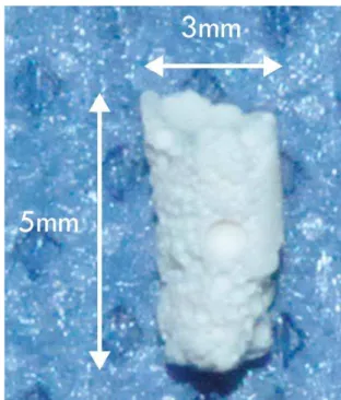
used in all exper im ent s w er e 3 m m diam et er and 5
m m lengt h ( Figur e 1) .
A com m er cially av ailab le r e- ab sor b ab le g elat in sponge ( GS, Cut anplast St andar d, SP, Brazil) was also
used in t he exper im ent s. Bot h t ypes of scaffolds w er e
analyzed by scanning elect ron m icroscopy ( SEM) , using
a m icr oscope Hit achi, m odel TM3000.
Com binat ion of ASCs t o biom at er ials
ASCs w er e a p p l i ed t o t h e sca f f o l d s b y st a t i c
seeding. Dr y D-TCP cylinder s ( 3 m m w idex5 m m deep) or a cor r esponding v olum e of gelat in sponge w er e
individually placed in w ells of a 24- w ell cult ur e plat e.
ASCs w er e suspended in DMEM at 5x10⁶ cells/ m L, and
Nj/ZHUHSODFHGRQHDFKVFDIIROG$GKHUHQFHRI$6&V
t o D-TCP scaffolds was det erm ined by incubat ion for 2 h
DW&ZLWKDGGLWLRQRINj/'0(0DWHYHU\PLQ DIWHUZKLFKNj/RI'0(0ZHUHDGGHGWRHDFKZHOODQG
t he scaffolds w er e t ransfer r ed t o new w ells w it h DMEM com plem ent ed w it h 10% FCS. The r em aining,
non-adhered cells were st ained wit h Giem sa and count ed, t o
det erm ine t he num ber of cells adhered t o each scaffold.
For analy sis of cell pr oliferat ion, t he scaffolds w er e m aint ained for 3 d in 5% CO2 at 37°C, and proliferat ion
was assessed using t he MTT t est9. Opt ical densit y was
read in a spect rophot om et er at a wavelengt h of 540 nm .
The sam e num ber of cells com bined w it h t he scaffolds was cult ivat ed in convent ional condit ions for 3 d and
analyzed w it h t he MTT t est .
Sur gical pr ocedur e and t r eat m ent
Sur gical pr ocedur es w er e per for m ed under general
anest hesia using int raper it oneal inj ect ions of 10 m g/
NJERG\ZHLJKW[\OD]LQH%D\HU1HZKDYHQ&7DQG PJNJERG\ZHLJKWNHWDPLQH3DUNH'DYLV$QQ$UERU
MI ) . As Figur e 2 show s, t w o bone defect s m easur ing
3.1 m m diam et er and 3.5 m m dept h w er e sur gically creat ed in t he right fem oral diaphysis, using a m ot orized
t r eph in e. I n 1 6 r at s, t h e t w o def ect s w er e t ot ally
¿OOHGZLWKHPSW\D-TCP or GS scaffolds ( E-D-TCP and E- GS, r espect ively) , and in anot her 16 anim als, w it h
ASC- loaded D-TCP or GS scaffolds ( L-D-TCP and L- GS,
respect ively) prepared as described above. The anim als
w er e obser v ed daily, accor ding t o usual v et er inar y post - operat ive car e.
On days seven, 14, 30, and 60 aft er t he sur gical
p r oced u r e, g r ou p s of eig h t r at s ( f ou r w it h em p t y
scaffolds and four w it h ASC- loaded scaffolds) w er e sacr i f i ced . Th e r i g h t f em u r s w er e d i ssect ed an d
r em oved for analysis.
Hist ology and hist om or phom et r y
,PSODQWHG ERQH DUHDV ZHUH FROOHFWHG GHFDOFL¿HG
w it h 5% nit r ic acid for 5 d, dehydrat ed w it h graded
et hanol and processed for hist ological analysis. Sect ions
perpendicular t o t he im plant and surrounding area were
st ained wit h hem at oxylin and eosin and analyzed wit h a
m icroscope ( Olym pus Opt ical Co, Miam i, FL) connect ed
t o an im age analyzer ( Olym pus, Qcolor 5, Coolet , RTV) ,
using t he Qcapt ur e soft war e ( Univer sit y of Texas) . The size of t he r em aining bone defect was m easur ed in
pixels w it h t he dist ance t ool of t he I m ageTool pr ogram
( Ver sion 2 . 8 1 , Qu an t it at iv e I m agin g Cor por at ion ) ,
and a zer o scor e was assigned t o fully r epair ed bone defect s. Repair ed defect s w er e cat egor ized and show n
as absolut e values, and non- r epair ed defect s w er e
st at ist ically analyzed as descr ibed below.
Th e am ou n t of n ew ly f or m ed b on e t issu e w as m easur ed in per cent age by draw ing a line fr om t he
out er sur face of a cor t ical layer, always w it h t he sam e
lengt h. Ot her boundar y lines w er e det er m ined at r ight angles t o t his one, for m ing a r ect angle. The sm allest
an d lar g est sid es of t h e r ect an g le m easu r ed 8 0 0
and 1200 pixels r espect ively. These m easur es w er e
GHWHUPLQHG WR NHHS D VWDQGDUGL]DWLRQ DQG IRU EHLQJ
t he lar gest ar ea of new bone for m at ion obser ved in t he
lit erat ur e. The ar ea of new ly for m ed bone t issue was
select ed, excluding int ert rabecular and vascular spaces
as w ell as t he r em aining cor t ical layer, generat ing pixel values t hat w er e conver t ed t o a per cent age value
T w o b l i n d e d r e s e a r c h e r s c o n d u c t e d
hist om or phom et r ic analyses, pr eviously calibrat ed as
show n by t he St udent ’s t - t est for pair ed sam ples and
by t he Bland- Alt m an plot .
St at ist ical analyses
Dat a are expressed as m ean and st andard deviat ion. Differ ences am ong t he gr oups w er e com par ed w it h t he
St udent ’s t - t est for independent sam ples, using t he
SPSS for Window s ver sion 19.0. A p value < 0.05 was
FRQVLGHUHGVWDWLVWLFDOO\VLJQL¿FDQW
Result s
Cult ivat ion and charact erizat ion of rat
adipose-der ived st em cells
As Figur e 3 show s, cult ivat ed rat ASCs show t he
ch ar act er i st i c f i b r o b l ast m o r p h o l o g y an d su r f ace
Figure 4-6FDQQLQJHOHFWURQPLFURVFRS\DQDO\VLVVKRZVWKHSRURXVVWUXFWXUHRIĮWULFDOFLXPSKRVSKDWHVFDIIROGVDDQGWKH¿EURXV
QHWZRUNVWUXFWXUHRIWKHJHODWLQVSRQJHE$IWHUWKUHHGD\VLQFXOWXUH$6&VFRPELQHGZLWKD7&3'SUROLIHUDWHVLJQL¿FDQWO\OHVVWKDQ FHOOVSODWHGLQFRQYHQWLRQDOFRQGLWLRQV'DVVKRZQE\077UHVXOWVFRPSDUHGZLWKWKHLQLWLDOFHOOQXPEHUFRQWUROS
Figure 3- Morphology, proliferation, immunophenotype, and differentiation potential of rat adipose-derived stem cells (ASCs). Rat
$6&VGLVSOD\WKHFKDUDFWHULVWLF¿EUREODVWPRUSKRORJ\DLQSKDVHFRQWUDVWEDIWHU*LHPVDVWDLQLQJDQGSUROLIHUDWLRQFDSDFLW\FRI PHVHQFK\PDOVWHPFHOOV$6&FXOWXUHVZHUHDEOHWRGLIIHUHQWLDWHLQWRDGLSRJHQLFDQGRVWHRJHQLFOLQHDJHVDVVKRZQE\VWDLQLQJZLWK2LO 5HG2DQG$OL]DULQ5HG6UHVSHFWLYHO\GDQGZHUHQHJDWLYHIRU&'EDQG&'DQGSRVLWLYHIRU&'DQG&'H8QGLIIHUHQWLDWHG FRQWURO$6&VDUHQRWPDUNHGZLWKWKHVHVWDLQV2ULJLQDOPDJQL¿FDWLRQ[
p h en ot y p e of m esen ch y m al- t y p e st em cells. Th e
cells expanded rapidly for a per iod of 40 d, w hen t he
ex pansion rat e show ed a decr ease ( Figur e 3 c) . As
expect ed, cult ur es differ ent iat ed int o adipogenic and ost eogenic lineages ( Figur e 3d) .
Pr oduct ion of
D
-TCP scaffolds and analysis of
cell adher ence and pr oliferat ion
The D-TCP scaffolds pr oduced w er e analy zed by
scan n in g elect r on m icr oscop y ( Fig u r e 4 a) , w h ich r evealed adequat e por ous st r uct ur e, w it h a por e size
EHWZHHQDQGNjP6(0RIVDPSOHVRIWKHJHODWLQ VSRQJH VKRZHG D ¿EURXV QHWZRUN VWUXFWXUH )LJXUH
Figure 5-7UHDWPHQWRIWKHERQHGHIHFWZLWK$6&ORDGHGD7&3VFDIIROGV/Į7&3UHVXOWVLQIDVWHUUHSDLUDVFRPSDUHGZLWKGHIHFWV
4b) . Rat ASCs had > 90% adher ence t o D-TCP ( not
show n) , and t he pr oliferat ion rat e of cells com bined
ZLWKWKHVFDIIROGVZDVVLJQL¿FDQWO\ORZHUWKDQWKDWRI
cells cult ivat ed in convent ional condit ions ( Figur e 4c) .
Hist ological analyses
$OOKLVWRORJLFDOHYDOXDWLRQVVKRZHGDPDUNHGUHJXODU
in t er r u pt ion of t h e ost ect om ized cor t ical bon e. As
pr esent ed in Figur e 5, t he t r eat m ent of bone defect s
wit h D-TCP scaffolds induced t he form at ion of increasing
am ount s of new bone, par t icular ly w hen t he scaffolds w er e loaded w it h ASCs. Mu lt in u cleat ed gian t cells
w er e obser ved ar ound t he bor der s of t he scaffolds,
SDUWLFXODUO\RQGD\$O\PSKRSODVPDF\WLFLQ¿OWUDWH
Figure 6- ([WHQVLYH ERQH IRUPDWLRQ LV REVHUYHG LQ ERQH GHIHFWV WUHDWHG ZLWK $6&ORDGHG JHO VSRQJH /*6 7UHDWPHQW ZLWK JHO
For b on e d ef ect s t r eat ed w it h t h e g el sp on g e, t he pr esence of ASCs also r educed t he m ean size of
t he defect on day seven, but t he differ ence was not
VLJQL¿FDQW2QHRIWKHGHIHFWVWUHDWHGZLWK/*6ZDV
alr eady com plet ely r epair ed on day seven. On day 60, t w o defect s t r eat ed w it h E- GS, and all four defect s
t r eat ed w it h L- GS, w er e com plet ely r epair ed.
The hist om or phom et r ic analysis show ed a gr eat er
percent age of newly form ed bone in t he t est cavit ies on days 7 and 60, but t he differ ences w er e no st at ist ically
VLJQL¿FDQW7DEOH
Discussion
7KLV ZRUN LQYHVWLJDWHG WKH HI¿FDF\ RI DGLSRVH
d e r i v e d st e m ce l l s co m b i n e d w i t h t w o t y p e s o f
biom at er ials, alpha- t r icalcium phosphat e and gelat in sponge, in t he r epair of bone defect s in a rat m odel.
Th e cells u sed in t h is st u d y w er e easily isolat ed
f r om t h e adipose t issu e an d ex pan ded in cu lt u r e,
VKRZLQJWKHFKDUDFWHULVWLF¿EUREODVWPRUSKRORJ\DQG
im m unophenot ype of m esenchym al- t ype st rom al cells.
They w er e also capable t o differ ent iat e int o adipocyt es
and ost eoblast s. Due t o t heir self- r enewal capacit y and plast icit y, ASCs r epr esent an im por t ant com ponent for
r egenerat ive m edicine and t issue engineer ing7.
Alt hough no consensus was reached about t he ideal
anim al m odel t o be used for t he invest igat ion of bone r egenerat ion5, t he exper im ent al pr ot ocol used in t his
st udy, est ablished by Puricelli, et al.10 ( 2010) , has been
ver y adequat e for t his t ype of st udy. The rat s w er e
DOOPDOHDJHG¿YHPRQWKVDQGZHLJKLQJDQDYHUDJH
could be obser ved in som e of t he sam ples. Wher eas in defect s t r eat ed w it h E-D-TCP t he new bone t issue was
spar ser and disor ganized, t he use of L-D-TCP r esult ed
in new bone t issue t hat sur r ounded and pr ogr essively
r eplaced t h e scaf f olds, or gan izin g an d closin g t h e surgical defect , wit h m ore frequent angiogenic regions.
Figur e 6 show s t he sequence of event s obser ved
aft er t r eat m ent of t he bone defect s w it h em pt y or
ASC- loaded GS scaffolds. I n defect s t reat ed w it h E- GS, t he scaffold was obser ved up t o 30 d; new bone t issue
ZDV¿UVWVHHQRQGD\DQGLQFUHDVHGXQWLOPRVWRI
t he defect was r epair ed on day 60. How ever, int ense
¿EURVLVZDVDOVRREVHUYHGLQWKHVHVDPSOHVSDUWLFXODUO\
on day s 14 and 30. The pr esence of ASCs induced
ext ensive bone for m at ion, obser ved on day seven and
pr ogr essing unt il t he bone defect was fully r epair ed.
On day seven, L- GS scaffolds w er e alr eady sur r ounded
E\ PDVVLYH WUDEHFXODU ERQH ZLWK DQ LQÀDPPDWRU\ LQ¿OWUDWHFRQWDLQLQJSUHGRPLQDQWO\SRO\PRUSKRQXFOHDU
cells. I n t hese sam ples, t he scaffolds w er e not visible
aft er day 14.
Hist om or phom et r ic analyses
Hist om or phom et r ic analyses evaluat ed t he size of
t he bone defect s on days seven and 60 aft er t reat m ent . As present ed in Table 1, t he presence of adipose- derived
VWHPFHOOVUHVXOWHGLQDVLJQL¿FDQWGHFUHDVHRIWKHPHDQ
size of bone defect s t reat ed wit h D-TCP scaffolds on day
seven ( p= 0.016) or day 60 ( p= 0.042) , as com par ed w it h defect s t r eat ed w it h t he scaffolds alone. On day
60, t w o of t he four defect s t r eat ed w it h ASC- loaded
D-TCP scaffolds w er e com plet ely r epair ed.
Day 7 Day 60
Empty Loaded p Empty Loaded p
D-TCP size
1,537±218 702±454 0.016 492±19 288±58 0.042
n rep 0 0 0 2
GS size
1,658±180 1,089±508 0.087 1,489±227 -
-n rep 0 1 2 4
Table 1-6L]HRIERQHGHIHFWLQSL[HOVVL]HDQGQXPEHURIFRPSOHWHO\UHSDLUHGERQHGHIHFWVQUHSDIWHUWUHDWPHQWZLWKD-tricalcium phosphate scaffolds (D7&3RUJHOVSRQJH*6DORQHHPSW\RUORDGHGZLWK$6&VORDGHG
Day 7 Day 60
Empty Loaded p Empty Loaded p
D-TCP 10.1±9.8 29.2±17.9 0.127 40.2±9.7 42.6±8.4 0.772 GS 7.3±2.5 19.2±19.2 0.303 36.4±11.8 36.8±6.5 0.959
Table 2-6L]HRIERQHGHIHFWLQSHUFHQWDJHDIWHUWUHDWPHQWZLWKD-tricalcium phosphate (D-TCP) or gel sponge (GS) scaffolds empty
of 304 g, and t he dim ension of t he sur gical cavit ies
ZHUHSURSRUWLRQDOWRWKHOHQJWKDQGWKLFNQHVVRIWKH
rat fem ur, m easur ing 3. 1 m m in diam et er and 3. 5
m m deep. The ost ect om ized cor t ical st r uct ur e show ed
PDUNHG UHJXODULW\ LQ DOO JURXSV DQG H[SHULPHQWDO
per iods inv est igat ed. I n addit ion , t est an d con t r ol
cav it ies w er e dist r ibu t ed in t h e f em u r of dif f er en t
anim als, prevent ing a possible effect of m igrat ing st em cells in cavit ies t hat did not r eceive t he cells.
Ou r r e su l t s sh o w e d t h a t t h e u se o f ASCs
com bined w it h D-TCP of GS scaffolds r esult ed in an
accelerat ion of bone repair. This effect was m ainly seen at seven and 60 days in t he gr oup t r eat ed w it h L-D-TCP
and on day 14 when L- GS was used, result ing in great er
bone neoform at ion and fast er repair of t he bone defect .
,QWHQVH¿EURSODVLDZDVREVHUYHGLQGHIHFWVWUHDWHGZLWK
gelat in sponge scaffolds alone, par t icular ly on days 14
DQG7KHKLJKHUHI¿FDF\RID-TCP scaffolds suggest an ost eoconduct ive pr oper t y t hat r esult s in a biological support t o t he cells10, whereas t he GS scaffold funct ions
j ust as a car r ier.
The gelat in sponge used in t his st udy is a chem ically
FURVVOLQNHG JHODWLQ RI KLJK SRUH GHQVLW\3, a n d h a s a l r e a d y b e e n su cce ssf u l l y u se d a l o n e o r i n
com binat ion w it h m esenchy m al st em cells for bone
t issu e en gin eer in g. How ev er, in t h ose st u dies, t h e
biom at er ial w as u sed j u st as a su ppor t f or t issu e im plant s11 or was com bined w it h cells t ransduced w it h
bone m or phogenic pr ot ein- 213.
$OWKRXJKWKHRVWHRFRQGXFWLYHSURSHUWLHVRIǃ7&3 VFDIIROGV DUH ZHOO NQRZQ ZLWK DQ XQGHUVWDQGLQJ RI
t he signaling pat hways t hr ough w hich t hey induce t he
different iat ion of st em cells int o ost eoblast ic cells12, less
LVNQRZQDERXWD-TCP. I n an in vit ro st udy, Wój t owicz, et al. ( 2014)14 invest igat ed t he capacit y of t hr ee t ypes of CPCs t o suppor t t he gr ow t h of hum an ost eoblast s. The
result s showed t hat , alt hough displaying cell- support ing
pr oper t ies, D7&3 VFDIIROGV ZHUH OHVV HI¿FLHQW WKDQ ca r b o n a t e h y d r o x y a p a t i t e a n d b i p h a si c ca l ci u m
phosphat e in inducing cell viabilit y and spr eading or
ost eogenic capacit y of t he ost eoblast s. I n t his st udy,
t he result s showed t hat ASCs have high adhesion t o t he cem ent scaffold. The sm aller pr oliferat ion rat e of t he
cells com bined w it h D-TCP t han in convent ional cult ur e
con d it ion s p ossib ly in d icat e in cr eased ost eog en ic
different iat ion, as shown by t he accelerat ed bone repair w hen t his com binat ion was used. The biocom pat ibilit y
of D-TCP, alr eady obser ved by Pur icelli, et al.10 ( 2010) ,
r esult ed in absence of for eign body r eact ion in t he
t r eat ed t issu es an d in cr eased r eab sor p t ion of t h e
biom at er ial, obser ved as ear ly as seven day s aft er
t r eat m ent , w it h new bone for m at ion at t he edges of
t he im plant s.
)RU WKLV VWXG\ PRGLILFDWLRQV WRRN SODFH LQ WKH
co m p o si t i o n o f t h e cem en t w i t h t h e a d d i t i o n o f
su r f act an t sod iu m lau r y l su lf at e, r esu lt in g in t h e
incor porat ion of por es in it s st r uct ur e and allow ing t he
JURZWKRIERQHWLVVXHLQWRWKHPDWHULDO7KHEORFNZDV
por ous and fragm ent ed in hist ological sect ions. This
m ay suggest t hat a larger area of cont act wit h t he bone
t issue accelerat es t he pr ocess of r epair, w hich could explain t he accelerat ed pr ocess of bone r epair in t his
st udy w hen com par ed w it h t he r esult s by Pur icelli, et
al.10 ( 2010) , in w hich cont r ol cavit ies r em ained open 60 d aft er t r eat m ent .
Hist om or phom et r ic r esult s ar e generally pr esent ed
as t he percent age of neoform ed t issue in relat ion t o t he
t ot al defect , but , in t his st udy, t he size of t he cavit ies were also m easured, on days 7 and 60. The percent age
RI QHZO\ IRUPHG ERQH WLVVXH VKRZHG QR VLJQL¿FDQW
differ ence in t r eat ed and cont r ol gr oups, but t he linear
DQDO\VLVRIFDYLW\RFFOXVLRQVKRZHGVLJQL¿FDQWUHVXOWV
w it h incr eased r epair of t he cavit ies w hen D-TCP was
com bined w it h ASCs.
7KHVHUHVXOWVVXJJHVWDQGFRQ¿UPWKHSRWHQWLDORI
ASCs in accelerat ing bone r epair in exper im ent al rat m odels. Several aut hor s agr ee t hat t he associat ion
of a ceram ic biom at er ial t o st em cells, fr om var ious
or igins, seem s t o favor and accelerat e t he pr ocess of
bone r epair, as obser ved in t his st udy in exper im ent al t im es seven and 60 d.
Conclusions
This st udy showed t hrough descript ive hist ological
analyses t hat t he com binat ion of adipose- derived st em
cells t o t w o differ ent t ypes of biom at er ials r esult ed in accelerat ion of bone r epair. Calcium cem ent scaffolds
ZHUH PRUH HI¿FLHQW WKDQ JHODWLQ VSRQJH VFDIIROGV
suggest ing an ost eoinduct ive funct ion in addit ion t o a
cell- car r ier funct ion. These r esult s should be fur t her explor ed in lar ger anim al m odels so t hat bone t issue
engineer ing m ay soon becom e par t of t he t herapeut ic
arsenal t o t reat an increasing num ber of pat ient s in t he
$FNQRZOHGJHPHQWV
Th is st u d y w as su p p or t ed b y DECI T/ SCTI E- MS
WKURXJK1DWLRQDO&RXQFLOIRU6FLHQWL¿FDQG7HFKQRORJLFDO
Developm ent ( CNPq) and FAPERGS.
Refer ences
1- Silva Meir elles L, Chagast elles PC, Nar di NB. Mesenchym al st em
cells r eside in vir t ually all post - nat al or gans and t issues. J Cell Sci.
2006; 119( Pt 11) : 2204- 13.
2- De Francesco F, Ricci G, D'Andr ea F, Nicolet t i GF, Fer rar o GA. Hum an
adipose st em cells: fr om bench t o bedside. Tissue Eng Par t B Rev.
2015; 21( 6) : 572- 84.
+DMRVFK 5 6XFNIXHOO 0 2HVVHU 6 $KOHUV 0 )OHFKVHQKDU .
Schlosshauer B. A novel gelat in sponge for accelerat ed hem ost asis. J
Biom ed Mat er Res B Appl Biom at er. 2010; 94( 2) : 372- 9.
,ZDWD7<DPDWR0,VKLNDZD,$QGR72NDQR77LVVXHHQJLQHHULQJ LQSHULRGRQWDOWLVVXH$QDW5HF+RERNHQ
5- Khoj ast eh A, Behnia H, Dasht i SG, St evens M. Cur r ent t r ends in
m esenchym al st em cell applicat ion in bone augm ent at ion: a r eview of
t he lit erat ur e. J Oral Maxillofac Sur g. 2012; 70( 4) : 972- 82.
/L-%DNHU%$0RX;5HQ14LX-%RXJKWRQ5,/LX+%LRSRO\PHU
calcium phosphat e scaffolds for bone t issue engineer ing. Adv Healt hc
Mat er. 2014; 3( 4) : 469- 84.
7- Meirelles LS, Nardi NB. Met hodology, biology and clinical applicat ions
RIPHVHQFK\PDOVWHPFHOOV)URQW%LRVFL/DQGPDUN(G
98.
8- Or yan A, Monazzah S, Bigham - Sadegh A. Bone inj ur y and fract ur e
healing biology. Biom ed Envir on Sci. 2015; 28( 1) : 57- 71.
9- Per es A, Bauer M, Cr uz I B, Nar di NB, Chies JA. I m m unophenot yping
an d T- cel l p r o l i f er at i v e cap aci t y i n a h eal t h y ag ed p o p u l at i o n .
Bioger ont ology. 2003; 4( 5) : 289- 96.
10- Pur icelli E, Cor set t i A, Ponzoni D, Mar t ins GL, Leit e MG, Sant os
LA. Charact er izat ion of bone r epair in rat fem ur aft er t r eat m ent w it h
calcium phosphat e cem ent and aut ogenous bone graft . Head Face
Med. 2010; 6: 10.
11- Sohn DS, Moon JW, Moon KN, Cho SC, Kang PS. New bone form at ion
in t he m axillar y sinus using only absor bable gelat in sponge. J Oral
Maxillofac Sur g. 2010; 68( 6) : 1327- 33.
6]SDOVNL&:HWWHUDX0%DUU-:DUUHQ60%RQHWLVVXHHQJLQHHULQJ
cur r ent st rat egies and t echniques - par t I : Scaffolds. Tissue Eng Par t
B Rev. 2012; 18( 4) : 246- 57.
13- Wei L, Lei GH, Yi HW, Sheng PY. Bone form at ion in rabbit 's leg m uscle
aft er aut ologous t ransplant at ion of bone m arrow- derived m esenchym al
st em cells expr essing hum an bone m or phogenic pr ot ein- 2. I ndian J
Or t hop. 2014; 48( 4) : 347- 53.
:yMWRZLF]-/HV]F]\ĔVND-&KUyĞFLFND$6OyVDUF]\N$3DV]NLHZLF]
Z, Zim a A, et al. Com parat ive in v it r o st udy of calcium phosphat e
ceram ics for t heir pot ency as scaffolds for t issue engineer ing. Biom ed
Mat er Eng. 2014; 24( 3) : 1609- 23.
15- Zhang J, Liu W, Schnit zler V, Tancret F, Bouler JM. Calcium phosphat e
cem ent s for bone subst it ut ion: chem ist r y, handling and m echanical


![Table 1-6L]HRIERQHGHIHFWLQSL[HOVVL]HDQGQXPEHURIFRPSOHWHO\UHSDLUHGERQHGHIHFWVQUHSDIWHUWUHDWPHQWZLWKD -tricalcium phosphate scaffolds ( D7&3RUJHOVSRQJH*6DORQHHPSW\RUORDGHGZLWK$6&VORDGHG](https://thumb-eu.123doks.com/thumbv2/123dok_br/14984638.511586/8.892.84.818.361.447/hrierqhghihfwlqsl-hdqgqxpehurifrpsohwho-uhsdluhgerqhghihfwvquhsdiwhuwuhdwphqwzlwkd-tricalcium-phosphate-rujhovsrqjh-dorqhhpsw-ruordghgzlwk.webp)
