Structure
−
Activity Relationships for Withanolides as Inducers of the
Cellular Heat-Shock Response
E. M. Kithsiri Wijeratne,
†,#Ya-Ming Xu,
†,#Ruth Scherz-Shouval,
§Marilyn T. Marron,
†Danilo D. Rocha,
†,‡Manping X. Liu,
†Leticia V. Costa-Lotufo,
‡Sandro Santagata,
§Susan Lindquist,
§,∥,⊥Luke Whitesell,
*
,§and A. A. Leslie Gunatilaka
*
,††
SW Center for Natural Products Research and Commercialization, School of Natural Resources and the Environment, College of
Agriculture and Life Sciences, University of Arizona, 250 East Valencia Road, Tucson, Arizona 85706, United States
‡
Laboratório de Oncologia Experimental, Departamento de Fisiologia e Farmacologia, Universidade Federal do Ceara
́, P.O. Box 3157,
Fortaleza, Ceará
60430-270, Brazil
§
Whitehead Institute for Biomedical Research, 9 Cambridge Center, Cambridge, Massachusetts 02142, United States
∥Department of Biology, Massachusetts Institute of Technology, Cambridge, Massachusetts 02142, United States
⊥Howard Hughes Medical Institute, Cambridge, Massachusetts 02142, United States
*
S Supporting InformationABSTRACT:
To understand the relationship between the
structure and the remarkably diverse bioactivities reported for
withanolides, we obtained withaferin A (WA;
1
) and 36
analogues (
2
−
37
) and compared their cytotoxicity to
cytoprotective heat-shock-inducing activity (HSA). By
analyz-ing structure
−
activity relationships for the series, we found
that the ring A enone is essential for both bioactivities.
Acetylation of 27-OH of 4-
epi
-WA (
28
) to
33
enhanced both activities, whereas introduction of
β-OH to WA at C-12 (
29
) and
C-15 (
30
) decreased both activities. Introduction of
β-OAc to 4,27-diacetyl-WA (
16
) at C-15 (
37
) decreased HSA without
a
ff
ecting cytotoxicity, but at C-12 (
36
), it had minimal e
ff
ect. Importantly, acetylation of 27-OH, yielding
15
from
1
,
16
from
14
,
and
35
from
34
, enhanced HSA without increasing cytotoxicity. Our
fi
ndings demonstrate that the withanolide sca
ff
old can be
modi
fi
ed to enhance HSA selectively, thereby assisting development of natural product-inspired drugs to combat protein
aggregation-associated diseases by stimulating cellular defense mechanisms.
■
INTRODUCTION
Withanolides, a class of steroidal lactones structurally based on
an ergostane skeleton, are abundant in plants of the family
Solanaceae.
1Plants of this family belonging to genera
Withania
,
Acnistus
, and
Physalis
have been extensively investigated, which,
in large part, is because they are used in many of the traditional
systems of medicine practiced throughout Asia and South
America.
2The bene
fi
cial e
ff
ects of many of these plants have
been attributed to the presence of withanolides. One of the best
studied of these withanolides is withaferin A (
1
, WA), a major
constituent of the plant
Withania somnifera
(L.) Dunal.
3,4Popularly known as Ashwagandha or Indian ginseng, it has
been used in Indian Ayurvedic medicine for over 3000 years.
5Various preparations of Ashwagandha are available as herbal
dietary supplements worldwide. The National Center for
Complementary and Alternative Medicines (NCCAM) of the
U.S. National Institutes of Health has recently recognized
Ashwagandha as a high-priority topic for mechanistic research.
6Numerous reports describe anticancer,
7neuroprotective,
8−10anti-in
fl
ammatory,
11immunomodulatory,
12,13and antioxidant
5activities for medicinal preparations of
W. somnifera
and its
constituent withanolides. Among these, the most extensively
studied has been the anticancer activity of WA.
14−16For
example, the Developmental Therapeutics Program (DTP) of
the U.S. National Cancer Institute has tested WA (NSC
101088) against its panel of 60 human cancer cell lines and
found a mean 50% growth inhibitory concentration (GI
50) of
620 nM.
17Other studies have demonstrated signi
fi
cant activity
for WA against human brain,
18prostate,
19pancreatic,
20and
breast cancer
21xenografts in mice.
In previous work, we used the heat-shock response (HSR) as
a biosensor to discover potential anticancer compounds that
target protein homeostasis. We found that WA and other
thiol-reactive natural products activate the heat shock factor 1
(HSF1)-dependent stress response as a prominent component
of their anticancer activity.
18,22Others have reported inhibition
of cell motility and angiogenesis,
23,24inhibition of NF-κB
activation,
25−29protein kinase C,
30and Notch-1.
31Reports also
describe induction of Par-4-dependent apoptosis,
19FOXO3a-and Bim-dependent
21apoptosis, and sensitization to
TRAIL-induced apoptosis.
32Despite the great diversity of biological
e
ff
ects reported for natural and semisynthetic withanolides,
Received: August 19, 2013 Published: March 13, 2014
structure
−
activity relationship studies have relied almost
exclusively on cytotoxicity as their end point for activity.
To begin probing how cytoprotective heat-shock-inducing
activity relates to the cytotoxic activity of withanolides, we
isolated
1
and natural withanolides
2
−
13
using the biomass
derived from aeroponically cultivated
W. somnifera
and
prepared 24 structurally related analogues,
14
−
37
, by
perform-ing chemical and microbial transformations of
1
that had been
isolated from the same material, providing a total of 36
analogues. We evaluated these compounds for their ability to
activate the heat-shock response using cell-based reporter
systems, whereas antiproliferative activity was measured in a
reporter cell line as well as two other cancer cell lines. This
approach allowed us to identify relatively modest structural
modi
fi
cations that alter the chemical reactivity of analogues
toward thiols and selectively enhance heat-shock-inducing
activity over cytotoxicity and vice versa. Importantly, we
con
fi
rmed these reporter-based cell culture results through
exploratory pharmacodynamic studies in mice. Our
fi
ndings
suggest that reporter assay-guided tuning of the withanolide
sca
ff
old provides a useful approach to improving the
therapeutic potential of this class and perhaps other
thiol-reactive natural products as anticancer or neuroprotective
agents.
Figure 1.Withanolides1−13obtained from aeroponically grownWithania somniferaand their derivatives14−26.
■
RESULTS AND DISCUSSION
Isolation and Semisynthesis of Withanolides 1
−
37.
We investigated the e
ff
ects of various substituents, their
position and stereochemistry, and their lipophilicity on
antiproliferative versus heat-shock-induction activity for 37
withanolides. The panel of compounds used in this study
included natural withanolides
1
−
13
obtained from
aeroponi-cally grown
W. somnifera
22,33and their derivatives
14
−
26
(Figure 1). Analogues
27
−
31
were obtained from WA by
chemical and microbial transformations. These compounds
were further derivatized to yield compounds
32
−
37
(Figure 2).
Semisynthesis of 4-
epi
-withaferin A (
28
) was e
ffi
ciently
achieved by the MnO2
oxidation of
1
to 4-dehydrowithaferin
A (
27
)
3followed by regio- and stereoselective reduction of its
C-4 carbonyl group with NaBH
4/CeCl
3. The use of lanthanoid
cations (such as Ce
3+) in reactions of enones with NaBH
4
is
known to cause 1,2-reduction of the carbonyl group with high
selectivity compared to 1,4-reduction caused by NaBH
4in the
absence of these cations,
34with the ratio of epimeric alcohols
formed being determined by steric factors.
35Conversion of
27
to
28
, however, constitutes the
fi
rst report of a steroselective
reduction of only one carbonyl group of an ene-dione with
NaBH
4/CeCl
3/MeOH/THF. The high degree of regio- and
stereoselectivity observed for
27
yielding
28
may be explained
as being due to the chelation of the boron atom of the reducing
species [NaBH
4−n(OMe)
n]
34to the oxygen atom of the ring-B
oxirane of
27
, delivering the hydride from the
β-phase. The
structure of
28
was elucidated by the analysis of its
1H,
13C, and
2D NMR spectroscopic data including HMBC. The
α-orientation of the 4-OH was con
fi
rmed by NOE experiments
(see Supporting Information Figure S17). The two O-sulfated
analogues of
1
, withaferin A-27-sulfate (
18
) and withaferin
A-4,27-disulfate (
19
), were prepared by the reaction of WA with
SO3-pyridine.
363-Azido-2,3-dihydrowithaferin A (
31
) was
obtained by treating
1
with NaN3/Et3N.
37Microbial biotransformation of
1
with the fungus
Cunning-hamella echinulata
a
ff
orded 12β-hydroxywithaferin A (
29
) and
15β-hydroxywithaferin A (
30
).
38Controlled acetylation
(Ac2O/pyridine) of
1
yielded 27-acetylwithaferin A (
15
) and
4,27-diacetylwithaferin A (
16
). Acetyl analogues
21
−
26
,
32
,
and
35
−
37
were obtained by the standard acetylation of their
corresponding alcohols using Ac
2O/pyridine. In contrast,
preparation of the 4-acetyl analogues, 4-acetylwithaferin A
(
14
) and 4-acetyl-4-
epi
-withaferin A (
34
), of
1
and
28
,
respectively, required protection of their more reactive
27-OH groups as
tert
-butyldimethylsilyl (TBDMS) ether
deriva-tives
39
(Scheme 1) and
42
(Scheme 2). Treatment of
39
and
42
with Ac2O/pyridine followed by deprotection (HCl/THF/
MeOH) a
ff
orded
14
and
34
, respectively.
Scheme 1. Conversion of 1 to 14
aaReagents and conditions: (a) TBDMS-Cl, 4-PP, DMF, 60°C; (b) Ac
2O, pyridine, 25°C; (c) 2 N HCl, THF, MeOH, 0°C.
Scheme 2. Conversion of 1 to 28 and Its Derivatives
aaReagents and conditions: (a) MnO
2, CHCl3, EtOAc, 25 °C; (b) NaBH4, CeCl3·7H2O, MeOH, THF, 0°C; (c) Ac2O, pyridine, 25°C; (d)
Cytotoxicity.
As an initial screen, we measured the acute
cytotoxicity of
1
and its analogues
2
−
37
at a single
concentration (4.0
μM) using the human Ewing’s sarcoma
cell line CHP-100 (Supporting Information, Figure S29). We
chose this very rapidly proliferating cell line to maximize
sensitivity over a short period of compound exposure. Cells
were incubated for 24 h with compounds, and the relative
viable cell number was measured by a standard dye-reduction
assay.
39Doxorubicin and DMSO were used as positive and
negative controls, respectively. WA and analogues
2
,
5
,
14
−
17
,
21
,
22
,
24
,
28
, and
31
−
37
inhibited the overall proliferation
and survival of CHP-100 cells over this short time interval by
>80%. In a follow-up experiment, we determined the IC
50for
each of these cytotoxic compounds using the same
method-ology (Table 1). Intriguingly, the most potent analogues,
namely,
15
and
33
, contained a 27-OAc in addition to the 4α/
β-hydroxy-2(3)-en-1-one moiety in ring A. These data are
consistent with previous reports that the 2(3)-en-1-one moiety
in ring A of withanolides is essential for their cytotoxic
activity.
40−47The absence of cytotoxic activity for 2,3-dihydrowithaferin
A-3β-
O
-sulfate (
4
) was expected on the basis of our previous
fi
nding that the conversion of
4
to
1
in cell culture media is a
slow process requiring longer than the 24 h incubation period
used for these experiments.
22In contrast, acetyl derivatives of
4
,
namely,
21
and
22
, were found to be highly active, suggesting
that acetylation of the OH groups of
4
assists in ready
conversion of the 1-oxo-3β-
O
-sulfate to a 2(3)-en-1-one system
in cell culture medium. Analogues
5
and
31
, which contain a
masked 2(3)-en-1-one system capable of generating this moiety
under physiological conditions, also exhibited cytotoxic activity
comparable to
1
. 3-Azido-1-ones such as
31
are known to
undergo elimination of HN
3under mildly basic conditions
(e.g., Al
2O
3) to produce their corresponding 2(3)-en-1-ones.
48A recent report, however, suggested that the nature of the
substituent at C-3 rather than the 2(3)-en-1-one group is
essential for the enhanced cytotoxicity of a series of
3-substituted withanolides including
31
.
37This report prompted
us to investigate the possibility that
31
could undergo
elimination of HN
3to produce
1
under physiological
conditions. Thus,
31
was incubated in cell culture medium
consisting of Dulbecco/Vogt modi
fi
ed Eagle’s minimal essential
medium (DMEM) supplemented with 10% fetal bovine serum
(FBS), and its conversion to
1
was monitored by HPLC.
22Over the course of 24 h at 37
°
C, nearly all of
31
disappeared
from the culture medium with the corresponding de novo
appearance of
1
(Supporting Information, Figure S30),
suggesting that the cytotoxic activity exhibited by
31
may be
partly due to its conversion to
1
under the experimental
conditions. Cytotoxic activity exhibited by
3β-uracyl-2,3-dihydrowithaferin A (
5
) may also be explained as a result of
its conversion to
1
in cell culture medium because the uracyl
anion is known to be a good leaving group.
49Our data indicate that the nature of the substituent at C-4
has a major e
ff
ect on the antiproliferative activity of
withanolides, at least the ones we examined. An
electron-withdrawing carbonyl group at C-4, as in
27
, reduced activity
when compared to
1
, supporting our previous observation that
the reactivity of the 2(3)-en-1-one moiety of withanolides
determines their ability to adduct thiols and their consequent
cytotoxicity.
184-Dehydrowithaferin A (
27
) has been previously
reported to be more cytotoxic than
1
,
47but di
ff
erent conditions
and cell lines used for cytotoxicity assays could easily account
for such discrepancy.
Comparing the potencies of
1
and
28
indicates that the
orientation of the 4-OH has very little e
ff
ect on cytotoxicity
toward CHP-100 cells. In our previous study of the
antiproliferative activities of
28
and
1
against pancreatic cancer
cell lines MIAPaCa-2 and BxPC-3, both withanolides also had
similar activities. In pancreatic cancer cell line PANC-1,
however,
1
demonstrated ca. 4-fold higher potency than
28
for reasons that are unclear, but this could be due to their
di
ff
erences in cellular uptake and/or metabolism.
16Several SAR
studies have noted the importance of the 5β,6β-epoxy group in
ring B for the cytotoxicity of withanolides.
41,46When this group
is replaced with a double bond. as in
24
, acute cytotoxicity was
retained against CHP-100 cells (Table 1 and Supporting
Information, Figure S29). This
fi
nding indicates that the
5β,6β-epoxy group is not required but can enhance the cytotoxicity of
withanolides. Our
fi
ndings with
2
and
15
agree with previous
reports that the presence of an OH group at C-27 of
withanolides leads to a reduction in their antiproliferative
activity.
46,47This
fi
nding together with the enhancement of
activity observed for acetyl derivatives
15
,
16
,
21
,
22
,
24
,
32
,
34
, and
35
−
37
as compared to their parent alcohols suggests
that increased lipophilicity for substituents at C-4, C-12, C-15,
or C-27 tends to enhance the cytotoxicity of withanolides.
However, our results using an alternative cell line (H929
myeloma cells) indicated that acetylation of the OH at C-27 of
1
,
14
, and
34
to
15
,
16
, and
35
, respectively, had very little
e
ff
ect on cytotoxicity in a more typical 3 day drug-exposure
design, but C-27 acetylation of
28
to provide
33
yielded over a
5-fold enhancement of cytotoxic activity under these conditions
(Table 2).
Heat-Shock Induction.
The heat-shock response plays a
critical role in maintaining protein homeostasis and helps cells
to cope with a wide range of proteotoxic insults.
50As a result,
the ability of WA and other electrophilic natural products to
activate this response could provide a valuable approach to
combating protein aggregation-associated neurodegenerative
disorders such as Parkinson’s disease and Alzheimer’s
disease.
51,52Previous SAR studies on the withanolide sca
ff
old,
however, have focused on cytotoxicity as their end point, not
the heat-shock response. To begin de
fi
ning structural features
contributing to heat-shock induction, we measured
concen-tration-dependent activation of the response by withanolides
1
−
37
. We applied serial dilutions of each compound in a
384-Table 1. Cytotoxicity of 1 and Its Active Analogues against
Ewing
’
s Sarcoma Cell Line CHP-100
withanolide IC50a withanolide IC50a
1 0.97±0.01 24 0.53±0.05
2 0.64±0.27 28 0.93±0.08
5 0.96±0.01 31 0.81±0.06
14 0.78±0.15 32 0.39±0.02
15 0.23±0.01 33 0.22±0.02
16 0.35±0.00 34 0.83±0.08
17 0.76±0.01 35 0.35±0.01
21 0.73±0.03 36 0.28±0.07
22 0.39±0.03 37 0.31±0.04
aConcentration required to inhibit cell proliferation/survival by 50%
after 24 h of compound exposure, with each measured in octuplicate. IC50 values were determined from dose−response curves using
well format to transduced reporter cells stably expressing a
green
fl
uorescent protein
−
fi
re
fl
y luciferase fusion protein under
transcriptional control of classical heat-shock promoter
elements.
53After overnight incubation, luciferase activity was
determined as a quantitative measure of relative heat-shock
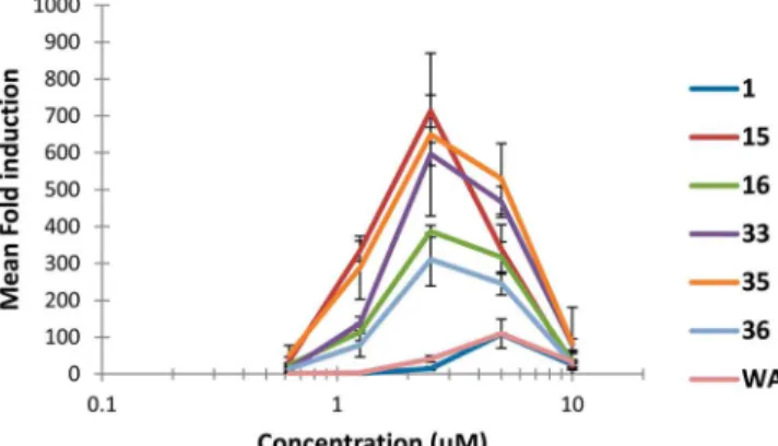
activation. As evident in Figure 3, for
1
and its
fi
ve most active
analogues (
15
,
16
,
33
,
35
, and
36
), reporter response was not a
monotonic function but rather peaked over a limited
concentration range for each active compound and then
declined, presumably as toxicity compromised the ability of
cells to respond. To quantitate this type of concentration
dependence in a way that would capture overall
heat-shock-inducing potential, we de
fi
ned a heat-shock index (HSI) for
each compound calculated as Log
2of the maximal response
(fold induction) divided by the concentration required to
stimulate this response (Table 2). Interestingly, all
fi
ve
withanolide analogues (
15
,
16
,
33
,
35
, and
36
) that
demonstrated a HSI more than 2-fold of that of
1
contained
acetyl substituents at C-12 and/or C-27 in addition to the
ring-A 2(3)-en-1-one and ring-B 5β,6β-oxirane moieties. Greater
heat-shock-induction activity for analogues
33
and
35
as
compared to WA was con
fi
rmed using an alternate heat-shock
reporter cell line, as previously described.
18Using this reporter
system in nontransformed cells instead of cancer cells and
fl
uorescence instead of luciferase activity as an end point,
heat-shock indices for
1
,
33
, and
35
were calculated as 2.1, 4.7, and
5.2, respectively (data not shown). Conservation of the rank
order for these compounds under di
ff
erent assay conditions
indicates that their heat-shock induction activity is an intrinsic
property, not an artifact of the particular system used to
measure it.
Heat-Shock Induction vs Cytotoxicity.
Next, to
determine whether the heat-shock-inducing activity and
cytotoxicity of withanolides
1
−
37
could be dissected on the
basis of structural features, we examined the correlation of these
two activities across all 37 compounds (Figure 4). To monitor
cytotoxicity in the most sensitive manner possible, we
incubated H929 myeloma cells for 3 days with serial dilutions
of each compound in a 384-well format. This human cell line,
as with most myeloma cell lines, is particularly sensitive to
agents that disrupt protein homeostasis, especially proteasome
inhibitors such as MG-132.
54If heat-shock induction was solely
a consequence of cytotoxicity arising from impairment of the
proteasome and HSP90 (previously reported targets for WA
20)
or other mediators of protein homeostasis, then we would have
expected to see a consistent correlation between these activities
across all analogues tested. Instead, we found a relatively poor
correlation (
r
2= 0.62), primarily because of a group of outlying
analogues (
15
,
16
,
35
, and
36
) that displayed greater
heat-shock induction than
1
at approximately the same level of
toxicity. In contrast, withanolide
33
was both more cytotoxic
and more heat-shock active than
1
, and the proteasome
inhibitor MG-132 used as a positive control showed greater
Table 2. Heat-Shock Index and Cytotoxicity of 1 and Its
Analogues 2
−
37
withanolide heat-shock indexa cytotoxicityb
1 1.35 0.25 (0.239−0.255)
2 0.52 0.88 (0.826−0.941)
3 0.00 >10
4 0.00 5.25 (4.689−5.881)
5 0.59 0.54 (0.447−0.657)
6 0.00 0.61 (0.564−0.656)
7 0.00 8.74 (8.147−9.372)
8 0.00 >10
9 0.00 >10
10 0.00 5.09 (4.430−5.841)
11 0.00 >10
12 0.00 >10
13 0.00 4.96 (4.486−5.489)
14 1.73 0.25 (0.242−0.261)
15 3.81 0.25 (0.243−0.266)
16 3.44 0.22 (0.202−0.232)
17 1.53 0.43 (0.387−0.475)
18 0.20 3.32 (3.115−3.528)
19 0.00 >10
20 0.00 >10
21 0.36 1.12 (1.076−1.163)
22 0.78 1.04 (0.933−1.642)
23 0.00 >10
24 0.90 1.63 (1.567−1.703)
25 0.74 2.29 (2.161−2.430)
26 0.00 >10
27 0.79 1.16 (0.914−1.475)
28 1.68 0.28 (0.268−0.298)
29 0.00 4.30 (3.950−4.686)
30 0.00 3.83 (3.085−4.755)
31 0.00 1.65 (1.580−1.715)
32 0.92 0.46 (0.408−0.516)
33 3.69 0.05 (0.044−0.054)
34 1.64 0.25 (0.222−0.272)
35 3.74 0.27 (0.255−0.287)
36 3.31 0.18 (0.156−0.200)
37 1.65 0.24 (0.221−0.252)
MG-132 0.71 0.17 (0.164−0.184)
aMeasured in the 293T reporter line and calculated as [Log
2(treated/
control]/concentration (μM). bConcentration resulting in 50%
reduction in relative viable myeloma (H929) cell number after 72 h exposure based on a nonlinear curvefit of the dose−response data in Prism 5.0 software (95% confidence interval).
toxicity than heat-shock induction compared to
1
. As noted
earlier, analogues with the greatest heat-shock activity all
contained acetyl substituents at C-4 and/or C-27 in addition to
the ring-A 2(3)-en-1-one and ring-B 5β,6β-oxirane moieties.
Withanolide
33
was acetylated at only C-27, whereas
35
carried
acetyl substituents at both C-4 and C-27.
Given the surprising nature of these
fi
ndings, we veri
fi
ed the
results by repeat testing of analogues
33
and
35
in a
low-throughput format.
1
and MG-132 were included for reference
(Figure 5). Although the absolute magnitude of e
ff
ects changed
with the alternate format, relative relationships were preserved.
Again,
35
demonstrated greater heat-shock-inducing activity
(Figure 5a) with less cytotoxicity. This was the case both in the
cell line used for the reporter assays (293T cells, Figure 5b) and
in H929 cells, the line used for our initial correlation analysis
(Figure 5c). MG-132 was more cytotoxic for H929 cells than
293T cells, consistent with the known hypersensitivity of
myeloma cells to proteasome inhibition. It is noteworthy that
the more potent cytotoxicity of
33
in these assays was
consistent with results from the acute toxicity assay we used to
generate the data summarized in Table 1. Here, using CHP-100
cells, 24 h compound exposure, and MTT dye reduction as the
end point,
33
(IC
50= 0.22
±
0.02) was also signi
fi
cantly more
potent than withanolide
35
(IC50
= 0.35
±
0.01).
Thiol Reactivity and Biological Activity.
Compound
1
contains three electrophilic sites that could play important roles
in its biological activity, including HSA and cytotoxicity. These
are C-3 of the ring-A 2(3)-en-1-one moiety, C-5/C-6 of the
ring-B 5β,6β-oxirane moiety, and C-24 of the ring-E
unsaturated lactone moiety. We have previously shown that
coincubation of our heat-shock reporter cells with
N
-acetylcysteine (NAC) and
1
leads to a near complete
suppression of heat-shock activation caused by
1
.
18Thus, it
was of interest to assess the chemical reactivity of
1
compared
to informative analogues using NAC as a representative thiol
nucleophile. Treatment of
1
with NAC a
ff
orded the product of
Michael addition at C-3 (
38
) in 66% yield. No addition was
observed at C-5, C-6, or C-24 by
1H NMR and HPLC. As
expected, 2,3-dihydrowithaferin A (
20
) was unreactive under
these conditions. Because
20
is devoid of both HSA and
cytotoxic activity, the presence of the ring-A 2(3)-en-1-one
moiety appears to be essential for many, if not all, of WA’s
biological activities. To probe the relationship between the thiol
reactivity of this enone moiety and HSA further, we monitored
reaction of
1
with 1 equiv of NAC in DMSO-
d
6and, in parallel,
reaction of NAC with
35
, our analogue with the highest
heat-shock index. The progress of these reactions was assessed by
1H
NMR using the disappearance of the signal from H-2 of the
ring-A 2(3)-en-1-one moiety as the end point (Supporting
Information, Figure S31). The ratio of the starting material to
product was used to determine the Gibb’s free energy (Δ
G
) of
the thiol addition/elimination processes.
55Reactions of
1
and
Figure 4. Correlation of heat-shock induction with cytotoxicity forcompounds1−37.The proteasome inhibitor MG-132 is included as a mechanistically and structurally distinct control compound. The solid line depicts a linear curve fit for all data points (r2 = 0.62), as performed in Microsoft Excel software. The heat-shock-active analogues lying furthest off the curve fit are circled in red. Heat-shock index (determined in 293T reporter cells) was calculated as [Log2(treated/control)]/concentration. Cytotoxicity (determined in
H929 myeloma cells) was calculated as Log2 IC50, where IC50 is
concentration (μM) resulting in 50% reduction in the relative viable cell number.
35
with NAC showed typical second-order reaction curves,
with calculated
Δ
G
values of
−
7.0 and
−
9.3 kcal/mol,
respectively, indicating that analogue
35
reacts more readily
with NAC than
1
. Therefore, in regards to the group appended
at C-4 of the withanolide sca
ff
old, its identity (OH or OAc),
orientation (α
or
β), or a combination of these factors can alter
the chemical reactivity of the ring-A 2(3)-en-1-one system. The
more reactive analogue,
35
, demonstrated greater HSA, but
additional experimentation will be required to de
fi
ne the
relationship between the relative chemical reactivity of this
enone system and HSA fully. We can conclude, however, that
orientation of the group at C-4 appears to exert little or no
e
ff
ect on acute cytotoxicity, as the pairs of analogues
1
and
28
,
15
and
33
,
16
and
35
, and
17
and
34
showed very similar IC
50values in limiting the growth and survival of CHP-100 cells
(Table 1).
Heat-Shock Induction In Vivo.
To determine whether the
heat-shock activity of withanolides demonstrated in cell culture
would translate to whole animals, we performed exploratory
pharmacodynamic studies in mice (Figure 6). Establishing a
biocompatible cyclodextrin-based formulation for these very
poorly water-soluble compounds, we compared the
dose-dependent ability of
1
and
35
to activate a systemic heat-shock
response after parenteral administration. Single doses up to 50
mg/kg were tolerated without overt acute toxicity. Choosing
spleen as a sentinel organ representative of the hematopoietic
compartment, we assayed lysates for upregulation of heat shock
protein 72 (HSP72), the most highly heat-inducible isoform of
the HSP70 family of molecular chaperones. At a dose of 25
mg/kg, several mice receiving
35
responded with a robust
increase in relative HSP72 level, leading to a highly signi
fi
cant
increase in variance for this group. In contrast,
1
caused a much
smaller, nonsigni
fi
cant e
ff
ect on variance, indicative of little
treatment e
ff
ect under these conditions. Increasing the dose of
35
to 40 mg/kg produced more uniform induction across the
treatment group and a signi
fi
cant di
ff
erence in the mean HSP72
level compared to the vehicle control group. Although much
work beyond the scope of this initial report obviously remains,
these exploratory
fi
ndings con
fi
rm that withanolides can
activate the heat-shock response in mice at systemically
tolerable exposures. Whether activation of the heat-shock
response per se will prove a key determinant of the therapeutic
bene
fi
ts ascribed to withanolides in diverse human diseases
remains to be determined. Equally important may be their
ability to form thiol adducts with a range of important
electrophile sensors in cells that are among the
fi
rst-line
defenses for launching adaptive transcriptional and
post-transcriptional responses.
56,57Nevertheless,
fi
ndings presented
here indicate that heat-shock induction can serve as a useful
biomarker for their activity in vivo.
■
CONCLUSIONS
same relative level of cytotoxicity, it does appear possible to
discriminate between these activities through speci
fi
c structural
modi
fi
cations to the withanolide core. Conversely, other
modi
fi
cations increased bioactivity in general, both heat-shock
activation and cytotoxicity, perhaps by enhancing cellular
uptake or limiting metabolic inactivation.
From our results, we conclude that the withanolide sca
ff
old
can be modi
fi
ed to shift its spectrum of bioactivity while
preserving potency. Enhancing heat-shock response while
minimizing cytotoxicity could provide a better therapeutic
index in pursuit of compounds that activate intrinsic cellular
defense mechanisms to combat protein aggregation-associated
neurodegenerative disorders. Conversely, minimizing activation
of the cytoprotective heat-shock response while maintaining
antiproliferative activity could provide more e
ff
ective anticancer
agents. Furthermore, reporter assay-guided tuning of the
withanolide core appears to provide a practical route to
realizing the full therapeutic potential of this versatile sca
ff
old.
■
MATERIALS AND METHODS
General Procedures.Melting points were determined in capillary tubes using a Mel-Temp apparatus and are uncorrected. Optical rotations were measured in MeOH or CHCl3 with a Jasco DIP-370
digital polarimeter. UV spectra were determined in MeOH on a Shimadzu UV-1601 spectrometer. One-dimensional and 2D NMR spectra were recorded in CDCl3, unless otherwise stated, using
residual solvents as internal standards on Bruker DRX-500, DRX-600, and Avance III 400 spectrometers at 500, 600, and 400 MHz for1H
NMR and 125, 150, and 100 MHz for13C NMR. The chemical shift
values (δ) are given in parts per million (ppm), and the coupling constants (Jvalues) are in Hz. LR−MS and HR−MS were recorded using Shimadzu LCMS 8000QPαand JEOL HX110A spectrometers, respectively. Analytical thin-layer chromatography was carried out on silica gel 60 F254aluminum-backed TLC plates (Merck). Preparative
thin-layer chromatography was performed on Analtech silica gel 500
μm glass plates. Compounds were visualized with short-wavelength UV and by spraying with anisaldehyde-sulfuric acid spray reagent and heating until the spots appeared. Silica gelflash chromatography was accomplished using 230−400 mesh silica gel. All yields refer to yields of isolated compounds. Unless otherwise stated, chemicals and solvents were of reagent grade and used as obtained from commercial sources without further purification. Purity of allfinal compounds was determined to be≥95% by HPLC and1H NMR analysis.
Isolation of Naturally Occurring Withanolides 1−13. With-aferin A (1), 27-deoxywithaferin A (2), viscosalactone B (3), 2,3-dihydrowithaferin A-3β-O-sulfate (4), 3α -(uracil-1-yl)-2,3-dihydrowi-thaferin A (5), 3β-(adenin-9-yl)-2,3-dihydrowithaferin A (6), 3β-O -butyl-2,3-dihydrowithaferin A (7), withanolide A (8), 27-hydroxywi-thanolide B (9), 4β,27-dihydroxy-1-oxo-22R-witha-2,5,24-trienolide (10), 2,3-didehydrosomnifericin (11), jaborasalactone D (12), and pubesenolide (13) were obtained from aeroponically grown W. somniferaas described previously.22,33
General Procedure for Acetylation of Withanolides. To a solution of the withanolide (2.0 mg) in anhydrous pyridine (0.5 mL) was added Ac2O (0.5 mL), and the mixture was stirred at 25°C until
the reaction was complete (judged by the disappearance of the starting material by TLC). The reaction mixture was poured into ice/water (10.0 mL), and the resulting solution was passed through a short column of RP (C18) silica gel (0.2 g). The column was washed with
water (30.0 mL) followed by elution with MeOH (10.0 mL). The MeOH fraction after evaporation was subjected to preparative TLC (silica gel) to yield the corresponding acetyl derivative.
Preparation of 27-Acetylwithaferin A (15) and
4,27-Diacetylwi-thaferin A (16).To a solution of1(10.0 mg) in pyridine (0.1 mL) was
added Ac2O (2.4μL), and the mixture was stirred at 25°C. After 2 h,
EtOH (15.0 mL) was added to the reaction mixture and evaporated under reduced pressure. The residue thus obtained was separated by
preparative TLC (silica gel) using 6% MeOH in CH2Cl2as eluant to
give15(2.1 mg, 19%) and16(8.5 mg, 72%).
27-Acetylwithaferin A (15).White solid; mp 218−220°C; [α]D25
+128 (c0.8, CHCl3);1H NMR (500 MHz, CDCl3)δ6.90 (dd,J= 9.9,
5.8 Hz, 1H, H-3), 6.18 (d,J= 9.9 Hz, 1H, H-2), 4.88 (d,J= 11.8 Hz, 1H, H-27a), 4.84 (d,J= 11.8 Hz, 1H, H-27b), 4.38 (dt,J= 13.6, 3.3 Hz, 1H, H-22), 3.74 (dd,J= 5.8, 2.1 Hz, 1H, H-6), 3.22 (s, 1H, H-4), 2.51 (dd,J= 13.2, 10.9 Hz, 2H), 2.12 (ddd,J= 14.9, 6.3, 2.6, 1H, H-7a), 2.05 (s, 3H, H3-28), 2.04 (s, 3H, OAc), 1.96 (m, 2H), 1.93 (dt,J=
9.6, 3.3 Hz, 1H), 1.82 (dt,J= 14.2, 3.6 Hz, 1H), 1.69−1.59 (m, 2H), 1.53−1.43 (m, 2H), 1.39 (s, 3H, H3-18), 1.25 (m, 3H), 1.18−1.01 (m,
2H), 0.98 (d,J= 6.6 Hz, 3H, H3-21), 0.91−0.82 (m, 2H), 0.69 (s, 3H,
H3-19); HRMS (ESI): [M + H]+calcd for C30H41O7, 513.2847; found,
513.2850.
4,27-Diacetylwithaferin A (16).White solid; mp 232−234°C;1H
NMR data were consistent with those reported;3APCI-MS (+)m/z:
[M + 1]+555.
Sulfation of Withaferin A.To a stirred solution1(10.0 mg) in pyridine (0.5 mL) was added SO3-pyridine complex (5.0 mg). and the
reaction mixture was heated at 80°C. After 1 h (TLC control), the reaction mixture was poured to ice water (30 mL), and the resulting solution was introduced to a short column of RP (C18) silica gel (5.0
g) and washed with water (50 mL) followed by elution with 40% MeOH(aq). The crude product obtained by evaporation of MeOH-(aq) eluents was finally purified by C18 preparative TLC (40% MeOH(aq)) to afford18(8.3 mg) and19(3.6 mg).
Withaferin A 27-Sulfate (18). Amorphous colorless solid; [α]D20
+55.9 (c0.78, MeOH);1H NMR (400 MHz, pyridine-d5)δ0.53 (s,
3H), 0.94 (d,J= 7.0 Hz, 3H), 1.84 (s, 3H), 2.09 (s, 3H), 2.30 (dd,J= 13.5, 17.5 Hz, 1H), 3.59 (brs, 1H), 4.02 (d,J= 6.5 Hz, 1H), 4.42 (brd,
J= 13.0 Hz, 1H), 5.31 (d,J= 11.0 Hz, 1H), 5.44 (d,J= 11.0 Hz, 1H), 6.41 (d,J= 9.5 Hz, 1H), 7.22 (dd,J= 6.5, 9.5 Hz, 1H);13C NMR
(125 MHz, pyridine-d5)δ11.7, 13.5, 17.3, 20.5, 21.8, 24.6, 27.3, 30.1, 30.4, 31.8, 39.1, 39.6, 42.7, 44.7, 48.6, 52.0, 56.1, 60.2, 61.2, 64.6, 70.4, 78.5, 122.7, 132.4, 145.2 158.2, 165.8, 202.6.
Withaferin A 4,27-Disulfate (19).Amorphous colorless solid; [α]D20
+158.5 (c0.2, MeOH);1H NMR (400 MHz, CD
3OD)δ0.74 (s, 3H),
1.00 (d,J= 6.5 Hz, 3H), 1.36 (s, 3H), 2.13 (s, 3H), 2.54 (dd,J= 13.0, 18.0 Hz, 1H), 3.29 (brs, 1H), 4.28 (d,J= 6.0 Hz, 1H), 4.45 (dt,J= 3.5, 13.0 Hz, 1H), 4.76 (d,J= 11.0 Hz, 1H), 4.85 (overlap with H2O
peak, 1H), 6.23 (d,J= 10.0 Hz, 1H), 7.17 (dd,J= 6.0, 10.0 Hz, 1H);
13C NMR (125 MHz, CD
3OD)δ 10.5, 12.2, 14.7, 19.3, 20.9, 24.0,
26.8, 29.5, 29.6, 31.0, 38.9, 39.1, 42.2, 44.2, 51.6, 55.8, 59.8, 60.8, 61.3, 75.6, 78.6, 121.2, 132.4, 134.6, 142.1, 156.9, 166.4, 201.7.
2,3-Dihydrowithaferin A (20). To a solution of 1(15.0 mg) in
EtOH (1.0 mL) were added Et3N (60μL) and 10% Pd/C (1.0 mg),
and the mixture was stirred under an atmosphere of H2for 1 h. The
reaction mixture wasfiltered, and the filtrate was evaporated under reduced pressure to give 2,3-dihydrowithaferin A (20) as a white solid (14.7 mg, 98%); mp 267−269°C;1H NMR data were consistent with
those reported;3APCI-MS (+)m/z: [M + 1]+473.
27-Acetyl-2,3-dihydrowithaferin A 4-Sulfate (21).Acetylation of4
by the general procedure afforded21as an amorphous colorless solid; [α]D20 +27.7 (c 0.33, MeOH); 1H NMR (400 MHz, pyridine-d5) δ
0.51(s, 3H), 0.97 (d,J= 6.0 Hz, 3H), 1.67 (s, 3H), 2.00 (d,J= 1.5 Hz, 3H), 2.02 (s, 3H), 2.41 (dd,J= 13.0, 16.0 Hz, 1H), 3.27 (dd,J= 6.0, 18.0 Hz, 1H), 3.60 (dd,J= 10.5, 14.0 Hz, 1H), 4.42 (brd,J= 13.0 Hz, 1H), 4.50 (brs, 1H), 5.12 (d,J= 12.0 Hz, 1H), 5.22 (d,J= 12.0 Hz, 1H), 5.59 (dd,J= 6.5, 8.0 Hz, 1H);13C NMR (125 MHz, pyridine-d5)
δ11.5, 13.5, 15.4, 20.3, 20.8, 21.3, 24.5, 27.3, 30.0, 30.2, 31.5, 39.2 (×2), 41.6, 42.8 (×2), 49.5, 52.0, 56.0, 56.8, 58.6, 64.7, 72.6, 76.4, 78.4, 122.0, 157.9, 165.3, 170.7, 209.1; HRMS (ESI): [M−H]−
calcd for C30H41O11S, 609.2375; found, 609.2365.
4,27-Diacetyl-2,3-dihydrowithaferin A 3β-O-Sulfate (22).
Amor-phous colorless solid; [α]D20 +42.4 (c 0.38, MeOH);1H NMR (400
8.5 Hz, 1H), 5.86 (brs, 1H);13C NMR (125 MHz, pyridine-d5)δ11.5,
13.5, 15.1, 20.3, 20.7, 20.8, 21.4, 24.4, 27.2, 29.6, 30.1, 31.0, 39.08, 39.13, 41.7, 42.7, 42.8, 50.0, 51.9, 55.9, 57.7, 58.6, 62.1, 69.7, 78.2, 78.4, 122.1, 157.9, 165.3, 170.4, 170.7, 208.2; HRMS (ESI): [M + H]+
calcd for C32H45O12S, 653.2626; found, 653.2626.
4,6,27-Triacetyl-2,3-didehydrosomnifericin (25). Amorphous
col-orless solid; [α]D20 +203.2 (c 0.03, MeOH); 1H NMR (400 MHz,
CDCl3)δ0.68 (s, 3H), 0.96 (d,J= 6.0 Hz, 3H), 1.28 (s, 3H), 2.02 (s,
3H), 2.04 (s, 3H), 2.06 (s, 3H), 2.18 (s, 3H), 2.49 (dd,J= 13.5, 17.6 Hz, 1H), 4.38 (dt,J= 3.4, 13.2 Hz, 1H), 4.85 (d,J= 19.0 Hz, 1H), 4.88 (d,J= 19.0 Hz, 1H), 5.06 (dd,J= 5.0, 12.1 Hz, 1H), 6.06 (dd,J= 2.2, 10.3 Hz, 1H), 6.29 (dd,J= 2.2, 10.3 Hz, 1H), 6.35 (t,J= 2.2 Hz, 1H); APCI-MSm/z: [M + H]+615.
4-Dehydrowithaferin A (27). To a solution of 1 (30 mg) in
CHCl3/EtOAc (5:7, 2.0 mL) was added freshly prepared manganese
dioxide (300 mg), and the mixture was stirred at 25°C. After 16 h, the reaction mixture wasfiltered, thefiltrate was evaporated under reduced pressure, and the residue was purified by preparative TLC (silica gel) using 8% MeOH in CH2Cl2as eluant to give27(18.4 mg, 62%) as a
white powder; mp 273−275°C (lit.3272−275°C); [α]
D
25+143 (c0.8,
MeOH) [lit.3+147 (c0.83, MeOH)]; APCI-MS (+)m/z: [M + 1]+
469;1H NMR data were consistent with those reported.3
27-Acetyl-4-dehydrowithaferin A (32).To a solution of27 (5.0
mg) in pyridine (0.1 mL) was added Ac2O (0.05 mL), and the mixture
was stirred at 25°C for 18 h. The reaction mixture was evaporated under reduced pressure and by adding EtOH, and the residue was purified by preparative TLC (silica gel) using 4% MeOH in CH2Cl2as eluant to give32(5.25 mg, 96%) as a white solid; mp 173−175°C;1H
NMR (500 MHz, CDCl3)δ6.85 (d,J= 10.3 Hz, 1H, H-3), 6.82 (d,J
= 10.3 Hz, 1H, H-2), 4.88 (d,J= 11.8 Hz, 1H, H-27a), 4.85 (d,J= 11.8 Hz, 1H, H-27b), 4.39 (dt,J= 13.2, 3.4 Hz, 1H, H-22), 3.41 (d,J
= 2.3 Hz, 1H, H-6), 2.51 (dd,J= 18.4, 13.2 Hz, 1H, H-23a), 2.15 (dt,J
= 15.2, 3.4 Hz, 1H, H-7a), 2.06 (s, 3H, H3-28), 2.04 (s, 3H, OAc),
2.03−1.95 (m, 3H), 1.70−1.57 (m, 5H), 1.47−1.39 (m, 2H), 1.35−
1.07 (m, 5H), 1.37 (s, 3H, H3-18), 1.00 (d,J= 6.7 Hz, 3H, H3-21),
0.71 (s, 3H, H3-19); APCI-MS (+)m/z: [M + 1]+511.
Withaferin A 27-tert-Butyldimethylsilyl Ether (39).To a solution
of1(25 mg) in DMF (1.5 mL) were addedt-BDMS-Cl (63 mg) and 4-PP (78 mg), and the mixture was stirred at 60°C for 3 h, after which the reaction mixture was diluted with EtOAc, washed with brine, dried over anhydrous Na2SO4, and evaporated under reduced pressure, and
the residue was separated on preparative TLC (silica gel) using 3% MeOH in CH2Cl2as eluant to give39as a white solid (28 mg, 90%);
mp 178−180°C;1H NMR (500 MHz, CDCl
3)δ6.90 (dd,J= 9.9, 5.8
Hz, 1H, H-3), 6.18 (d,J= 9.9 Hz, 1H, H-2), 4.48 (d,J= 11.6 Hz, 1H, H-27a), 4.37 (d,J= 11.6 Hz, 1H, H-27b), 4.36 (dt,J= 16.6, 3.3 Hz, 1H, H-22), 3.73 (d,J= 5.8 Hz, 1H, H-4), 3.21 (s, 1H, H-6), 2.44 (dd,
J= 17.4, 13.5 Hz, 1H, H-23a), 2.13 (m, 1H, H-7a), 2.04 (s, 3H, H3
-28), 1.97−1.91 (m, 2H), 1.80 (dq,J= 14.2, 3.6 Hz, 1H), 1.68−1.58 (m, 3H), 1.52−1.43 (m, 2H), 1.39 (s, 3H, H3-18), 1.37−0.99 (m,
11H), 0.95 (d,J= 8.8 Hz, 3H, H3-21), 0.87 (s, 9H, 3 x CH3), 0.68 (s,
3H, H3-19), 0.07 (s, 3H, SiCH3), 0.06 (s, 3H, SiCH3); APCI-MS (+) m/z: [M + 1]+585.
4-Acetylwithaferin A 27-tert-Butyldimethylsilyl Ether (40).
Acetylation of39(20.0 mg) by the usual procedure (Ac2O/pyridine)
afforded40as a white solid (21.0 mg, 98%); APCI-MS (+)m/z: [M + 1]+627.
4-Acetylwithaferin A (14). Deprotection of 40 (19.0 mg) was
carried out by treating a solution of it in THF (0.3 mL) and MeOH (0.05 mL) with 2 N HCl (0.05 mL) at 0°C for 1 h. The reaction mixture was diluted with H2O, evaporated under reduced pressure,
and extracted with EtOAc. The EtOAc layer was evaporated under reduced pressure, and the residue was purified by preparative TLC (silica gel) using 5% MeOH in CH2Cl2as eluant to give14as a white
solid (15 mg, 96%); mp 192−194°C;1H NMR data were consistent
with those reported;58 HRMS (ESI): [M + H]+calcd for C 30H41O7,
513.2847; found, 513.2852.
4-epi-Withaferin A (28).To a stirred solution of 27(6.0 mg) in
MeOH (1.0 mL) and THF (0.5 mL) was added CeCl3·7H2O (17 mg).
The reaction mixture was cooled to 0°C in an ice bath, and NaBH4
(2.0 mg) was added. After 30 min at 0°C, the reaction mixture was evaporated, and the residue was separated on preparative TLC (silica gel) using 6% MeOH in CH2Cl2as eluant to give28as a white solid
(4.2 mg, 70%); mp 227−228 °C; [α]D25 +29.9 (c 1.0, CHCl3);1H
NMR (500 MHz, CDCl3+ CD3OD)δ6.80 (dd,J= 10.1, 1.5 Hz, 1H,
H-3), 5.97 (dd,J= 10.1, 2.5 Hz, 1H, H-2), 4.64 (brs, 1H, H-4), 4.37 (dt,J= 13.5, 3.3 Hz, 1H, H-22), 4.32 (d,J= 12.5 Hz, 1H, H-27a), 4.27 (d,J= 12.5 Hz, 1H, H-27b), 3.65 (brs, 1H, H-6), 2.45 (dd,J= 13.6, 7.2 Hz, 1H, H-23a), 2.10 (brd, 1H, H-7a), 2.00 (s, 3H, H3-28),1.96−
1.89 (m, 4H), 1.78 (brs, 1H), 1.67−1.58 (m, 2H), 1.49−1.42 (m, 2H), 1.31 (m, 1H), 1.18 (s, 3H, H3-18), 1.15−1.00 (m, 4H), 0.94 (d,J= 6.6
Hz, 3H, H3-21), 0.88 (m, 1H), 0.66 (s, 3H, H3-19);13C NMR (125
MHz, CDCl3+ CD3OD)δ201.4, 167.1, 153.3, 148.0, 128.4, 125.6,
78.7, 65.8, 64.4, 57.0, 55.9, 55.4, 51.9, 47.6, 45.5, 42.6, 39.4, 38.8, 30.7, 29.8, 27.2, 24.2, 22.2, 20.0, 13.8, 13.2, 12.0; HRMS (ESI): [M + H]+
calcd for C28H39O6, 471.2747; found, 471.2764.
4,27-Diacetyl-4-epi-withaferin A (35).Acetylation of28(1.0 mg)
by the usual procedure (Ac2O/pyridine) followed by purification on
preparative TLC (silica gel) using 6% MeOH in CH2Cl2 as eluant
afforded 35 (1.1 mg, 93%); mp 214−216 °C; [α]D25 +36.8 (c 1.1,
CHCl3);1H NMR (600 MHz, CDCl3)δ6.66 (dd,J= 10.1, 1.5 Hz,
1H, H-3), 6.05 (dd,J= 10.1, 2.4 Hz, 1H, H-2), 5.87 (brs, 1H, H-4), 4.88 (d,J= 11.8 Hz, 1H, H-27a), 4.84 (d,J= 11.8 Hz, 1H, H-27b), 4.38 (dt,J= 13.1, 3.3 Hz, 1H, H-22), 3.53 (brs, 1H, H-6), 2.50 (dd,J= 17.6, 13.3 Hz, 1H, H-23a), 2.09 (s, 3H, OAc), 2.05 (s, 3H, H3-28),
2.03 (s, 3H, OAc), 2.01−1.92 (m, 4H), 1.67−1.33(m, 6H), 1.28 (s, 3H, H3-18), 1.23−1.01 (m, 4H), 0.98 (d, J = 6.6 Hz, 3H, H3-21),
0.94−0.81 (m, 2H), 0.70 (s, 3H, H3-19); 13C NMR (100 MHz,
CDCl3)δ201.1, 170.9, 169.8, 165.3, 157.0, 144.9, 129.4, 121.9, 78.2,
65.8, 63.3, 58.0, 55.9, 55.8, 52.0, 48.1, 45.5, 42.5, 39.5, 38.8, 34.6, 31.8, 30.8, 30.1, 29.6, 27.3, 24.2, 22.6, 20.9, 20.8, 20.5, 14.6, 14.1, 13.3, 11.7; HRMS (ESI): [M + H]+ calcd for C
32H43O8, 555.2952; found,
555.2950.
27-Acetyl-4-epi-withaferin A (33).To a stirred solution of32(3.0
mg) in THF (0.2 mL) and MeOH (0.2 mL) at 0 °C were added CeCl3·7H2O (65 mg) and NaBH4 (small portion), and the mixture
was stirred at 0°C. After 10 min, a small ice cube was added to the reaction mixture, the solvent and water were evaporated under reduced pressure, and the residue was partition between H2O and
EtOAc. The EtOAc layer was dried over anhydrous Na2SO4 and
evaporated under reduced pressure, and the residue was separated on preparative TLC (silica gel) using 2% MeOH in CH2Cl2as eluant to
give33(2.5 mg, 70%) as a white solid; mp 188−190°C; [α]D25+ 42.4
(c1.0, CHCl3);1H NMR (500 MHz, CDCl3)δ6.83 (dd,J= 10.2, 1.4
Hz, 1H, H-3), 6.00 (d,J= 10.2, 2.5 Hz, 1H, H-2), 4.88 (d,J= 11.9 Hz, 1H, H-27a), 4.85 (d,J= 11.9 Hz, 1H, H-27b), 4.71 (s, 1H, H-4), 4.38 (dt,J= 13.2, 3.3 Hz, 1H, H-22), 3.63 (s, 1H, H-6), 2.50 (dd,J= 17.6, 14.5 Hz, 1H, H-23a), 2.12 (m, 1H, H-7a), 2.05 (s, 3H, H3-28), 2.03 (s,
3H, OAc), 1.99 (dd,J= 13.2, 3.3 Hz, 1H), 1.93 (brd,J= 9.9 Hz, 1H), 1.68−1.45 (m, 4H), 1.34 (m, 1H), 1.28−1.22 (m, 3H), 1.21 (s, 3H, H3-18), 1.18−1.03 (m, 4H), 0.98 (d,J= 6.7 Hz, 3H, H3-21), 0.94−
0.81 (m, 2H), 0.69 (s, 3H, H3-19);13C NMR (100 MHz, CDCl3) δ
201.2, 170.9, 165.3, 156.9, 147.6, 141.5, 129.0, 121.9, 78.2, 65.6, 64.6, 58.0, 55.3, 52.0, 47.6, 45.8, 42.5, 35.4, 38.8, 30.8, 30.1, 27.7, 27.3, 24.3, 22.6, 22.2, 20.9, 20.6, 14.1, 13.7, 13.3, 11.8, 11.6; HRMS (ESI): [M + H]+calcd for C
30H41O7, 513.2847; found, 513.2854.
4-Dehydrowithaferin A 27-tert-Butyldimethylsilyl Ether (41).To a
solution of27(11.3 mg) in DMF (0.5 mL) were addedt-BDMS-Cl (36.4 mg) and 4-pp (42.9 mg), and the mixture was stirred under an atmosphere of N2for 1 h at 60°C. The reaction mixture was then
diluted with EtOAc, washed with brine, and evaporated under reduced pressure, and the residue was separated on preparative TLC using CH2Cl2as eluant to give41(9.5 mg). APCI-MS (+)m/z: [M + 1]+
583.
4-epi-Withaferin A 27-tert-Butyldimethylsilyl Ether (42). To a
solution of41(9.5 mg) in THF (0.2 mL) and MeOH (0.2 mL) at 0
°C was added CeCl3·7H2O (125 mg), and the mixture was stirred at 0 °C for 5 min. To this solution was then added NaBH4(small portion),
under reduced pressure, and the residue was partition between H2O
and EtOAc. The EtOAc layer was dried over anhydrous Na2SO4and
evaporated under reduced pressure, and the residue was separated on preparative TLC (silica gel) using 2% MeOH in CH2Cl2as eluant to
give42(7.5 mg, 70%) as a white solid; APCI-MS (+)m/z: [M + 1]+
585.
4-Acetyl-4-epi-withaferin A 27-tert-Butyldimethylsilyl Ether (43).
A solution of42(7.5 mg) in pyridine (0.3 mL) and Ac2O (0.2 mL)
was stirred at 25°C for 4 h. The reaction mixture was evaporated under reduced pressure to give43(8.0 mg) as a white solid; APCI-MS (+)m/z: [M + 1]+627.
4-Acetyl-4-epi-withaferin A (34).To a solution of43(8.0 mg) in
THF (0.5 mL) and MeOH (0.3 mL) at 0°C was added 2 N HCl (0.15 mL), and the mixture was stirred at 0°C. After 1 h, the reaction mixture was diluted with H2O, MeOH and THF were evaporated
under reduced pressure and extracted with EtOAc (3×15 mL), the combined EtOAc layer was washed with H2O, dried over anhydrous
Na2SO4, and evaporated under reduced pressure, and the residue was
separated on preparative TLC (silica gel) using 5% MeOH in CH2Cl2
as eluant to give34as a white solid (5.3 mg, 70%); mp 236−38°C; [α]D25+ 29.7 (c1.2, CHCl3);1H NMR (600 MHz, CDCl3)δ6.66 (dd, J= 10.4, 1.5 Hz, 1H, H-3), 6.05 (dd,J= 10.4, 2.4 Hz, 1H, H-2), 5.87 (brs, 1H, H-4), 4.39 (brd,J= 13.4, 3.3 Hz, 1H, H-22), 4.37 (d,J= 12.5 Hz, 1H, H-27a), 4.32 (d,J= 12.5 Hz, 1H, 27b), 3.53 (brs, 1H, H-6), 2.48 (dd,J= 16.2, 13.9 Hz, 1H, H-23a), 2.11 (brd, 1H, H-7a), 2.09 (s, 3H, OAc), 2.01 (s, 3H, H3-28), 1.97−1.93 (m, 4H), 1.54−1.45 (m,
2H), 1.34 (m, 1H), 1.28 (s, 3H, H3-18), 1.23−1.00 (m, 6H), 0.98 (d,J
= 6.6 Hz, 3H, H3-21), 0.94−0.84 (m, 2H), 0.69 (s, 3H, H3-19);13C
NMR (100 MHz, CDCl3)δ201.1, 169.9, 166.0, 152.8, 144.8, 129.4,
125.8, 78.7, 65.8, 63.3, 57.5, 55.9, 55.8, 52.0, 48.1, 45.5, 42.6, 39.5, 38.8, 30.6, 29.9, 27.3, 24.2, 22.9, 20.8, 20.0, 14.5, 13.3, 11.6; HRMS (ESI): [M + H]+calcd for C
30H41O7, 513.2847; found, 513.2850. Microbial Biotransformation of 1.Small-scale fermentation of
Cunninghamella echinulata (ATCC 10028B) was performed in an Erlenmeyerflask (125 mL) containing soybean meal-glucose medium (25 mL) on a rotary shaker operating at 220 rpm at 28°C for 24 h. Large-scale fermentation was performed under the same conditions in Erlenmeyerflasks (3×250 mL) holding 50 mL of the medium in each flask, which was inoculated with 15% of the 1 day old inoculum. A solution of 1(5 mg in 0.5 mL of DMF) was added to each flask containing 24 h old second cultivation. After 72 h, fermentation broths were combined, and mycelium wasfiltered offand washed with H2O (100 mL), which was combined withfiltrate and extracted with EtOAc (3×200 mL). The combined organic layer was washed with H2O (2 ×200 mL), dried over anhydrous Na2SO4, and evaporated to give
EtOAc extract (42 mg). Gel-permeation chromatography of this extract over a column of Sephadex LH-20 (3.0 g) followed by preparative TLC (silica gel) gave 12β-hydroxywithaferin A (29, 3.8 mg, 24%) and 15β-hydroxywithaferin A (30, 4.9 mg, 32%).
12β-Hydroxywithaferin A (29).mp 120−121 °C (lit.38 119−120
°C);1H NMR data were consistent with those reported.38
15β-Hydroxywithaferin A (30).mp 271−273 °C (lit.38 270−274
°C);1H NMR data were consistent with those reported.38
12β-Acetoxy-4,27-diacetylwithaferin A (36).To a solution of 29
(1.0 mg) in pyridine (0.05 mL) was added Ac2O (0.05 mL), and the
mixture was stirred at 25 °C for 18 h. The reaction mixture was evaporated under reduced pressure and by adding EtOH, and the residue was purified on preparative TLC (silica gel) using 4% MeOH in DCM as eluant to give36(1.2 mg, 95%) as a white amorphous solid.1H NMR data were consistent with those reported.38
15β-Acetoxy-4,27-diacetylwithaferin A (37).To a solution of 30
(1.0 mg) in pyridine (0.05 mL) was added Ac2O (0.05 mL), and the
mixture was stirred at 25 °C for 18 h. The reaction mixture was evaporated under reduced pressure and by adding EtOH, and the residue was purified on preparative TLC (silica gel) using 4% MeOH in DCM as eluant to give37(1.2 mg, 95%) as a white amorphous solid.1H NMR data were consistent with those reported.38
Withaferin A N-Acetylcysteine Adduct (38). To a solution of1
(40.0 mg) in MeOH (4.0 mL) was addedN-acetylcysteine (NAC) (80.0 mg), and the solution was stirred at 25°C. After 48 h, MeOH
was removed under reduced pressure, and the residue was separated by HPLC [Phenomenex Luna C18 (5μ) 10 ×250 mm, gradient solvent system from 60 to 80% in 20 min, 3 mL/min flow rate, detection at 230 nm] to afford38(22.0 mg; 66% based on recovered
1) (tR = 13.0 min) and 1(9.4 mg) (tR = 14.8 min). 38: off-white
amorphous solid;1H NMR (400 MHz, CDCl
3)δ4.36 (1H, brd,J=
12.8 Hz, H-22), 4.32 and 4.27 (1H each, d,J= 12.4 Hz, H-27), 3.43 (1H, brs, H-4), 3.29 (1H, brs, H-6), 3.00 (2H, brs, Cys-CH2), 2.76 and
2.40 (1H each, m, H2-2), 2.00 (6H, s, Cys-CH3and H3-28), 1.23 (3H,
s, H3-19), 0.93 (3H, d,J= 6.4 Hz, H3-21), 0.61 (3H, s, H3-18);13C
NMR (100 MHz, pyridine-d5) δ 209.8 (C-1), 174.3 (Cys-CON), 170.6 (Cys-CO2H), 166.7 26), 154.2 3), 128.9 2), 121.9
(C-25), 88.1 (C-17), 80.9 (C-14), 79.5 (C-22), 78.9 (C-20), 65.3 (C-18), 64.3 (C-24), 127.8 (C-25), 78.7 (C-22), 77.2 (C-4), 65.4 (C-5), 59.5 (C-6), 56.6 (C-27), 56.3 (C-14), 54.0 (Cys-CH), 52.3 (C-17), 51.4 (C-10), 44.8 (C-3), 43.8 (C-9), 43.0 (C-13), 41.1 (C-2), 39.6 (C-16), 39.5 (C-20), 34.9 (Cys-CH2), 31.9 (C-23), 30.4 (C-8), 30.3 (C-7),
27.6 (C-12), 24.8 (C-15), 23.4 (Cys-CH3), 22.1 (C-11) 20.5 (C-28),
16.0 (C-19), 13.9 (C-21), 11.9 (C-18); LR-APCIMS (+):m/z[M + Na]+656, [M + H]+634, [M + H-NAC]+471.
Cytotoxicity Assays. Ewing’s sarcoma cell line CHP-100 and myeloma cell line H929 were cultured at 37°C under 6% CO2 in
RPMI 1640 media supplemented with 10% fetal bovine serum (FBS), and 293T cells were grown in DMEM supplemented with 10% FBS. All cell lines were tested and found to be negative for Mycoplasma
contamination. Cultures were passaged twice weekly, and cells in exponential growth were used for experiments. Stock solutions of compounds were formulated in DMSO and maintained at −20°C protected from light. To measure acute toxicity, CHP-100 cells were seeded inflat-bottom 96-well plates (7500 cells/well) and allowed to adhere overnight. Serial dilutions of compounds or DMSO vehicle control (not exceeding 0.2%) were added, and the relative viable cell number was determined 24 h later by dye-reduction assay using the substrate [3-(4,5-dimethylthiazol-2-yl)-2,5-diphenyltetrazolium bro-mide] (MTT). Cytotoxicity over 3 days against H929 and 293T cells was measured in 384-well format (3000 cells/well), and the relative viable cell number was determined by reduction of the dye resazurin (Alamar Blue) as previously described.40
Heat-Shock Reporter Assays.Reporter cells were generated by infecting 293T cells (American Type Culture Collection) with a previously reported lentiviral vector encoding a fusion protein consisting of enhanced GFP fused tofirefly luciferase under control ofHSP70B′promoter elements.54 The plasmid encoding the fusion
protein was generously provided by Khalid Shah (Massachusetts General Hospital, Boston, MA, USA). To isolate a homogeneous population of high responding cells, a transduced culture was heat-shocked at 42°C for 1 h and then processed 8 h later byfl uorescence-activated cell sorting (FACS). Prior to use, cells were reverse-selected by FACS to eliminate a minority population of cells constitutively expressing the reporter in the absence of induction. To evaluate compounds, cells were seeded in white 384-well plates (20 000 cells/ well). The following day, serial compound dilutions were added to quadruplicate wells, and incubation was continued overnight. Measurement of relative luciferase activity was achieved using an Envision plate luminometer (PerkinElmer) and Steady-Glo reagent (Promega) per the manufacturer’s recommendations. As a con-firmatory assay for some compounds, 3T3-Y9-B12 reporter cells were seeded in blackflat-bottom 96-well plates (20 000/well) and allowed to adhere overnight as previously reported.23 Cells were then incubated for 24 h in the presence of WA or analogues (1, 2, or 4
μM). After washing with PBS, fluorescence was quantified using an Analyst AD (LJL Biosystems) plate reader with excitation and emissionfilters set at 485 and 530 nm, respectively.