DOI: 10.14260/jemds/2015/1476
CASE REPORT
J of Evolution of Med and Dent Sci/ eISSN- 2278-4802, pISSN- 2278-4748/ Vol. 4/ Issue 58/ July 20, 2015 Page 10249
A CASE OF EXTRASKELETAL
EWING’
S SARCOMA/PNET PRESENTING AS A
MEDIASTINAL MASS
Mahendra Singh , S. )ram Arshad , Sweta Shekhar , Chayanika Pantola
HOW TO CITE THIS ARTICLE:
Mahendra Singh, S. )ram Arshad, Sweta Shekhar, Chayanika Pantola. A Case of Extraskeletal Ewing’s Sarcoma/PNET Presenting as a Mediastinal Mass . Journal of Evolution of Medical and Dental Sciences 2015; Vol. 4, Issue 58, July 20; Page: 10249-10251, DOI: 10.14260/jemds/2015/1476
ABSTRACT: The Ewing’s sarcoma/PNET family of tumors comes under small round blue cell tumors
SRBCT’s . These are high grade relatively rare malignant neoplasms affecting children and young
adults predominantly in 2nd decade. Most of these are seen affecting major long bones like femur, pelvis and ribs. Cases of extraskeletal Ewing’s saroma (EES) involving chest wall, pleura, mediastinum, retroperitoneum, paravertebral region, lower extremities and even very few cases of isolated maxillary sinus Ewing’s sarcoma have been reported.[1] These malignancies are characterized by a chromosome 22 rearrangement.[2] They are highly aggressive tumors with a high incidence of local recurrence and distant metastases.[3] Recent studies suggest that this tumour is likely to originate from primitive stem cells and degree of malignancy depends upon the stage of stem cell arrest during differentiation. PNET shows variable degree of neuroectodermal differentiation.[4]
CASE REPORT: We report a very rare case of primary mediastinal Ewing’s sarcoma. An 18 year old, previosly healthy, female presented in the OPD with left sided constant dull-aching chest pain, dysphagia and generalised weakness for two months. A CT scan was done which revealed a large increased attenuating space occupying lesion in left upper and middle zones having solid as well as cystic components, pulmonary deposits, and displacement of mediastinum and raised left lobe of diaphragm. CT-guided FNAC was performed from the left mediastinal mass and it revealed a dimorphic population of cells in the prepared smears. One population of cells showed clusters of uniform round cells with single hyperchromatic nuclei, dense chromatin and scanty basophilic cytoplasmic rim.
The other population showed larger round-to-oval light staining cells with fine chromatin and one or two small nucleoli having a moderate amount of vacuolated cytoplasm. These cells were tending to form rosettes at places suggesting a possibility of PNET/ Ewing’s family of tumors. Pneumonectomy of the patient was done on the basis of CT findings and CT-guided FNAC and after the histopathological processing of the specimen, it revealed similar bimodal population of cells, one of them being small darkly staining blue cells and the other being large light cells with moderate amount of cytoplasm tending to form rosettes at places. It was further confirmed by immunohistochemistry. Tumour cells displayed diffuse cytoplasmic CD99 positivity.
DOI: 10.14260/jemds/2015/1476
CASE REPORT
J of Evolution of Med and Dent Sci/ eISSN- 2278-4802, pISSN- 2278-4748/ Vol. 4/ Issue 58/ July 20, 2015 Page 10250
Rhabdomyosarcoma can be positive for CD99 but myogenin and desmin negativity ruled it out. Neuroblastomas are usually negative for CD99 and positive for neuron specific enolase (NSE) and CD56. Hence, it was also ruled out.
The most common types of non-hodgkin’s lymphomas seen in children are T and B cell
lymphoblastic, Burkitt and Anaplastic large cell lymphoma. This presents like SRBCT’S as the
lymphoblastic lymphomas also presents in young teenage boys, very commonly, as a mediastinal mass. Most of these are positive for Terminal deoxynucleotidyl transferase (TdT). TdT negativity in our case did not support this diagnosis. Also, lymphomas usually present as showing singly dispersed monotonous population of lymphoblasts while in our case, there was seen a tendency of rosette-formation. Negativity for CD3, CD20, CD45 and ALK also ruled out lymphomas.[6]
Thus, with the use of a variety of IHC markers and MIC-2 gene product (CD99) positivity, we could establish the diagnosis of Ewing’s Sarcoma/PNET. It also shows FLI-1 positivity.[7]
CONCLUSION: Although a rare entity, extraskeletal ES/PNET should be considered in the differential diagnosis of primary mediastinal neoplasms, especially in children and young adults. Further, if cytology and histology reveals small-round-cell-tumour-like picture, we should go in for immunocytochemistry, immunohistochemistry, cytogenetic analysis and other molecular tests to identify chromosomal translocations. 90% of the molecular analyses results show t (11; 22) (q24; q12) translocation. All these investigations are invaluable to establish a specific diagnosis.
REFERENCES:
1. Kuzucu A, Erkal (S, Soysal O, Serin M. Extraskeletal Ewing’s sarcoma presenting with multifocal intrathoracic mass lesions associated with Mediastinal Shift. Ann Thorac Surg. 2006; 81: 1487–8.
2. Manduch M, Dexter DF, Ellis PM, Reid K, Isotalo P A. Extraskeletal Ewing’s Sarcoma/primitive neuroectodermal tumor of the posterior mediastinum with t (11;22) (q24;q12) Tumori. 2008; 94: 888-91.
3. Applebaum MA, Worch J, Matthay KK, Goldsby R, Neuhaus J, West DC, et al.Clinical features and outcomes in patients with extraskeletal Ewing’s Sarcoma.Cancer. ; : -32.
4. Sculman H, Heinman-Neuman N, Kurtzbart E, Maor E, Zirkin H. Thoracoabdominal peripheral primitive neuroectodermal tumours in childhood: Radiological features. Eur Radiol 2000; 10: 1649-52.
5. Bubasi B, Cyriac S, Tenali SG. Clinicopathological analysis and outcome of primary mediastinal malignancies-a report of 91 cases from a single institute. Ann Thorac Med 2009; 4: 140-2. 6. El Weshi A, Allam A, Ajarim D, Al Dayel F, Pant R, Bazarbashi S, et al. Extraskeletal Ewing’s
sarcoma family of tumours in adults: Analysis of 57 patients from a single institution. Clin Oncol (R Coll Radiol) 2010; 22: 374–81.
DOI: 10.14260/jemds/2015/1476
CASE REPORT
J of Evolution of Med and Dent Sci/ eISSN- 2278-4802, pISSN- 2278-4748/ Vol. 4/ Issue 58/ July 20, 2015 Page 10251
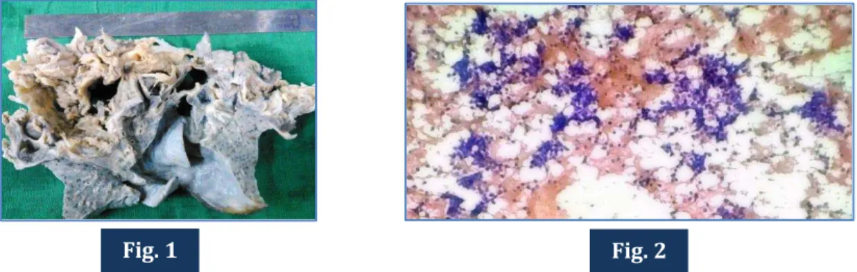
Fig. 1: Gross image of the pneumonectomy specimen received showing gray white friable areas representing the mass.
Fig. 2: FNAC from the mass showing small round blue cells tending to form rosettes at places (100x H & E).
Fig. 3: Histopathology showing bimodal population of small blue round cells and lighter staining larger cells. (400x H&E).
Fig. 4: Immunohistochemistry showing diffuse cytoplasmic CD 99 positivity.
AUTHORS:
1. Mahendra Singh 2. S. )ram Arshad 3. Sweta Shekhar 4. Chayanika Pantola
PARTICULARS OF CONTRIBUTORS:
1. Professor, Department of Pathology, GSVM Medical College, Kanpur.
2. Junior Resident, Department of Pathology, GSVM Medical College, Kanpur.
3. Junior Resident, Department of Pathology, GSVM Medical College, Kanpur.
FINANCIAL OR OTHER
COMPETING INTERESTS: None
4. Assistant Professor, Department of Pathology, GSVM Medical College, Kanpur.
NAME ADDRESS EMAIL ID OF THE CORRESPONDING AUTHOR:
Dr. S. Iram Arshad, Room No. 65, New PG Girls Hostel, GSVM Medical College,
Swaroopnagar, Kanpur-208002, Uttar Pradesh.
E-mail: [email protected]
Date of Submission: 13/06/2015. Date of Peer Review: 15/06/2015. Date of Acceptance: 13/07/2015. Date of Publishing: 20/07/2015.
Fig. 1 Fig. 2